- Division of Neurosurgery, Department of Neurosciences, Philippine General Hospital, College of Medicine, University of the Philippines Manila, Manila, Philippines.
Correspondence Address:
Juan Silvestre Grecia Pascual, Division of Neurosurgery, Department of Neurosciences, Philippine General Hospital, College of Medicine, University of the Philippines Manila, Manila, Philippines.
DOI:10.25259/SNI_405_2021
Copyright: © 2021 Surgical Neurology International This is an open-access article distributed under the terms of the Creative Commons Attribution-Non Commercial-Share Alike 4.0 License, which allows others to remix, tweak, and build upon the work non-commercially, as long as the author is credited and the new creations are licensed under the identical terms.How to cite this article: Juan Silvestre Grecia Pascual, Kevin Ivan Peñaverde Chan, Kathleen Joy Ong-Lopez Khu. Severe and persistent coronavirus disease 2019 cough resulting in bone flap displacement and pseudomeningocele. 12-Jul-2021;12:348
How to cite this URL: Juan Silvestre Grecia Pascual, Kevin Ivan Peñaverde Chan, Kathleen Joy Ong-Lopez Khu. Severe and persistent coronavirus disease 2019 cough resulting in bone flap displacement and pseudomeningocele. 12-Jul-2021;12:348. Available from: https://surgicalneurologyint.com/surgicalint-articles/10960/
Abstract
Background: Cough is one of the most common symptoms of coronavirus disease 2019 (COVID-19) infection. This relatively benign symptom may lead to serious sequelae, especially in postoperative neurosurgical patients.
Case Description: Here, we report a case of bone flap displacement, pseudomeningocele formation, and consequent cerebrospinal fluid leak in a patient with COVID-19 infection who recently underwent craniotomy for excision of cerebral metastasis. We highlight the pathophysiologic mechanisms of cough that may cause increased intracranial pressure (ICP), leading to the postoperative morbidity.
Conclusion: Aside from additional risks to the patient’s health and increased treatment costs, these complications also lead to subsequent delays in the management of the underlying disease. Symptomatic treatment of cough is advised to prevent complications resulting from increased ICP.
Keywords: Cancer, Complication, Coronavirus disease 2019, Cough, Pseudomeningocele
INTRODUCTION
Coronavirus disease 2019 (COVID-19) is a global pandemic affecting 132 million people worldwide as of this writing.[
CASE DESCRIPTION
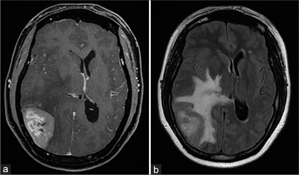
The patient is a 40-year-old female diagnosed with breast cancer in 2019. She underwent modified radical mastectomy in July 2019 and was on trastuzumab chemotherapy. She was referred to the neurosurgical service for a right parietal tumor on metastatic workup [
One week after discharge, the patient experienced severe and persistent cough that kept her awake at night. She self-medicated with antitussives with minimal relief. Three days later, she noted a gradually bulging fluid-filled mass at her postoperative site, as well as the sensation that there was something moving underneath the fluid collection. After a 2 more days, fluid leaked from the surgical incision, prompting the patient to consult at the emergency department.
On examination, the patient was awake, oriented, and able to follow commands. She did not have fever or nuchal rigidity. She had a pseudomeningocele over the postoperative site, with dehiscence of a portion of the inferior limb of the surgical incision and watery fluid draining from it. On palpation, the bone flap was found to be mobile and displaced inferiorly. As part of the hospital protocol for admission, the patient underwent a COVID-19 PCR test, which was positive. She was thus transferred to the COVID-19 isolation unit.
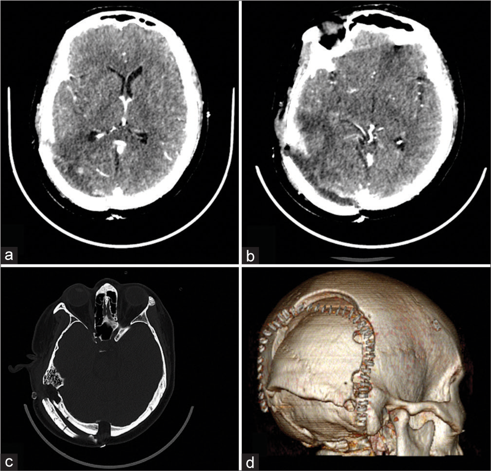
A contrast cranial computed tomography (CT) scan showed postoperative changes at the right parietal area and no evidence of enhancing tumor. There was also no evidence of hydrocephalus, subdural empyema, or brain abscess. The bone flap was displaced inferiorly [
Figure 2:
(a) Postoperative contrast cranial computed tomography (CT) showing postoperative changes at the right parietal area, no evidence of enhancing tumor, and no evidence of hydrocephalus; (b) same CT showing pseudomeningocele formation and outward displacement of the bone flap; (c) cranial CT, bone window, showing dislodged bone flap; (d) 3D reconstruction of the cranial CT highlighting the dislodged bone flap.
She was started on acetazolamide and mannitol to decrease CSF production and ICP. The leak site was sutured, and a lumbar drain was inserted to divert CSF and keep the postoperative site dry. The opening pressure at the time of lumbar drain insertion was normal at 7 cm H2O, likely due to the administration of mannitol, the CSF leak at the postoperative site, and the pseudomeningocele formation. Antibiotics were also given. The patient was quarantined in the COVID unit for 2 weeks and was only transferred to the regular ward after a negative COVID-19 PCR test.
The pseudomeningocele recurred after the lumbar drain was clamped, indicating failure of treatment. Thus, she underwent debridement, craniectomy, and duraplasty using fascia lata graft. Intraoperatively, the bone flap was found to be unsecured and displaced inferiorly, and the sutures securing the bone flap had become unraveled. A 3 cm × 4 cm dural defect was found along the margins of the previous dural repair, and a fascia lata graft was used to repair this. The bone flap was not reimplanted since the brain was swollen and herniating slightly past the craniectomy defect, likely due to cerebral edema from the infection as a consequence of the CSF leak. A new lumbar drain was inserted, then removed after a week. The patient’s postoperative site remained dry and flat, and she was discharged home.
On follow-up after 1 month, the patient was well, with no recurrence of the pseudomeningocele or CSF leak. She had no neurologic deficits and her cough had resolved completely.
DISCUSSION
COVID-19 infection has been shown to result in adverse outcomes in surgical patients, especially in terms of respiratory complications.[
Coughing is the sudden forceful expulsion of air through the large airways to clear them of irritants, particles, and fluids.[
In our case, we postulate that the repetitive increase in ICP caused by the severe persistent COVID-19-induced coughing led to a breakdown in the initial dural repair, causing CSF to leak out of the dura and accumulate under the scalp to form a pseudomeningocele. The severe and persistent coughing may have exerted pressure on the dural repair and unraveled the sutures. The continuous coughing further increased the size of the dural tear until it reached the dimensions seen during the surgery. This led to CSF leakage into the epidural space and accumulation beneath the bone flap. Subsequently, the increased pressure of the CSF probably exerted pressure on the overlying bone flap, unraveling the sutures securing the flap, and dislodging it inferiorly. In normal tissue, the pressures brought about by coughing would be transmitted evenly along the brain tissue and skull, but in a postoperative patient, pressures would intuitively be transmitted disproportionately toward the weakest point in the skull – that is, the postoperative site – resulting in the sequelae we have observed.[
Our case is important because it illustrates that COVID-19 infection and the cough associated with it may still cause problems even after the surgical procedure. Coughing may not be a benign symptom and may lead to similar types of adverse events. Symptomatic treatment of cough, especially those associated with COVID-19, is advised in postoperative neurosurgical patients to prevent adverse sequelae stemming from increased ICP.
CONCLUSION
Although cough may be thought of as a benign symptom of COVID-19, it may have adverse sequelae related to increase ICP. Symptomatic treatment of cough is advised in the postoperative neurosurgical patient to prevent complications resulting from increased ICP.
Ethics approval
Not applicable.
Declaration of patient consent
The authors certify that they have obtained all appropriate patient consent.
Financial support and sponsorship
Nil.
Conflicts of interest
There are no conflicts of interest.
Acknowledgments
None.
References
1. Donnelly J, Czosnyka M, Harland S, Varsos GV, Cardim D, Robba C. Increased ICP and its cerebral haemodynamic sequelae. Acta Neurochir Suppl. 2018. 126: 47-50
2. Jiang F, Deng L, Zhang L, Cai Y, Cheung CW, Xia Z. Review of the clinical characteristics of Coronavirus disease 2019 (COVID-19). J Gen Intern Med. 2020. 35: 1545-9
3. Knisely A, Zhou ZN, Wu J, Huang Y, Holcomb K, Melamed A. Perioperative morbidity and mortality of patients with COVID-19 who undergo urgent and emergent surgical procedures. Ann Surg. 2021. 273: 34-40
4. Williams B. Cerebrospinal fluid pressure: Changes in response to coughing. Brain. 1976. 96: 331-46
5. Weekly Operational Update on COVID-19. Available from: http://www.who.int/publications/m/item/weekly-update-on-covid-19 [Last assessed on 2020 Oct 16].


