- Department of Neurosurgery, Christian Medical College, Ludhiana, Punjab, India
DOI:10.25259/SNI_310_2020
Copyright: © 2020 Surgical Neurology International This is an open-access article distributed under the terms of the Creative Commons Attribution-Non Commercial-Share Alike 4.0 License, which allows others to remix, tweak, and build upon the work non-commercially, as long as the author is credited and the new creations are licensed under the identical terms.How to cite this article: Ashish Acharya, Sarvpreet Singh Grewal, Paul Sudhakar John, Ravindra Kumar Bind, Ankita Khurana. Solitary osteochondroma of dorsal spine causing canal stenosis with myelopathy – A case report with review of literature. Surg Neurol Int 18-Jul-2020;11:199
How to cite this URL: Ashish Acharya, Sarvpreet Singh Grewal, Paul Sudhakar John, Ravindra Kumar Bind, Ankita Khurana. Solitary osteochondroma of dorsal spine causing canal stenosis with myelopathy – A case report with review of literature. Surg Neurol Int 18-Jul-2020;11:199. Available from: https://surgicalneurologyint.com/surgicalint-articles/10143/
Abstract
Background: Osteochondroma is a common benign tumor arising from the long bones. It rarely arises in the spine, where it can cause mild symptoms such as backache all the way up to compressive myelopathy. Malignant transformation has also been reported. Here, the authors present a 52-year-old male with myelopathy attributed to a rare thoracic solitary osteochondroma.
Case Description: A 52-year-old male presented back pain radiating into both lower extremities with paresthesia to the toes of 1 year’s duration. On examination, he exhibited hyperactive bilateral lower extremity reflexes with bilateral Babinski signs, and focal sensory changes to pin, and touch appreciation in the left L5S1 distributions. Computed tomography and magnetic resonance imaging showed an abnormal bony mass arising from the posterior arch of T10 with protrusion into the spinal canal resulting in marked canal/cord compression. Surgery included a D10 laminectomy with en bloc resection of the lesion. Postoperatively, the patient’s symptoms resolved. Histologically, the lesion was an osteochondroma.
Conclusion: When patients present with myelopathy, one should include osteochondromas among the differential diagnostic possibilities.
Keywords: Osteochondroma, Spine, Spinal cord compression
INTRODUCTION
Solitary osteochondromas are the most common benign skeletal neoplasms arising from the long bones.[
CASE REPORT
A 52-year-old male presented with 1-year history of bilateral lower extremity radiculopathy with paraesthesia extending to the toes. The neurological examination revealed bilateral lower extremity hyperreflexia and Babinski responses plus a 10% loss of light touch in the left S1 distribution.
Thoracic Computed tomography (CT) and magnetic resonance (MR) Studies
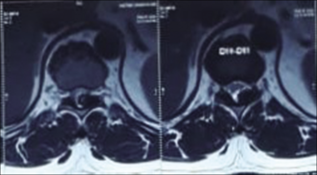
CT and MR examinations showed an abnormal bony mass arising from the posterior arch of T10 that protruded into the spinal canal, resulting in marked canal stenosis and cord compression. The cortex and medullary portions of the lesion were continuous with the T10 vertebral body (i.e., exostosis) [
Surgery
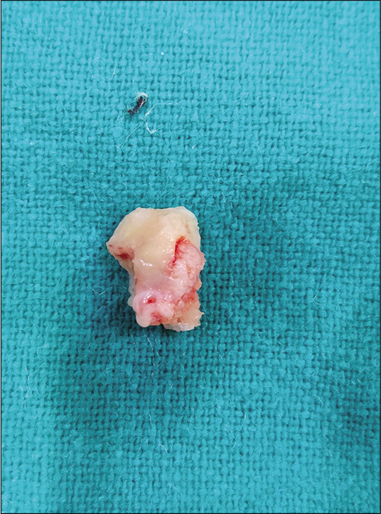
Surgery consisted of a bilateral T10 laminectomy for en bloc resection of the lesion, a predominantly bony mass originating from the left lamina of T10 that was (e.g., continuous with the inner surface of the T10 lamina on the left) [
Histopathology
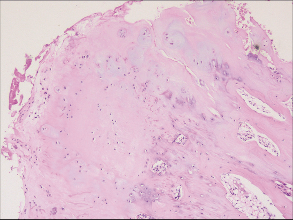
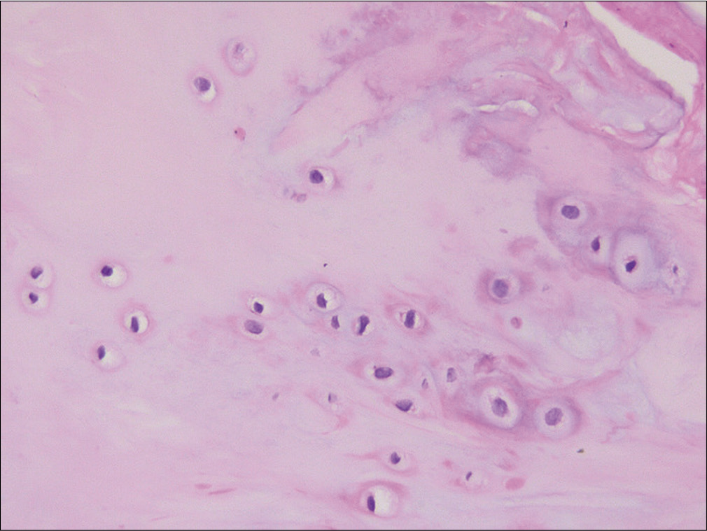
The histopathologic revealed a bony lesion with a cartilaginous cap covered with perichondrium, endochondral ossification continuous with bony trabeculae, and benign chondrocytes consistent with the diagnosis of osteochondroma [
DISCUSSION
Osteochondromas/exostoses comprise 2.6% of all benign tumors of the spine.[
Etiology of osteochondromas
Osteochondromas are developmental lesions rather than true neoplasms. They result from the separation of a portion of the epiphyseal growth plate cartilage, which herniate through the periosteum around the growth plate.[
Location and diagnosis of spinal osteochondromas with CT/MR
Spinal involvement is more common in HME, typically involving the laminae of the thoracic and lumbar vertebrae. Rarely, they exhibit extension into the spinal canal resulting in canal stenosis and myelopathy.
CONCLUSION
Here, we presented a patient with a rare left-sided T10 thoracic osteochondroma contributing to back pain, thoracic canal stenosis, and myelopathy. A decompressive laminectomy resulted in full resolution of the patient’s myelopathic deficit.
Declaration of patient consent
The authors certify that they have obtained all appropriate patient consent.
Financial support and sponsorship
Nil.
Conflicts of interest
There are no conflicts of interest.
References
1. Hassankhani EG. Solitary lower lumbar osteochondroma (spinous process of L3 involvement): A case report. Cases J. 2009. 2: 9359-
2. Lotfinia I, Vahedi P, Tubbs RS, Ghavame M, Meshkini A. Neurological manifestations, imaging characteristics, and surgical outcome of intraspinal osteochondroma: Clinical article. J Neurosurg Spine. 2010. 12: 474-89
3. Mehrian P, Karimi MA, Kahkuee S, Bakhshayeshkaram M, Ghasemikhah R. Solitary osteochondroma of the thoracic spine with compressive myelopathy; a rare presentation. Iran J Radiol. 2013. 10: 77-80
4. Murphey MD, Choi JJ, Kransdorf MJ, Flemming DJ, Gannon FH. Imaging of osteochondroma: Variants and complications with radiologic-pathologic correlation. Radiographics. 2000. 20: 1407-34
5. Shim JH, Park CK, Shin SH, Jeong HS, Hwang JH. Solitary osteochondroma of the twelfth rib with intraspinal extension and cord compression in a middle-aged patient. BMC Musculoskelet Disord. 2012. 13: 57-


