- Department of Neurosurgery, University of Oklahoma Health Sciences Center, Oklahoma City, Oklahoma, United States.
DOI:10.25259/SNI_3_2020
Copyright: © 2020 Surgical Neurology International This is an open-access article distributed under the terms of the Creative Commons Attribution-Non Commercial-Share Alike 4.0 License, which allows others to remix, tweak, and build upon the work non-commercially, as long as the author is credited and the new creations are licensed under the identical terms.How to cite this article: Camille K. Milton, Kyle P. O’Connor, Adam D. Smitherman, Andrew K. Conner, Michael D. Martin. Solitary osteochondroma of the cervical spine presenting with quadriparesis and hand contracture. Surg Neurol Int 21-Mar-2020;11:51
How to cite this URL: Camille K. Milton, Kyle P. O’Connor, Adam D. Smitherman, Andrew K. Conner, Michael D. Martin. Solitary osteochondroma of the cervical spine presenting with quadriparesis and hand contracture. Surg Neurol Int 21-Mar-2020;11:51. Available from: https://surgicalneurologyint.com/surgicalint-articles/9918/
Abstract
Background: Spinal osteochondromas are rare, benign tumors arising from the cartilaginous elements of the spine that may appear as solitary lesions versus multiple lesions in patients with hereditary multiple exostoses. Here, we present a 15-year-old female with a solitary C3-C4 osteochondroma who presented with a progressive quadriparesis and hand contracture successfully managed with a laminectomy/posterior spinal fusion.
Case Description: A 15-year-old female presented with a 3-month history of progressive quadriparesis and hand contracture secondary to a magnetic resonance (MR) documented C3-C4 cervical spine osteochondroma. The MR imaging revealed a solitary osseous extramedullary outgrowth arising from the left laminar cortex of the C-3 vertebral body extending to C-4. Due to the marked resultant canal stenosis, the patient underwent a cervical laminectomy of C3- C4 with posterior spinal fusion. Gross total resection was achieved, and the pathology confirmed an osteochondroma. The patient’s myelopathy resolved, and 2 years later, she demonstrated no residual deficits or tumor recurrence.
Conclusion: Here, we report the successful management of a 15-year-old female with a C3-C4 osteochondroma and progressive quadriparesis through cervical laminectomy/fusion.
Keywords: Hand contracture, Posterior spinal fusion, Quadriparesis, Spinal osteochondroma
INTRODUCTION
Osteochondromas of the cervical spine rarely contribute to spinal cord compression and resultant paresis.[
Here, we report the case of a 15-year-old female whose progressive quadriparesis, due to a solitary C3-C4 osteochondroma, was successfully managed with a laminectomy and posterior spinal fusion.
CASE REPORT
A 15-year-old female with a history of scoliosis presented with 3 months of progressive quadriparesis and sphincter dysfunction. On examination, she had a contracture of the left hand, and mild left upper extremity weakness (4/5 bicep/tricep, 3/5 wrist and handgrip). The right upper extremity (4+/5 bicep/tricep, 4/5 wrist and handgrip) and bilateral lower extremity function remained largely intact (4+/5–5/5). The patient reported diminished sensation to light touch in bilateral hands (C6-8 dermatomes), abdomen (T4-12 dermatomes), and the left lower extremity (L2-3 dermatomes).
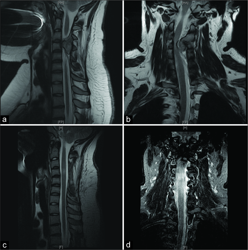
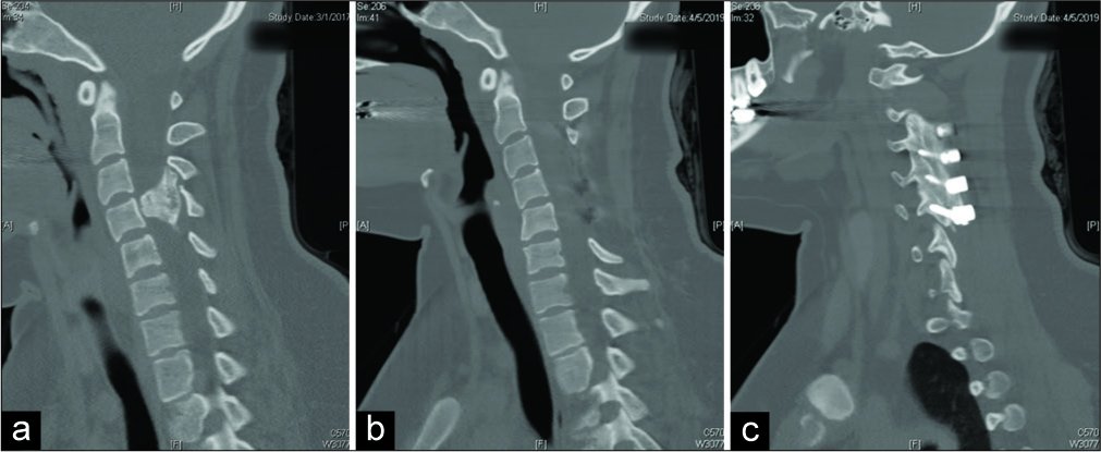
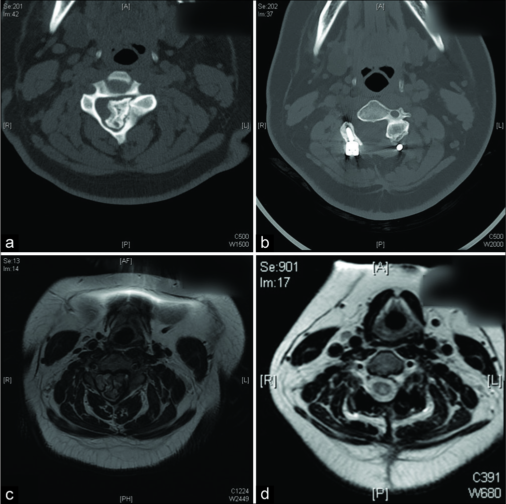
The cervical magnetic resonance imaging (MRI) showed an osseous extramedullary growth arising from the left laminar cortex of the C3 extending inferiorly to C4, resulting in marked cord/left foraminal compromise. At the level of C4, the marked T2 signal change of the cord confirmed significant edema. The patient successfully underwent a C3-C4 laminectomy with gross total tumor removal. The pathological evaluation demonstrated cartilaginous components consistent with osteochondroma. Postoperatively, she regained full function [
Figure 1:
Sagittal preoperative and postoperative magnetic resonance imaging (MRI). Interval cervical laminectomy of C3-5 with posterior spinal fusion of C2-3, C3-4, and C4-5 was performed. (a) Sagittal view of preoperative T2 MRI. (b) Coronal view of preoperative T2 MRI. Outgrowth from the left laminar cortex of the C3 vertebral body with inferior extension to the level of C4, resulting in severe spinal cord stenosis, compression of the thecal sac/cord, and moderate left foraminal narrowing is demonstrated. (c) Sagittal view of 3-month postoperative T2 MRI (left). (d) Coronal view of 3-month postoperative T2-STIR MRI (right). Resolution of cord compression is demonstrated.
DISCUSSION
Frequency and location of osteochondromas
Osteochondromas represent the most common benign tumor of bone and are only rarely found outside the appendicular skeleton.[
MR/computed tomography (CT) studies
MRI is the best imaging modality for these lesions, providing additional information regarding surrounding soft tissues and the degree of spinal cord compression. A CT scan may further demonstrate the osseous component of the tumor and the relationship between the tumor and the vertebrae.
Surgery
When these lesions are symptomatic, gross total resection should be performed to avoid malignant transformation or recurrence.[
CONCLUSION
A 15-year-old female presented with progressive quadriparesis and hand contracture secondary to a solitary osteochondroma of the cervical spine. Symptomatic and radiographic resolution was achieved following gross total resection of the mass with cervical laminectomy and fusion.
Declaration of patient consent
The authors certify that they have obtained all appropriate patient consent.
Financial support and sponsorship
Nil.
Conflicts of interest
There are no conflicts of interest.
References
1. Albrecht S, Crutchfield JS, SeGall GK. On spinal osteochondromas. J Neurosurg. 1992. 77: 247-52
2. Lotfinia I, Vahedi P, Tubbs RS, Ghavame M, Meshkini A. Neurological manifestations, imaging characteristics, and surgical outcome of intraspinal osteochondroma. J Neurosurg Spine. 2010. 12: 474-89
3. Schomacher M, Suess O, Kombos T. Osteochondromas of the cervical spine in atypical location. Acta Neurochir (Wien). 2009. 151: 629-33
4. Sciubba DM, Macki M, Bydon M, Germscheid NM, Wolinsky JP, Boriani S. Long-term outcomes in primary spinal osteochondroma: A multicenter study of 27 patients. J Neurosurg Spine. 2015. 22: 582-8
5. Veeravagu A, Li A, Shuer LM, Desai AM. Cervical osteochondroma causing myelopathy in adults: Management considerations and literature review. World Neurosurg. 2017. 97: 752.e5-752.e13
6. Zaijun L, Xinhai Y, Zhipeng W, Wending H, Quan H, Zhenhua Z. Outcome and prognosis of myelopathy and radiculopathy from osteochondroma in the mobile spine: A report on 14 patients. J Spinal Disord Tech. 2013. 26: 194-9


