- Department of Neurosurgery, Hospital Civil “Dr. Juan I. Menchaca,” Mexico.
- Department of Health Sciences, Autonomous University of Guadalajara, Guadalajara, Mexico.
- Department of Neurosurgery, Hospital Regional Valentin Gomez Farias Instituto de Seguridad y Servicios Sociales de los Trabajadores del Estado (ISSSTE), Zapopan, Mexico.
- Department of Radiology, Imaging and Diagnosis Center (CID), Guadalajara, Mexico.
Correspondence Address:
Sergio Valente Esparza Gutiérrez, Department of Neurosurgery, Hospital Civil “Dr. Juan I. Menchaca,” Guadalajara, Mexico.
DOI:10.25259/SNI_1127_2022
Copyright: © 2023 Surgical Neurology International This is an open-access article distributed under the terms of the Creative Commons Attribution-Non Commercial-Share Alike 4.0 License, which allows others to remix, transform, and build upon the work non-commercially, as long as the author is credited and the new creations are licensed under the identical terms.How to cite this article: Adrián Santana Ramírez1, Sergio Valente Esparza Gutiérrez1, Luz Monserrat Almaguer Ascencio1, Pedro Avila Rodríguez1, Omar Alejandro Santana Ortiz2, Rodrigo Fraga González3, Jesús Nicolás Serrano Heredia4. Solitary plasmacytoma of the sacrum treated with microwave ablation in conjunction with high dose of dexamethasone: A case report and review of the literature. 21-Apr-2023;14:145
How to cite this URL: Adrián Santana Ramírez1, Sergio Valente Esparza Gutiérrez1, Luz Monserrat Almaguer Ascencio1, Pedro Avila Rodríguez1, Omar Alejandro Santana Ortiz2, Rodrigo Fraga González3, Jesús Nicolás Serrano Heredia4. Solitary plasmacytoma of the sacrum treated with microwave ablation in conjunction with high dose of dexamethasone: A case report and review of the literature. 21-Apr-2023;14:145. Available from: https://surgicalneurologyint.com/surgicalint-articles/12278/
Abstract
Background: Plasma cell neoplasms are characterized by the neoplastic proliferation of a single clone of plasma cells. Solitary plasmacytomas most often occur in bone, but they can also be found in soft tissues.
Case Description: A 53-year-old male presented with localized sacral pain and urinary incontinence. His radiographic studies showed a solitary sacral plasmacytoma (i.e., involving the bone). He was successfully managed with high-dose dexamethasone and microwave ablation (MWA).
Conclusion: Plasmacytomas of bone can be occasionally successfully managed with MWA, adjuvant cytoreduction therapy, and high doses of dexamethasone.
Keywords: Dexamethasone cytoreduction, Microwave ablation, Plasmacytoma, Sacral plasmacytoma, Sacral tumor
INTRODUCTION
Plasma cell neoplasms are characterized by the neoplastic proliferation of a single clone of plasma cells. They may present as single (solitary plasmacytoma) or as multiple lesions multiple myeloma (MM). Solitary plasmacytomas most often occur in bone, but can also be found in soft tissues (i.e., extramedullary plasmacytoma). Sacral bony lesions add to the complexity of managing these tumors due to their attendant intrinsic/extrinsic structural, neural, and/or vascular concerns. Here, a 53-year-old male with a L5–S1–S2 plasmacytoma successfully underwent microwave ablation (MWA) and high dose of dexamethasone treatment.
CASE STUDY
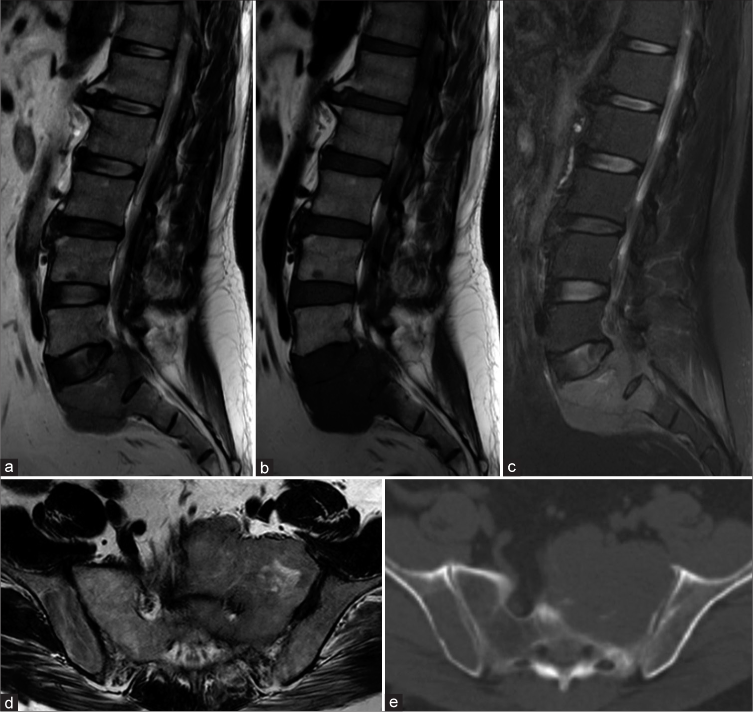
A 53-year-old male presented with lumbosacral pain for approximately 6 months’ duration, followed by the acute onset of a cauda equina syndrome (i.e., hypoesthesia in the genitals, paraparesis, and urinary incontinence). Both computed tomography and magnetic resonance imaging studies documented an erosive lytic L5–S1–S2 lesion that appeared to infiltrate and extension into the presacral space. On axial studies, tumor extended along the pelvic trajectory of the upper gluteal vessels and left iliac artery and vein. On the lateral images, it involved the first two sacral vertebrae, (S1–S2). There appeared to be no metastases [
Figure 1:
Preoperative sagittal magnetic resonance images (MRI) enhanced in T2 (a), T1 (b) and short-tau inversion recovery (STIR) (c); axial image displayed in T2 (d); and axial tomography image with a bone window image (e), where an infiltrative lesion is visualized in the S1 and S2 vertebrae as an expansive lytic bone lesion that is hypointense on T1 and T2 and hyperintense on STIR; the MRI presents an extraosseous soft-tissue component projected in the spinal canal and presacral space, encompassing and noticeably affecting the left L5 and S1 roots (d); note the characteristic absence of periosteal reaction in the tomography images (e).
Figure 2:
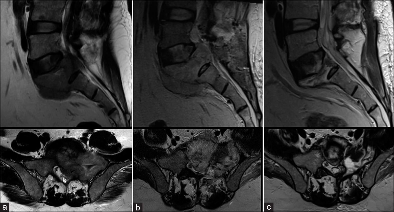
Sequential T2-weighted sagittal and axial magnetic resonance images; (a) Before treatment, (b) At 48 h postmicrowave ablation and (c) 4 months post-ablation. In the initial image before treatment, (a) the lesion is hypointense on images at 48 h and (b) the tumor with a slight increase in its size and a total change in its potency when it became hyperintense; in its control at 4 months, disappearance of the lytic lesion and the soft tissue component can be observed, with a noticeable decrease in the height of the S1 vertebra and formation of sclerotic areas in the bone.
Figure 3:
(a) Ablation antenna used in the patient’s surgical procedure; 14.5 gauge; Med Waves. (b) Image of the surgical procedure where the introduction of the antenna toward the sacral region can be seen. (c) Image obtained by fluoroscopy at the moment of the introduction of the antenna in one of the 5 vectors directed toward the sacral lesion.
Pathology
The histopathology revealed a proliferation of plasma cells suggestive of a plasmacytic plasmacytoma. This was further supported by special immunohistochemical studies; positive CD138 and CD45 membranes in neoplastic cells with negative CD20.
Postoperative course
Following the ablation, the patient was managed with high-dose dexamethasone (i.e., 3 cycles of 4 days of 40 mg/day with a 4-day break between cycles). He was also referred to oncology/hematology for further multidisciplinary management [
DISCUSSION
Plasma cell neoplasms are characterized by the neoplastic proliferation of a single plasma cell clone that typically produces monoclonal immunoglobulin. Single lesions occur most frequently in bones, but they can also be found in soft tissues (extramedullary plasmacytoma).[
MWA
MWA has become an important minimally invasive treatment option for different types of tumors, including bone tumors.[
CONCLUSION
Plasmacytoma is a rare lesion that can respond to microsurgical assisted ablation and adjuvant cytoreduction therapy using high-dose dexamethasone.
Declaration of patient consent
Patient’s consent not required as patient’s identity is not disclosed or compromised.
Financial support and sponsorship
Nil.
Conflicts of interest
There are no conflicts of interest.
Disclaimer
The views and opinions expressed in this article are those of the authors and do not necessarily reflect the official policy or position of the Journal or its management. The information contained in this article should not be considered to be medical advice; patients should consult their own physicians for advice as to their specific medical needs.
References
1. Avilés A, Huerta-Guzmán J, Delgado S, Fernández A, Díaz-Maqueo JC. Improved outcome in solitary bone plasmacytomata with combined therapy. Hematol Oncol. 1996. 14: 111-17
2. Finsinger P, Grammatico S, Chisini M, Piciocchi A, Foà R, Petrucci MT. Clinical features and prognostic factors in solitary plasmacytoma. Br J Haematol. 2016. 172: 554-60
3. Katodritou E, Speletas M, Pouli A, Tsitouridis J, Zervas K, Terpos E. Successful treatment of extramedullary plasmacytoma of the cavernous sinus using a combination of intermediate dose of thalidomide and dexamethasone. Acta Haematol. 2007. 117: 20-3
4. Khan MA, Deib G, Deldar B, Patel AM, Barr JS. Efficacy and safety of percutaneous microwave ablation and cementoplasty in the treatment of painful spinal metastases and myeloma. AJNR Am J Neuroradiol. 2018. 39: 1376-83
5. Kilciksiz S, Karakoyun-Celik O, Agaoglu FY, Haydaroglu A. A review for solitary plasmacytoma of bone and extramedullary plasmacytoma. Sci World J. 2012. 2012: 895765
6. Luna LP, Sankaran N, Ehresman J, Sciubba DM, Khan M. Successful percutaneous treatment of bone tumors using microwave ablation in combination with Zoledronic acid infused PMMA cementoplasty. J Clin Neurosci. 2020. 76: 219-25
7. Mignot F, Schernberg A, Arsène-Henry A, Vignon M, Bouscary D, Kirova Y. Solitary plasmacytoma: LenalidomideDexamethasone combined with radiation therapy improves progression-free survival and multiple myeloma-free survival. Int J Radiat Onco Biol Phys. 2019. p.
8. Rajkumar V. Diagnosis and Management of Solitary Extramedullary. Available from: https://www.uptodate.com/contents/diagnosis-and-management-of-solitary-extramedullary-plasmacytoma/printwww.uptodate.com [Last accessed on 2023 Apr 06].
9. Ramírez AS, Rodriguez PA, Gutiérrez SV, Ávila OG. Preliminary experience in the management of brain and skull-base tumors with microwave ablation; feasibility guided by ultrasound, report from 23 cases. Surg Neurol Int. 2019. 10: 17
10. Richard WT, Mary KG, Melania P, Andrea B, Woodrow W, David CH. Solitary plasmacytoma treated with radiotherapy: impact of tumor size on outcome. Int J Radiat Oncol Biol Phys. 2001. 50: 113-120
11. Richardson PG, Sonneveld P, Schuster MW, Irwin D, Stadtmauer EA, Facon T. Bortezomib or high-dose dexamethasone for relapsed multiple myeloma. N Engl J Med. 2005. 352: 2487-98
12. Sekiguchi Y, Asahina T, Shimada A, Imai H, Wakabayashi M, Sugimoto K. A case of extramedullary plasmablastic plasmacytoma successfully treated using a combination of thalidomide and dexamethasone and a review of the medical literature. J Clin Exp Hematop. 2013. 53: 21-8
13. Simon CJ, Dupuy DE, Mayo-Smith WW. Microwave ablation: Principles and applications. Radiographics. 2005. 25: S69-83
14. Wang Y, Li H, Liu C, Chen C, Yan J, editors. Solitary Plasmacytoma of Bone of the Spine. SPINE. 2018. 44: E117-E125


