- Department of Neurosurgery, All India Institute of Medical Sciences, Raipur, Chhattisgarh, India.
- Department of Pediatric Endocrinology, Bai Jerbai Wadia Hospital for Children, Mumbai, Maharashtra, India.
Correspondence Address:
Charandeep Singh Gandhoke
Department of Pediatric Endocrinology, Bai Jerbai Wadia Hospital for Children, Mumbai, Maharashtra, India.
DOI:10.25259/SNI_142_2021
Copyright: © 2021 Surgical Neurology International This is an open-access article distributed under the terms of the Creative Commons Attribution-Non Commercial-Share Alike 4.0 License, which allows others to remix, tweak, and build upon the work non-commercially, as long as the author is credited and the new creations are licensed under the identical terms.How to cite this article: Anil Kumar Sharma1, Charandeep Singh Gandhoke1, Simran Kaur Syal2. Spontaneous regression of herniated cervical disc: A case report and literature review. 08-Apr-2021;12:141
How to cite this URL: Anil Kumar Sharma1, Charandeep Singh Gandhoke1, Simran Kaur Syal2. Spontaneous regression of herniated cervical disc: A case report and literature review. 08-Apr-2021;12:141. Available from: https://surgicalneurologyint.com/surgicalint-articles/10702/
Abstract
Background: We have reviewed 75 cases plus our own single instance of spontaneous regression of herniated cervical discs.
Methods: We searched PubMed and EMBASE databases (until September 2020) utilizing the following keywords; “spontaneous regression,” “herniated cervical disc,” and “Magnetic Resonance Imaging (MRI) studies.”
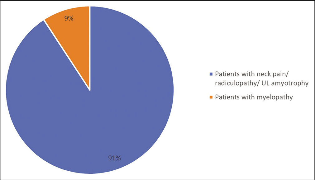
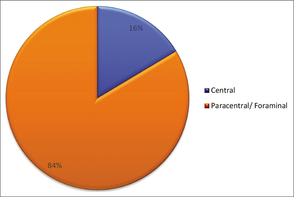
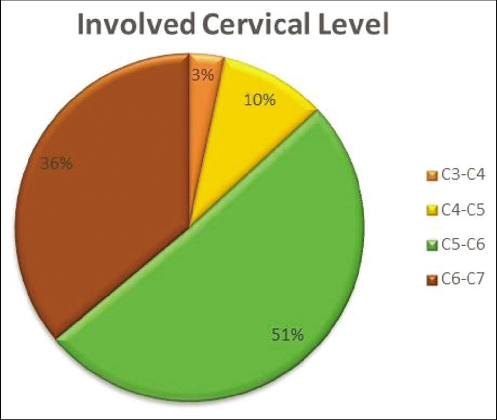
Results: In the literature, we found 75 cases of herniated cervical discs which spontaneously regressed; to this, we added our case. Patients averaged 40.95 years of age. Discs were paracentral or foraminal in 84% of the cases, with most occurring at the C5-C6 (51%) and C6-C7 (36%) levels. Symptoms included neck pain/radiculopathy (91%) or myelopathy (9%). The average interval between initial presentation and spontaneous regression of herniated discs on MRI was 9.15 months. Interestingly, on MRI, extruded/sequestrated discs were more likely to undergo spontaneous regression versus protruding discs.
Conclusion: Successive MRI studies documented the spontaneous regression of herniated cervical discs over an average of 9.15 months. Although this may prompt greater consideration for conservative treatment in younger patients without neurologic deficits, those with deficits should be considered for surgery.
Keywords: Extruded, Foraminal, Herniated cervical disc, Paracentral, Spontaneous regression
INTRODUCTION
Spontaneous regression of herniated lumbar disc has been well established in the literature, but, the phenomenon of spontaneous regression of herniated cervical discs has not been as thoroughly documented. Here, we focused on the 75 cases of spontaneous regression of herniated cervical discs from the literature and added our own experience with one patient.
CASE ILLUSTRATION
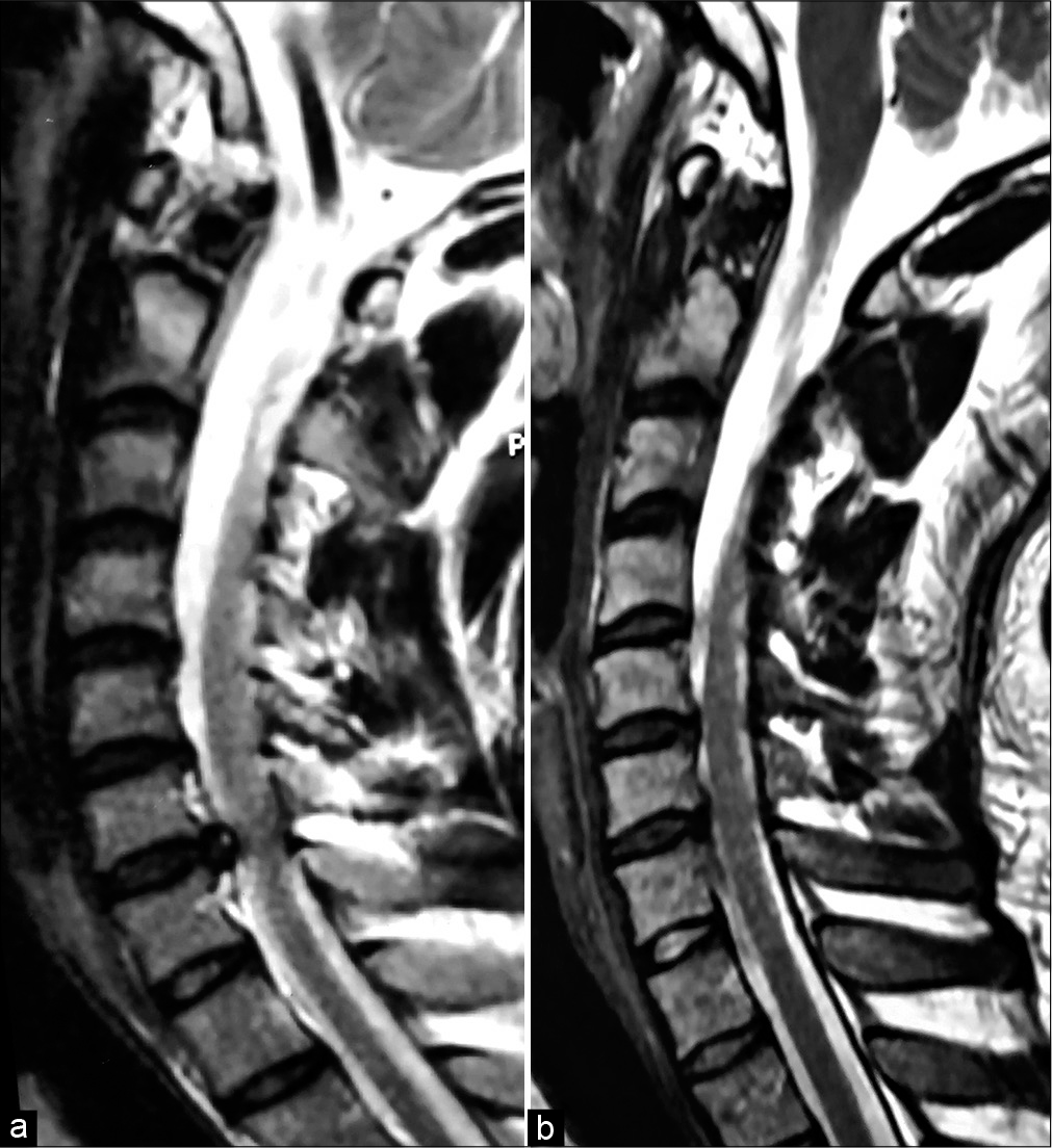
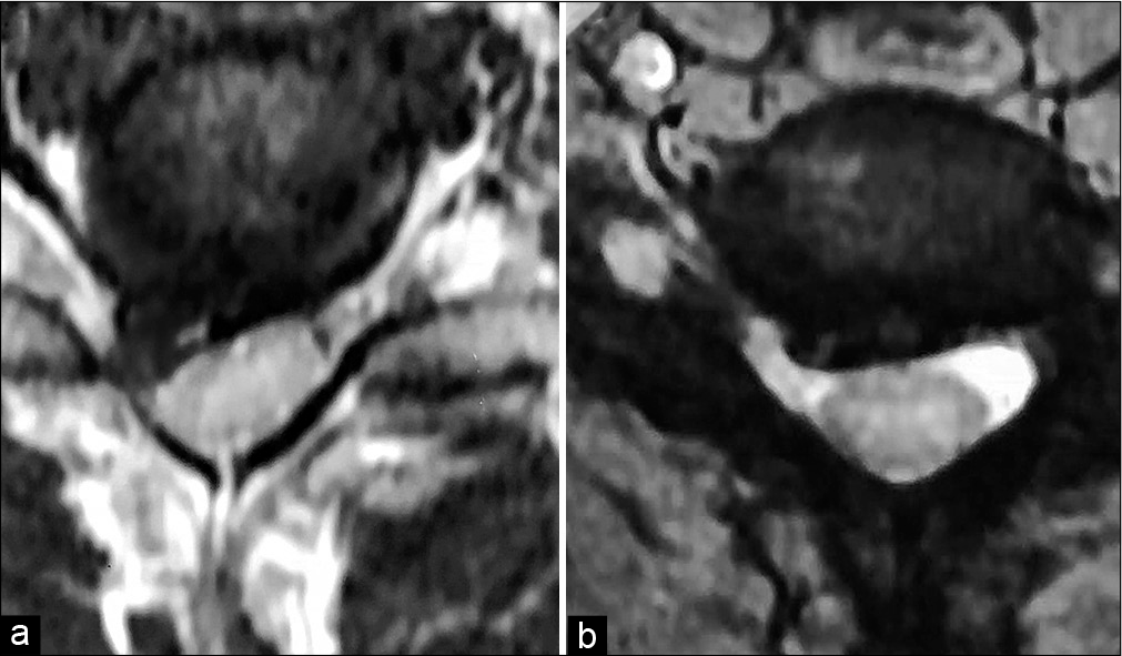
A 24-year-old male presented with 3 weeks’ duration of severe neck pain, right upper extremity radicular pain, and right C7 distribution weakness/numbness. The cervical MRI showed a right paracentral disc extrusion at the C6-C7 level resulting in the anterolateral cord and right C7 root compression [
Figure 2:
(a) MRI cervical spine, axial view, suggestive of a posterior disc extrusion at the C6-C7 level in the right paracentral location indenting the cervical spinal cord and the exiting right C7 root. (b) Follow-up MRI cervical spine, axial view, 3 months later revealed significant spontaneous regression of the C6-C7 intervertebral disc extrusion.
LITERATURE REVIEW
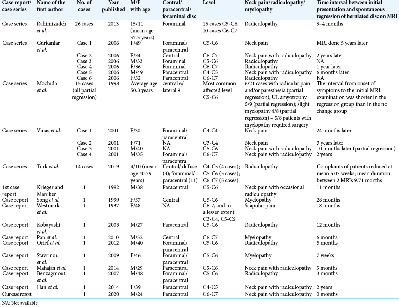
A literature search utilizing PubMed and EMBASE (i.e., until September 2020); using the keywords; “spontaneous regression,” “herniated cervical disc,” and “MRI studies” was performed. We identified 75 cases of the spontaneous regression of cervical disc herniations (CDH) to which we added our one case based on successive MRI studies [
Typical clinical presentation of patients with cervical disc herniations that resorbed
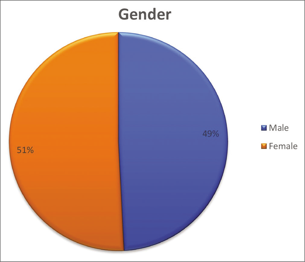
Here, we have summarized the typical clinical presentations of 76 patients with cervical disc herniations that regressed. Patients averaged 40.95 years of age and included equal numbers of males and females [
DISCUSSION
Mechanism of cervical disc resorption
There are three proposed mechanisms for spontaneous regression of CDH. The first involves dehydration and shrinkage of the herniated nucleus pulposus.[
MRI studies in cervical disc resorption
Extruded/sequestrated cervical discs on MRI showing rim enhancement with gadolinium are the most likely to regress.[
CONCLUSION
We have evaluated 76 patients with cervical disc herniations that regressed on successive MRI studies over an average period of 9.15 months. Those CDHs most likely to regress were extruded or sequestrated lesions, paracentral or foraminal in location, that demonstrated peripheral rim enhancement on gadolinium-enhanced MRI studies.
Declaration of patient consent
Patient’s consent not required as patients identity is not disclosed or compromised.
Financial support and sponsorship
Nil.
Conflicts of interest
There are no conflicts of interest.
References
1. Autio RA, Karppinen J, Niinimäki J, Ojala R, Kurunlahti M, Haapea M. Determinants of spontaneous resorption of intervertebral disc herniations. Spine (Phila Pa 1976). 2006. 31: 1247-52
2. Burke JG, Watson RW, McCormack D, Dowling FE, Walsh MG, Fitzpatrick JM. Spontaneous production of monocyte chemoattractant protein-1 and interleukin-8 by the human lumbar intervertebral disc. Spine (Phila Pa 1976). 2002. 27: 1402-7
3. Doita M, Kanatani T, Ozaki T, Matsui N, Kurosaka M, Yoshiya S. Influence of macrophage infiltration of herniated disc tissue on the production of matrix metalloproteinases leading to disc resorption. Spine (Phila Pa 1976). 2001. 26: 1522-7
4. Gurkanlar D, Yucel E, Er U, Keskil S. Spontaneous regression of cervical disc herniations. Minim Invasive Neurosurg. 2006. 49: 179-83
5. Han SR, Choi CY. Spontaneous regression of cervical disc herniation: A case report. Korean J Spine. 2014. 11: 235-7
6. Kobayashi N, Asamoto S, Doi H, Ikeda Y, Matusmoto K. Spontaneous regression of herniated cervical disc. Spine J. 2003. 3: 171-3
7. Krieger AJ, Maniker AH. MRI-documented regression of herniated cervical nucleus pulposus: A case report. Surg Neurol. 1992. 37: 457-9
8. Mochida K, Komori H, Okawa A, Muneta T, Haro H, Shinomiya K. Regression of cervical disc herniation observed on magnetic resonance images. Spine (Phila Pa 1976). 1998. 23: 990-7
9. Orief T, Orz Y, Attia W, Almusrea K. Spontaneous resorption of sequestrated intervertebral disc herniation. World Neurosurg. 2012. 77: 146-52
10. Pan H, Xiao LW, Hu QF. Spontaneous regression of herniated cervical disc fragments and its clinical significance. Orthop Surg. 2010. 2: 77-9
11. Rahimizadeh A, Hamidifard A, Rahimizadeh S. Spontaneous regression of the sequestrated cervical discs: A prospective study of 26 cases and review of the literature. World Spinal Column J. 2013. 4: 32-41
12. Song JH, Park HK, Shin KM. Spontaneous regression of a herniated cervical disc in a patient with myelopathy. Case report. J Neurosurg. 1999. 90: 138-40
13. Stavrinou LC, Stranjalis G, Maratheftis N, Bouras T, Sakas DE. Cervical disc, mimicking nerve sheath tumor, with rapid spontaneous recovery: A case report. Eur Spine J. 2009. 18: 176-8
14. Turk O, Yaldiz C. Spontaneous regression of cervical discs: Retrospective analysis of 14 cases. Medicine (Baltimore). 2019. 98: e14521
15. Vinas FC, Wilner H, Rengachary S. The spontaneous resorption of herniated cervical discs. J Clin Neurosci. 2001. 8: 542-6
16. Westmark RM, Westmark KD, Sonntag VK. Disappearing cervical disc. Case report. J Neurosurg. 1997. 86: 289-90
17. Yamashita K, Hiroshima K, Kurata A. Gadolinium-DTPA-enhanced magnetic resonance imaging of a sequestered lumbar intervertebral disc and its correlation with pathologic findings. Spine (Phila Pa 1976). 1994. 19: 479-82


