- Lab of Medical Physics School of Medicine, Faculty of Health Sciences, Thessaloniki, Greece
- Department of Neurosurgery, AHEPA University General Hospital, Aristotle University of Thessaloniki, Thessaloniki, Greece
DOI:10.25259/SNI-91-2019
Copyright: © 2019 Surgical Neurology International This is an open-access article distributed under the terms of the Creative Commons Attribution-Non Commercial-Share Alike 4.0 License, which allows others to remix, tweak, and build upon the work non-commercially, as long as the author is credited and the new creations are licensed under the identical terms.How to cite this article: Alkinoos Athanasiou, Ioannis Magras. Syringomyelia resolution after anterior cervical discectomy: A case report and literature review. 26-Mar-2019;10:42
How to cite this URL: Alkinoos Athanasiou, Ioannis Magras. Syringomyelia resolution after anterior cervical discectomy: A case report and literature review. 26-Mar-2019;10:42. Available from: http://surgicalneurologyint.com/surgicalint-articles/9242/
Abstract
Background: Syringomyelia is rarely associated with cervical disc herniations and/or spinal stenosis.
Case Description: A 62-year-old male presented with a 4-month history of right brachial pain and hyposensitivity in the C5 distribution. The cervical magnetic resonance (MR) imaging scan revealed a C5–C6 right anterolateral disc herniation with syringomyelia extending from C5–C6 to T1. Following a C5–C6 anterior cervical discectomy and fusion (ACDF), the patient’s symptoms resolved. The 3-month postoperative MR documented total resolution of the syrinx. Notably, due to residual neuropathic pain, the patient required a subdural spinal cord stimulator which was placed without any complications.
Conclusion: Syringomyelia rarely occurs in conjunction with cervical disc disease and stenosis, and even more infrequently resolves following an ACDF. Future research should focus on the etiology of syrinx formation in these patients and should explore their response to various treatment modalities.
Keywords: Anterior cervical discectomy and fusion, cervical disc herniation, cervical spondylosis, spondylotic myelopathy, syringomyelia
INTRODUCTION
Syringomyelia is usually attributed to Chiari malformations, spinal arachnoiditis, intramedullary tumors, and trauma. It is rarely due to cervical spondylosis, stenosis, or disc disease associated with myelopathy and/or radiculopathy.[
CASE REPORT
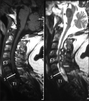
A 62-year-old male presented with a 4-month history of right brachial/shoulder pain and numbness with hyposensitivity in the C5 distribution. The magnetic resonance imaging (MRI) scan showed: (a) degenerative cervical spondylosis from the C3–C4 to C5–C6 levels, (b) a C5–C6 right anterolateral disc herniation with foraminal stenosis, and (c) syringomyelia extending from the C5–C6 to the T1 level [
Figure 1
Preoperative magnetic resonance imaging of the cervical spine of the patient (left: sagittal unenhanced T1-weighted, right: sagittal T2-weighted) showing right centrolateral C5–C6 disc herniation, cervical spondylosis from C3–C4 to C6–C6, and a syringomyelia cavity (marked “S”) extending from C6–C7 to T1 levels.
Surgery
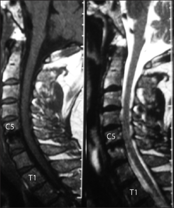
Following a C5–C6 anterior cervical discectomy and fusion (ACDF) with placement of a polyetheretherketone cage, the patient’s symptoms markedly improved. 3 months later, the MRI scan confirmed not only adequate decompression of the spinal cord but also the total resolution of the syrinx [
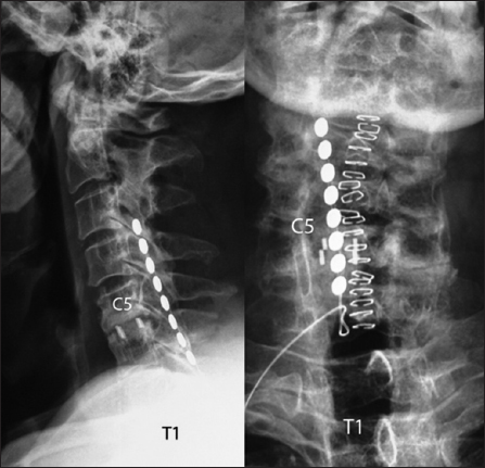
Nevertheless, for residual, non-dermatomal right cervical neuropathic pain, the patient underwent placement of an eight-electrode subdural spinal cord stimulator placed from the C3 to C4 through the C5–C6 levels; the patient significantly improved [
DISCUSSION
Syringomyelia is usually attributed to Chiari malformations, spinal arachnoiditis, intramedullary tumors, and trauma. It is seldom associated with cervical disc disease or spondylosis.[
CONCLUSION
Syringomyelia associated with cervical disc disease/spondylosis is rare. Here, we present a patient, who following a C5–C6 ACDF achieved complete resolution of the accompanying syrinx along with significant symptomatic improvement.
Declaration of patient consent
The authors certify that they have obtained all appropriate patient consent forms. In the form the patient has given his consent for his images and other clinical information to be reported in the journal. The patients understand that their names and initials will not be published and due efforts will be made to conceal their identity, but anonymity cannot be guaranteed.
Financial support and sponsorship
Nil.
Conflicts of interest
There are no conflicts of interest.
References
1. Ball JR, Little NS. Chiari malformation, cervical disc prolapse and syringomyelia-always think twice. J Clin Neurosci. 2008. 15: 474-6
2. Butteriss DJ, Birchall D. A case of syringomyelia associated with cervical spondylosis. Br J Radiol. 2006. 79: e123-5
3. Kimura R, Park YS, Nakase H, Sakaki T. Syringomyelia caused by cervical spondylosis. Acta Neurochir (Wien). 2004. 146: 175-8
4. Klekamp J. Surgical treatment of multilevel cervical spondylosis in patients with or without a history of syringomyelia. Eur Spine J. 2017. 26: 948-57
5. Landi A, Nigro L, Marotta N, Mancarella C, Donnarumma P, Delfini R. Syringomyelia associated with cervical spondylosis: A rare condition. World J Clin Cases. 2013. 1: 111-5
6. Pillich D, El Refaee E, Mueller JU, Safwat A, Schroeder HW, Baldauf J. Syringomyelia associated with cervical spondylotic myelopathy causing canal stenosis. A rare association. Neurol Neurochir Pol. 2017. 51: 471-5
7. Venkata SS, Arimappamagan A, Lafazanos S, Pruthi N. Syringomyelia secondary to cervical spondylosis: Case report and review of literature. J Neurosci Rural Pract. 2014. 5: S78-82
8. Yaman ME, Eylen A, Ayberk G. Resolution of isolated syringomyelia after treatment of cervical disc herniation: Association or coincidence?. Bratisl Lek Listy. 2012. 113: 500-2


