- Department of Neurosurgery, Allen Hospital, Unitypoint Clinic, Waterloo, Iowa, United States.
- Department of Pathology, Allen Hospital, Unitypoint Clinic, Waterloo, Iowa, United States.
Correspondence Address:
Nikolay L. Martirosyan, Department of Neurosurgery, Allen Hospital, Unitypoint Clinic, Waterloo, Iowa, United States.
DOI:10.25259/SNI_643_2021
Copyright: © 2021 Surgical Neurology International This is an open-access article distributed under the terms of the Creative Commons Attribution-Non Commercial-Share Alike 4.0 License, which allows others to remix, tweak, and build upon the work non-commercially, as long as the author is credited and the new creations are licensed under the identical terms.How to cite this article: Daniel Buchanan1, Nikolay L. Martirosyan1, Wei Yang2, Russell I. Buchanan1. Thoracic meningioma with ossification: Case report. 06-Oct-2021;12:505
How to cite this URL: Daniel Buchanan1, Nikolay L. Martirosyan1, Wei Yang2, Russell I. Buchanan1. Thoracic meningioma with ossification: Case report. 06-Oct-2021;12:505. Available from: https://surgicalneurologyint.com/?post_type=surgicalint_articles&p=11155
Abstract
Background: The incidence of spinal meningiomas is 0.33/100000 population, and ossified spinal meningiomas are even less commonly encountered.
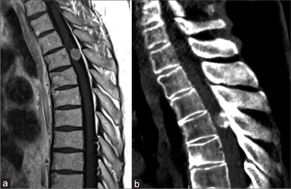
Case Description: A 64-year-old male presented with a progressive T4-level thoracic myelopathy. MR imaging revealed an intradural extramedullary mass that significantly compressed the spinal cord. The accompanying CT demonstrated hyperdensities within the lesion consistent with punctate calcification vs. ossification (i.e. consistent with histological bone formations within tumor). The patient underwent complete resection of the tumor resulting in a full recovery of neurological function within 6 postoperative weeks. The pathological specimen showed findings consistent with an ossified spinal meningioma.
Conclusion: Here, we identified a rare case of an ossified thoracic T4 meningioma occurring in a 64-year-old male.
Keywords: Myelopathy, Ossification, Spinal meningioma, Spine, Tumor
INTRODUCTION
A quarter of spinal tumors are meningiomas, and over 90% of them are benign.[
CASE REPORT
A 64-year-old male presented with a progressive T4-level paraparesis characterized by progressive numbness below the waistline, weakness in both lower extremities, and ataxia of gait. His neurological examination showed diffuse 4/5 bilateral lower extremity weakness with a relative T4-sensory level to pin appreciation.
Imaging
The thoracic MRI revealed a large right-sided dorsal intradural extramedullary lesion contributing to severe compression of the spinal cord at T4-level. The CT scan confirmed the lesion was hyperdense, consisting of intratumoral ossification [
Surgery
Under neuromonitoring and following a T4-T5 laminectomy, a midline durotomy was performed. This revealed an intradural extramedullary tumor with a base adherent to the right lateral dura. The tumor was dissected off the dura allowing for gross total resection (GTR); the sensory rootlets enmeshed in the tumor capsule were easily dissected off the lesion and preserved. A watertight closure followed, and there were no intraoperative neuromonitoring changes. Within 6 postoperative months, the patient was neurologically intact except for some mild residual gait ataxia, (i.e. requiring a cane to ambulate).
Surgical pathology
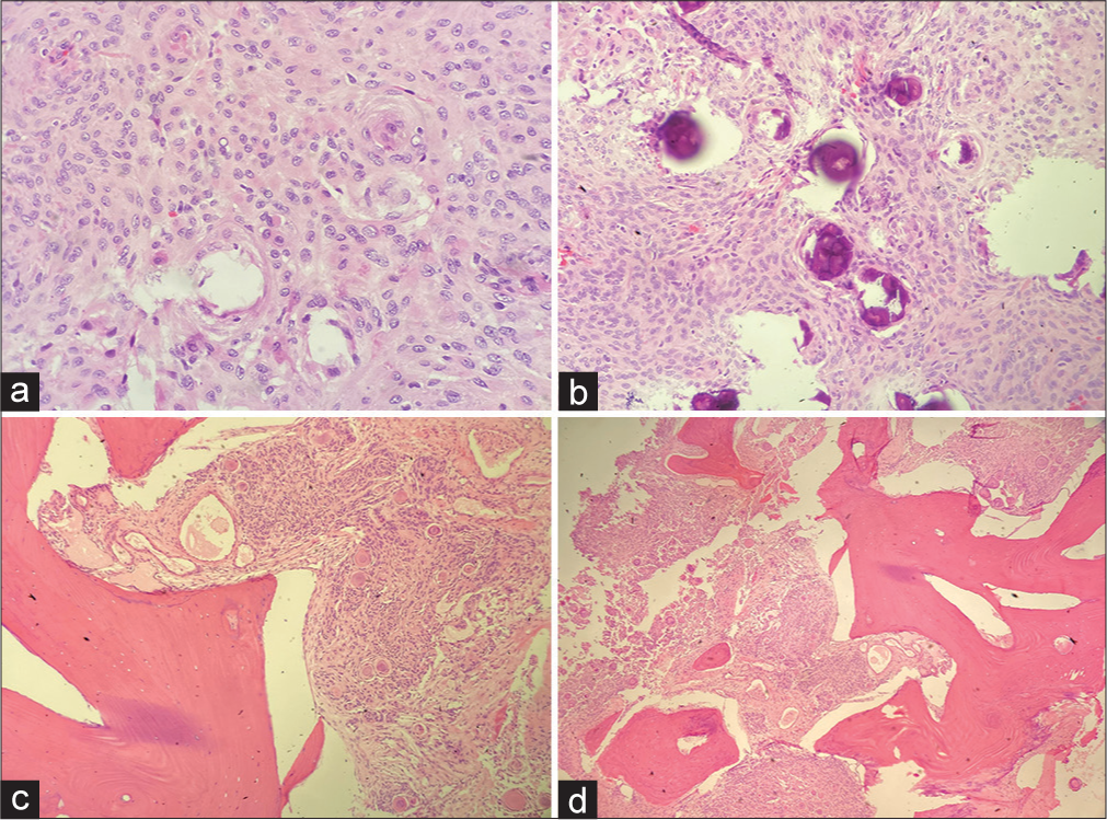
Gross pathology showed the lesion was irregular, tan, and rubbery, measuring 9 × 10 × 13 mm. On microscopy, there were meningothelial cells with oval to spindle-shaped nuclei containing occasional intranuclear pseudoinclusions. Frequent swirls of psammoma bodies were also seen. Additional areas showed more extensive “ossification” (i.e. bone formation, osseous metaplasia). As the tumor showed little mitotic activity, and there were no areas of hypercellularity, the final diagnosis was for a WHO grade I (benign) meningioma [
DISCUSSION
One percent of spinal meningiomas are ossified. The majority occur in females[
Incidence and prognosis for ossified spinal meningiomas
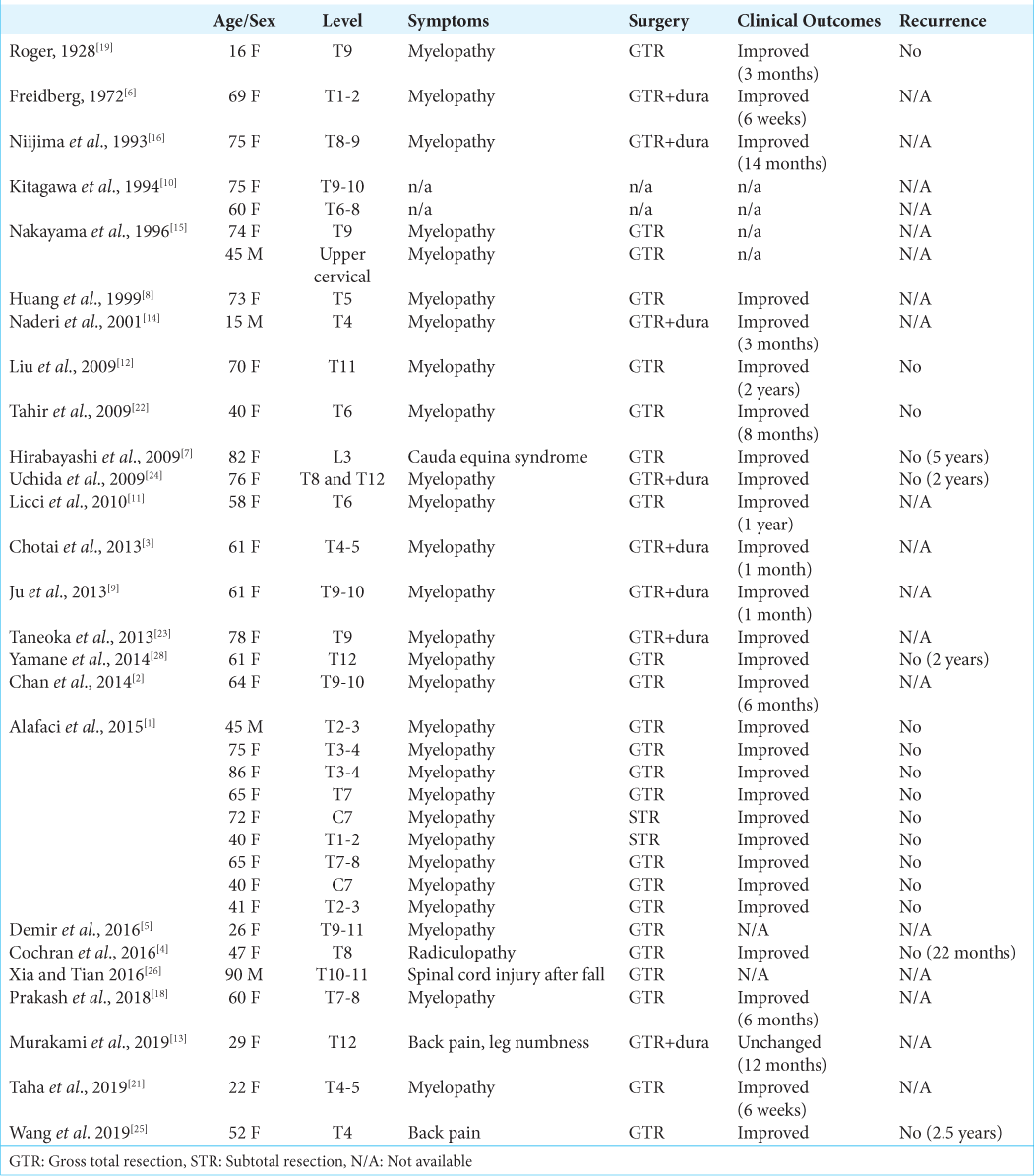
Of the 35 ossified meningiomas identified in the literature, only four occurred in males[
Although most cases reported favorable outcomes, others reported major perioperative morbidities, including paraplegia, complete sensory loss, cerebrospinal fluid leakage, and stroke. Despite these complications, following appropriate treatment/ medication, many patients sustained adequate recoveries.[
Symptom onset of ossified spinal meningiomas
Most of the 35 cases of ossified meningiomas presented with progressive myelopathy that worsened over a prolonged period. The clinical presentation was nonspecific and slow-progressing, therefore raise patients’ concern only when severely symptomatic. MRI identifies the size/extent of the mass, and CT studies are utilized to identify small calcifications/ossification. However, imaging modalities are unable to differentiate between ossification and calcification. The final diagnosis is made based on histopathologic evaluation.
Prediction of local recurrence
Only 17 cases had reported long-term follow up, and none of the patients had a recurrence. Interestingly, there was no recurrence in patients with subtotal resection [
CONCLUSION
Only 1% of spinal meningiomas are ossified, and few occur in males. Here we present a 64-year-old male with a T4 ossified meningomas responsible for a thoracic paraparesis that resolved following gross total tumor resection.
Declaration of patient consent
The authors certify that they have obtained all appropriate patient consent.
Financial support and sponsorship
Nil.
Conflicts of interest
There are no conflicts of interest.
Declaration of patient consent
The authors certify that they have obtained all appropriate patient consent.
Financial support and sponsorship
Nil.
Conflicts of interest
There are no conflicts of interest.
References
1. Alafaci C, Grasso G, Granata F, Salpietro FM, Tomasello F. Ossified spinal meningiomas: Clinical and surgical features. Clin Neurol Neurosurg. 2016. 142: 93-7
2. Chan TT, Lau VW, Chau TK, Lee YL. Ossified thoracic spinal meningioma with lamellar bone formation presented with paraparesis. J Orthop Trauma Rehabil. 2014. 18: 106-9
3. Chotai SP, Mrak RE, Mutgi SA, Medhkour A. Ossification in an extra-intradural spinal meningioma-pathologic and surgical vistas. Spine J. 2013. 13: e21-6
4. Cochran EJ, Schlauderaff A, Rand SD, Eckardt GW, Kurpad S. Spinal osteoblastic meningioma with hematopoiesis: Radiologic-pathologic correlation and review of the literature. Ann Diagn Pathol. 2016. 24: 30-4
5. Demir MK, Yapicier Ö, Toktaş ZO, Akakin A, Yilmaz B, Konya D. Ossified-calcified intradural and extradural thoracic spinal meningioma with neural foraminal extension. Spine J. 2016. 16: e35-7
6. Freidberg SR. Removal of an ossified ventral thoracic meningioma. Case report. J Neurosurg. 1972. 37: 728-30
7. Hirabayashi H, Takahashi J, Kato H, Ebara S, Takahashi H. Surgical resection without dural reconstruction of a lumbar meningioma in an elderly woman. Eur Spine J. 2009. 18: 232-5
8. Huang TY, Kochi M, Kuratsu JI, Ushio Y. Intraspinal osteogenic meningioma: Report of a case. J Formos Med Assoc. 1999. 98: 218-21
9. Ju CI, Hida K, Yamauchi T, Houkin K. Totally ossified metaplastic spinal meningioma. J Korean Neurosurg Soc. 2013. 54: 257-60
10. Kitagawa M, Nakamura T, Aida T, Iwasaki Y, Abe H, Nagashima K. Clinicopathologic analysis of ossification in spinal meningioma. Noshuyo Byori. 1994. 11: 115-9
11. Licci S, Limiti MR, Callovini GM, Bolognini A, Gammone V, di Stefano D. Ossified spinal tumour in a 58-year-old woman with increasing paraparesis. Neuropathology. 2010. 30: 194-6
12. Liu CL, Lai PL, Jung SM, Liao CC. Thoracic ossified meningioma and osteoporotic burst fracture: Treatment with combined vertebroplasty and laminectomy without instrumentation-case report. J Neurosurg Spine. 2006. 4: 256-9
13. Murakami T, Tanishima S, Takeda C, Kato S, Nagashima H. Ossified metaplastic spinal meningioma without psammomatous calcification: A case report. Yonago Acta Med. 2019. 62: 232-5
14. Naderi S, Yilmaz M, Canda T, Acar Ü. Ossified thoracic spinal meningioma in childhood: A case report and review of the literature. Clin Neurol Neurosurg. 2001. 103: 247-9
15. Nakayama N, Isu T, Asaoka K, Harata T, Hayashi S, Aoki T. Two cases of ossified spinal meningioma. Neurol Surg. 1996. 24: 351-5
16. Niijima K, Huang YP, Malis LI, Sachdev VP. Ossified spinal meningioma en plaque. Spine (Phila Pa 1976). 1993. 18: 2340-3
17. O’Brien EJ, Frank CB, Shrive NG, Hallgrímsson B, Hart DA. Heterotopic mineralization (ossification or calcification) in tendinopathy or following surgical tendon trauma. Int J Exp Pathol. 2012. 93: 319-31
18. Prakash A, Mishra S, Tyagi R, Bhatnagar A, Kansal S, Attri P. Thoracic psammomatous spinal meningioma with osseous metaplasia: A very rare case report. Asian J Neurosurg. 2015. 12: 270-2
19. Rogers L. A spinal meningioma containing bone. Br J Surg. 1928. 15: 675-7
20. Solero CL, Fornari M, Giombini S, Lasio G, Oliveri G, Cimino C. Spinal meningiomas: Review of 174 operated cases. Neurosurgery. 1989. 25: 153-60
21. Taha MM, Alawamry A, Abdel-Aziz HR. Ossified spinal meningioma: A case report and a review of the literature. Surg J (NY). 2019. 5: e137-41
22. Tahir M, Usmani N, Ahmad FU, Salmani S, Sharma MS. Spinal meningioma containing bone: A case report and review of literature. BMJ Case Rep. 2009. 2009: bcr11.2008.1186
23. Taneoka A, Hayashi T, Matsuo T, Abe K, Kinoshita N, Yasui H. Ossified thoracic spinal meningioma with hematopoiesis: A case report and review of the literature. Case Rep Clin Med. 2013. 2: 24-8
24. Uchida K, Nakajima H, Yayama T, Sato R, Kobayashi S, Mwaka ES. Immunohistochemical findings of multiple ossified en plaque meningiomas in the thoracic spine. J Clin Neurosci. 2009. 16: 1660-2
25. Wang C, Chen Y, Zhang L, Ma X, Chen B, Li S. Thoracic psammomatous meningioma with osseous metaplasia: A controversial diagnosis of a case report and literature review. World J Surg Oncol. 2019. 17: 150
26. Xia T, Tian JW. Entirely ossified subdural meningioma in thoracic vertebral canal. Spine J. 2016. 16: e11
27. Xu F, Tian Z, Qu Z, Yao L, Zou C, Han W. Completely ossified thoracic intradural meningioma in an elderly patient: A case report and literature review. Medicine (Baltimore). 2020. 99: e20814
28. Yamane K, Tanaka M, Sugimoto Y, Ichimura K, Ozaki T. Spinal metaplastic meningioma with osseous differentiation in the ventral thoracic spinal canal. Acta Med Okayama. 2014. 68: 313-6


