- Department of Neurosurgery, Bahria University Medical and Dental College, Karachi, Sindh, Pakistan.
- Department of Neurosurgery, Aga Khan University Hospital, Karachi, Sindh, Pakistan.
- Department of Radiology, Fauji Foundation Hospital, Rawalpindi, Punjab, Pakistan.
Correspondence Address:
Syed Sarmad Bukhari
Department of Neurosurgery, Aga Khan University Hospital, Karachi, Sindh, Pakistan.
DOI:10.25259/SNI_205_2020
Copyright: © 2020 Surgical Neurology International This is an open-access article distributed under the terms of the Creative Commons Attribution-Non Commercial-Share Alike 4.0 License, which allows others to remix, tweak, and build upon the work non-commercially, as long as the author is credited and the new creations are licensed under the identical terms.How to cite this article: Muhammad Junaid1, Syed Sarmad Bukhari2, Majid Ismail1, Anisa Kulsoom3. Transcranial resection of a juvenile psammomatoid ossifying fibroma of the orbit: A case report with 2-year follow-up. Surg Neurol Int 18-Sep-2020;11:293
How to cite this URL: Muhammad Junaid1, Syed Sarmad Bukhari2, Majid Ismail1, Anisa Kulsoom3. Transcranial resection of a juvenile psammomatoid ossifying fibroma of the orbit: A case report with 2-year follow-up. Surg Neurol Int 18-Sep-2020;11:293. Available from: https://surgicalneurologyint.com/surgicalint-articles/10269/
Abstract
Background: Juvenile psammomatoid ossifying fibromas (JPOFs) are benign, locally invasive lesion of the craniofacial skeleton that may undergo rapid growth resulting in damage to cranial and facial structures. They usually occur before the age of 15 years and should be carefully treated as their diagnosis may be confused with other lesions such as psammomatous meningioma.
Case Description: A 14-year-old male presented to the clinic with a history of progressive left proptosis. Imaging studies revealed a well-circumscribed lesion involving the left orbital roof and showing internal areas of calcification and sclerosis. He underwent a transcranial resection of the lesion and follow-up imaging revealed no evidence of recurrence.
Conclusion: JPOFs are locally invasive lesions that require careful diagnosis and meticulous excision to prevent recurrence.
Keywords: Fibro-osseous lesions, Juvenile ossifying fibroma, Orbit, Psammomatoid
INTRODUCTION
Juvenile psammomatoid ossifying fibromas (JPOFs) are among a group of ossifying fibrous lesions that also includes conventional ossifying fibroma (COF), JPOF, and juvenile trabecular ossifying fibroma (JTOF).[
JPOF typically affects people younger than 15 years of age and usually arises in the bones of the paranasal sinuses, orbit, and frontoethmoidal complex. It is notorious for recurrence following surgery and requires en bloc removal for complete extirpation. Radiologically, it is characterized by being a well-defined osteolytic lesion with a sclerotic rim.[
We present a case of a JPOF in a 14-year-old male who presented to our clinic with left sided painless proptosis. He underwent resection through a transcranial left frontal approach.
CASE PRESENTATION
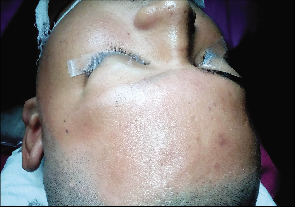
A 14-year-old male patient presented to the outpatient department with a 3-month history of painless proptosis of the left eye [
Figure 2:
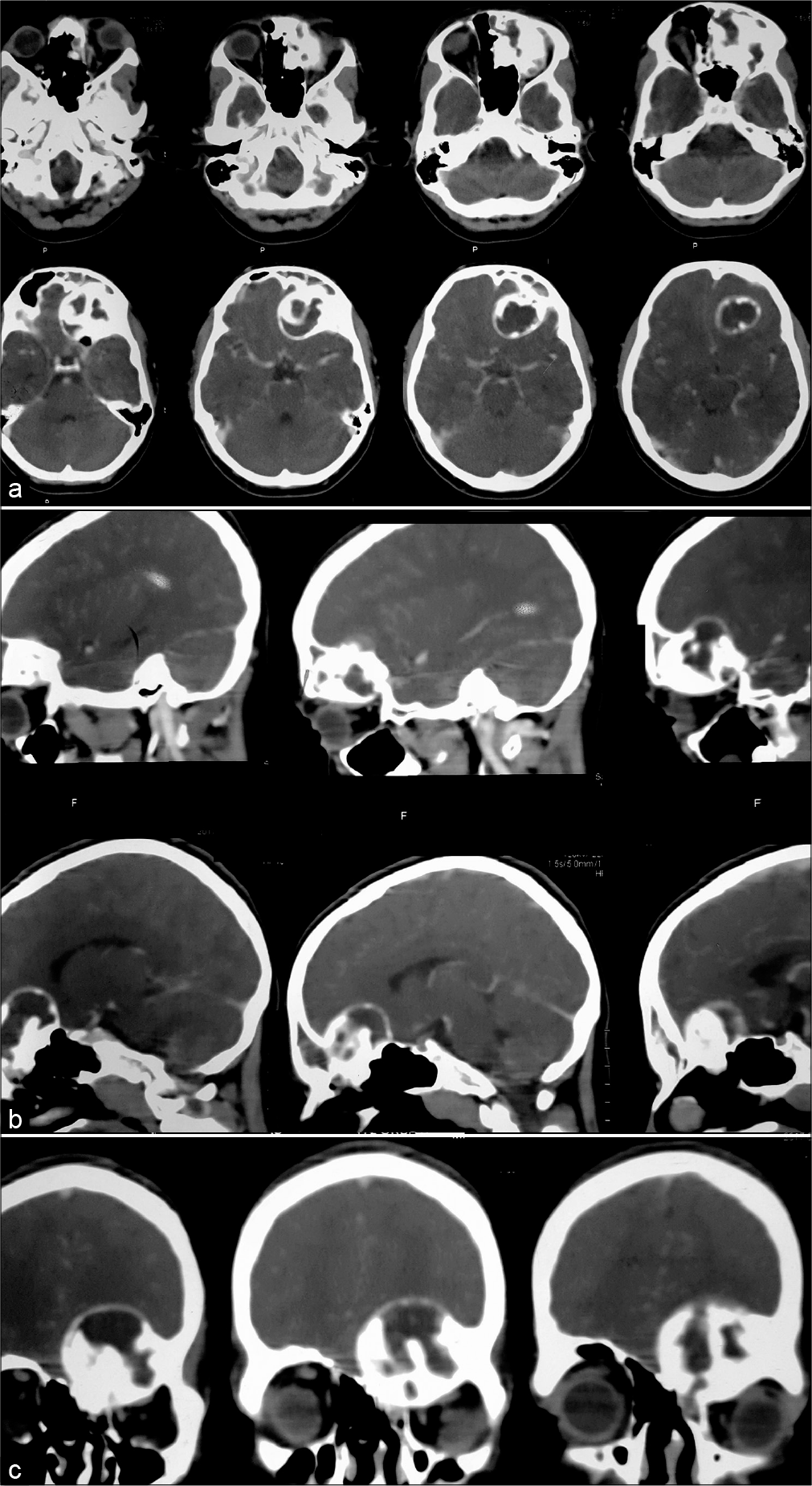
(a) Pre-operative CT brain with contrast axial sections demonstrating an anterior cranial fossa space occupying lesion with involvement of the orbital roof. There was a sclerotic contrast enhancing rim as well. (b) Preoperative CT brain with contrast sagittal sections demonstrating the lesion to be involving the orbital roof and causing mass effect and forward pushing the contents of the orbit. (c) Preoperative CT brain with contrast coronal sections through the orbit demonstrating the extent of the lesion to the midline and possible involvement of the cribriform plate.
Figure 3:
Pre-operative MRI Brain without contrast, T2 weighted axial images (top and middle row), demonstrating the extra axial nature of the lesion and mass effect on the frontal lobes with midline shift towards the right. T1 weighted post contrast coronal images (bottom row) demonstrates areas of heterogenous enhancement.
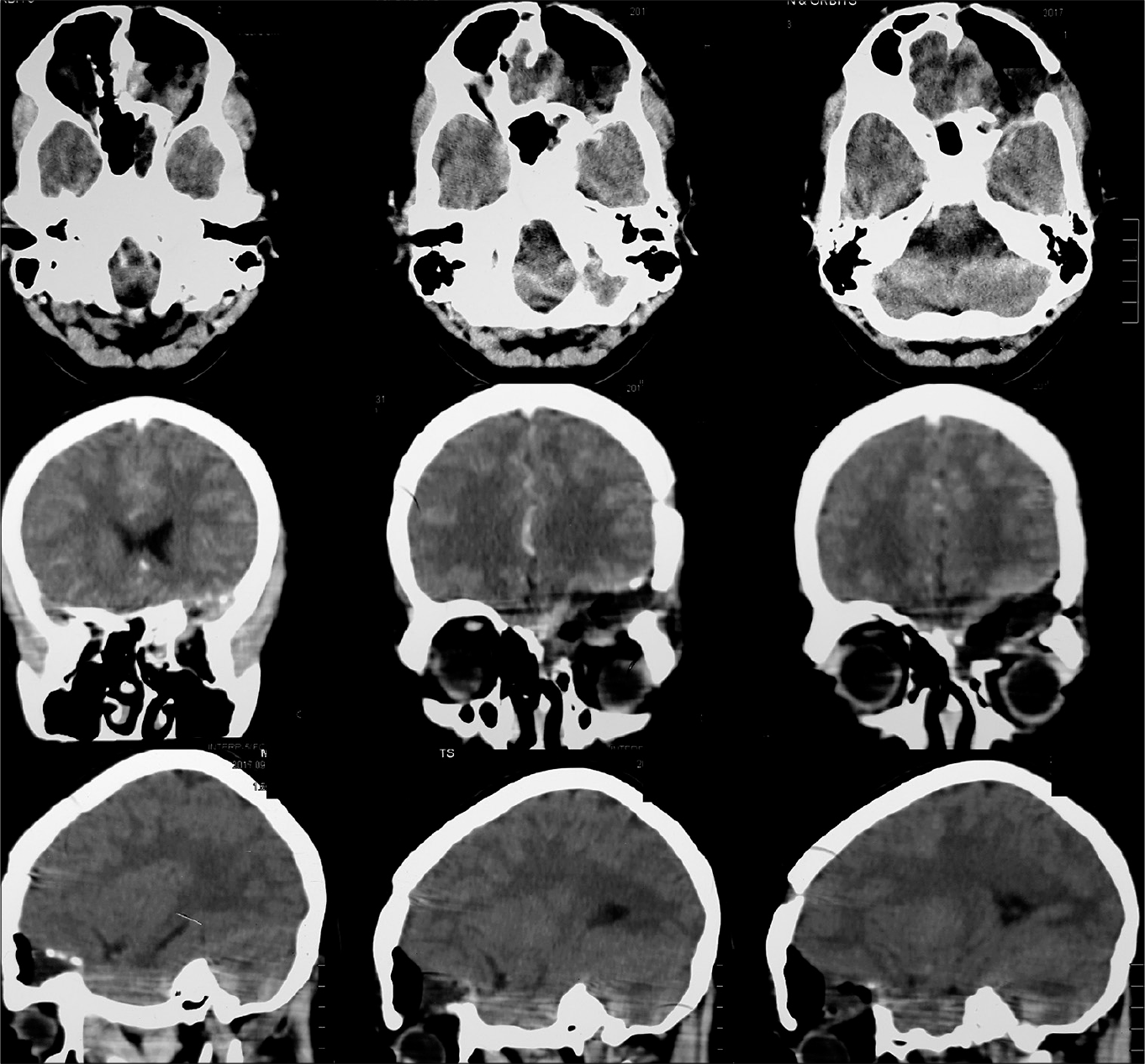
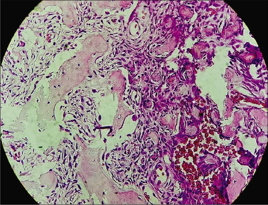
He underwent a left frontal craniotomy and subfrontal approach to the lesion with minimal retraction of the frontal lobe. The lesion was moderately vascular and firm in consistency. It appeared to be arising from the roof of the orbit and hence during excision, the periorbita was exposed which appeared to be disease free. Careful examination was performed to ensure maximum possible excision. An immediate postoperative CT with contrast showed complete removal of the lesion as well as the roof of the orbit [
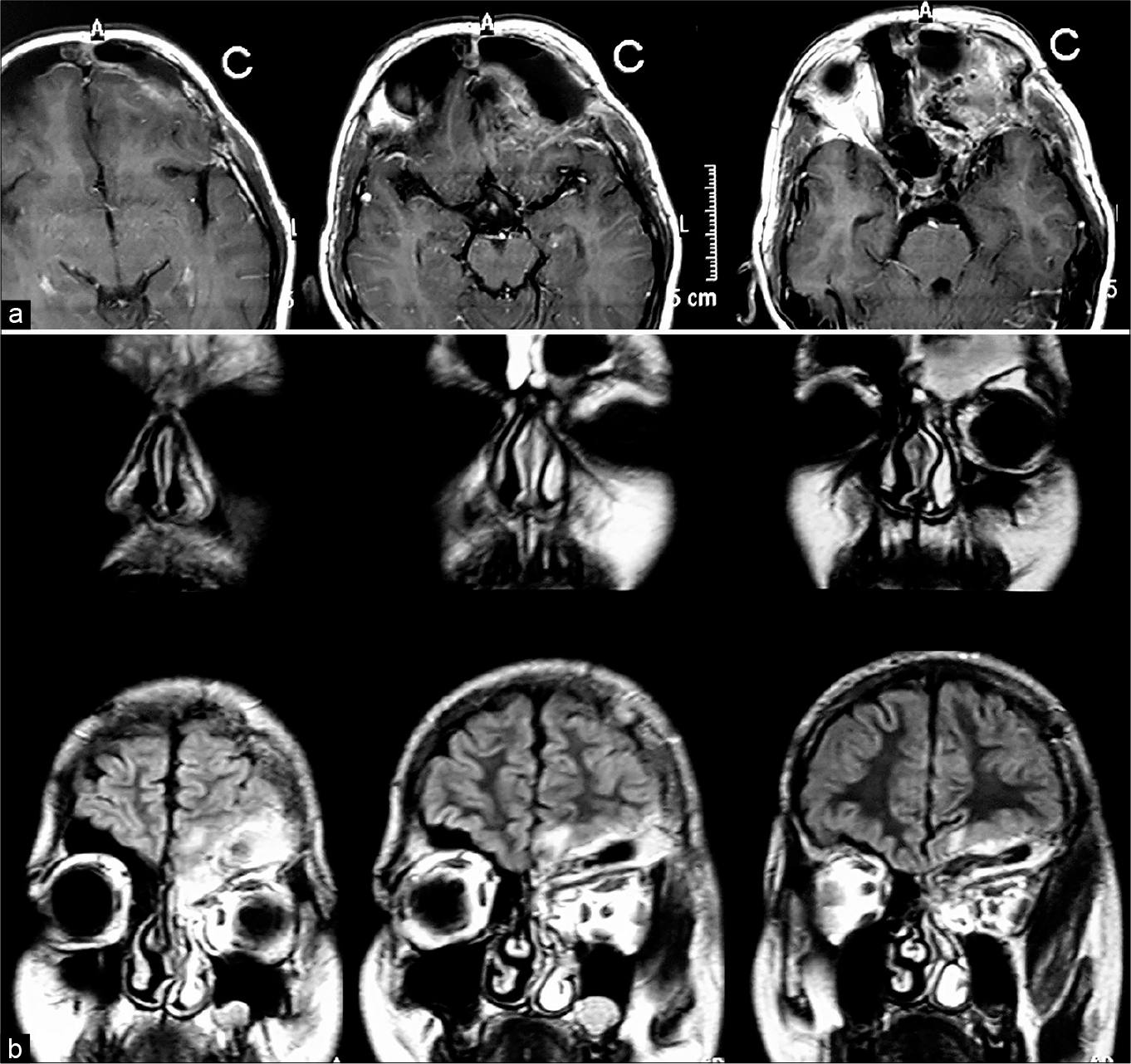
The patient’s recovery was uncomplicated and 1 year follow-up showed good scar healing. There was some residual ptosis with restriction of extraocular movements. Postoperative MRI brain with contrast [
DISCUSSION
Some authors have recommended that a pretreatment diagnosis of JPOF be established before undertaking surgery. This is due to the difference in management and prognosis between the similar lesions in the group. JPOF mandates complete excision while other variants such as COF require simple curettage. Even histopathological similarities are present in the spectrum, and hence, a complete review of imaging and clinical history must be considered.[
JPOF can occur in either the craniofacial skeleton including the orbit and paranasal but never in tooth bearing areas such as the maxilla or mandible. They are generally unencapsulated and can have growth spurts with rapid local invasion and skeletal destruction.[
Although prior confirmation through a biopsy before surgery is recommended by some authors, this may not always be possible as the lesion is rare and prone to being missed. We did not perform a biopsy in this case before attempting resection. Unfortunately, we do not have neuronavigation or frozen section available at our center and multiple surgeries would have placed significant financial pressure on the family. Hence, the plan of attempting maximum safe resection with available information was undertaken. Complete surgical excision should be the goal and is usually curative if successful. Neuronavigation may be useful in helping to achieve maximal resection. Recurrence following surgery has been reported to be upward of 50% and is due to incomplete excision. Complete excision may be challenging due to the complex anatomy of the regions involved and the ability of the lesion to involve and damage contiguous structures. In our case, the lesion involved the left supraorbital region and was amenable to an open approach that exposed the entire extent of the lesion. Wide open approaches may be preferred to ensure complete resection; however, several authors have reported that minimally invasive endoscopic approached may be advised. We would, however, express concern that since most of these tumors are firm to hard in consistency, an open approach with aggressive resection would be better. Patients may be followed up closely to detect any recurrence.[
Outcomes are good in cases of JPOF considering their benign nature but they can be fatal if untreated due to their unchecked growth. Cosmetic issues may also arise secondary to their growth and treatment. It is recommended that a multidisciplinary approach be used depending on the involvement of several areas of the face. In our case, the lesion was involving the roof of the orbit. The case was discussed with ophthalmology but they did not have sufficient exposure in such a case. We recommend that cases be followed closely following surgery with imaging ever 3–6 months for at least 2 years to monitor for recurrence.
Declaration of patient consent
The authors certify that they have obtained all appropriate patient consent.
Financial support and sponsorship
Nil.
Conflicts of interest
There are no conflicts of interest.
References
1. Brannon RB, Fowler CB. Benign fibro-osseous lesions: A review of current concepts. Adv Anat Pathol. 2001. 8: 126-43
2. El-Mofty S. Psammomatoid and trabecular juvenile ossifying fibroma of the craniofacial skeleton: Two distinct clinicopathologic entities. Oral Surg Oral Med Oral Pathol Oral Radiol Endod. 2002. 93: 296-304
3. Han MH, Chang KH, Lee CH, Seo JW, Han MC, Kim CW. Sinonasal psammomatoid ossifying fibromas: CT and MR manifestations. AJNR Am J Neuroradiol. 1991. 12: 25-30
4. Noudel R, Chauvet E, Cahn V, Mérol JC, Chays A, Rousseaux P. Transcranial resection of a large sinonasal juvenile psammomatoid ossifying fibroma. Childs Nerv Syst. 2009. 25: 1115-20
5. Post G, Kountakis SE. Endoscopic resection of large sinonasal ossifying fibroma. Am J Otolaryngol. 2005. 26: 54-6
6. Slootweg PJ, Panders AK, Koopmans R, Nikkels PG. Juvenile ossifying fibroma. An analysis of 33 cases with emphasis on histopathological aspects. J Oral Pathol Med. 1994. 23: 385-8
7. Speight PM, Carlos R. Maxillofacial fibro-osseous lesions. Curr Diagn Pathol. 2006. 12: 1-10
8. Thompson L. World Health Organization classification of tumours: Pathology and genetics of head and neck tumours. Ear Nose Throat J. 2006. 85: 74


