- Department of Neurosurgery, University of New Mexico Hospital, Albuquerque, New Mexico, United States of America.
DOI:10.25259/SNI_573_2019
Copyright: © 2020 Surgical Neurology International This is an open-access article distributed under the terms of the Creative Commons Attribution-Non Commercial-Share Alike 4.0 License, which allows others to remix, tweak, and build upon the work non-commercially, as long as the author is credited and the new creations are licensed under the identical terms.How to cite this article: Omar Saleh Akbik, Zoya A. Voronovich, Andrew P. Carlson. Treatment of unusually located traumatic intracranial aneurysms and severe vasospasm following a gunshot wound to the head: A case report. 28-Mar-2020;11:57
How to cite this URL: Omar Saleh Akbik, Zoya A. Voronovich, Andrew P. Carlson. Treatment of unusually located traumatic intracranial aneurysms and severe vasospasm following a gunshot wound to the head: A case report. 28-Mar-2020;11:57. Available from: https://surgicalneurologyint.com/surgicalint-articles/9927/
Abstract
Background: Traumatic intracranial aneurysms (TICAs) represent up to 1% of all intracranial aneurysms. They can be the result of non-penetrating and penetrating brain injury (PBI). Approximately 20% of TICA are caused by PBI. Endovascular treatments as well as surgical clipping are reported in the literature. Other vascular complications of PBI include vasospasm although the literature is lacking on this topic.
Case Description: The authors present a unique case of multiple TICAs after a PBI in a 15-year-old patient who sustained a gunshot wound to the head. The patient sustained injury through the middle cranial fossa and was taken emergently for a right-sided decompressive hemicraniectomy. Diagnostic cerebral angiogram (DCA) identified multiple TICAs along the right internal carotid artery (ICA) terminus and right middle cerebral artery as well as severe vasospasm. The patient was taken for clipping of those aneurysms and intraoperative treatment of vasospasm. Intraoperative blood flow measurements were taken before and after administration of intracisternal papaverine and arterial soft tissue dissection showing a significant increase in blood flow and improvement of vasospasm.
Conclusion: While the literature has shifted towards endovascular treatment for TICAs, surgery still offers a safe and efficacious treatment strategy especially when TICAs present at large vessel bifurcation points where parent vessel sacrifice and stent assisted coiling are less favorable strategies. Severe flow limiting vasospasm can be seen in post-traumatic setting specifically PBI. Vasospasm can be treated during open surgery with intracisternal papaverine and arterial soft dissection as confirmed in this case report with intraoperative micro-flow probe measurements.
Keywords: Penetrating brain injury, Traumatic intracranial aneurysm, Vasospasm
INTRODUCTION
Traumatic intracranial aneurysms (TICAs) represent up to 1% of all intracranial aneurysms.[
CASE REPORT
History
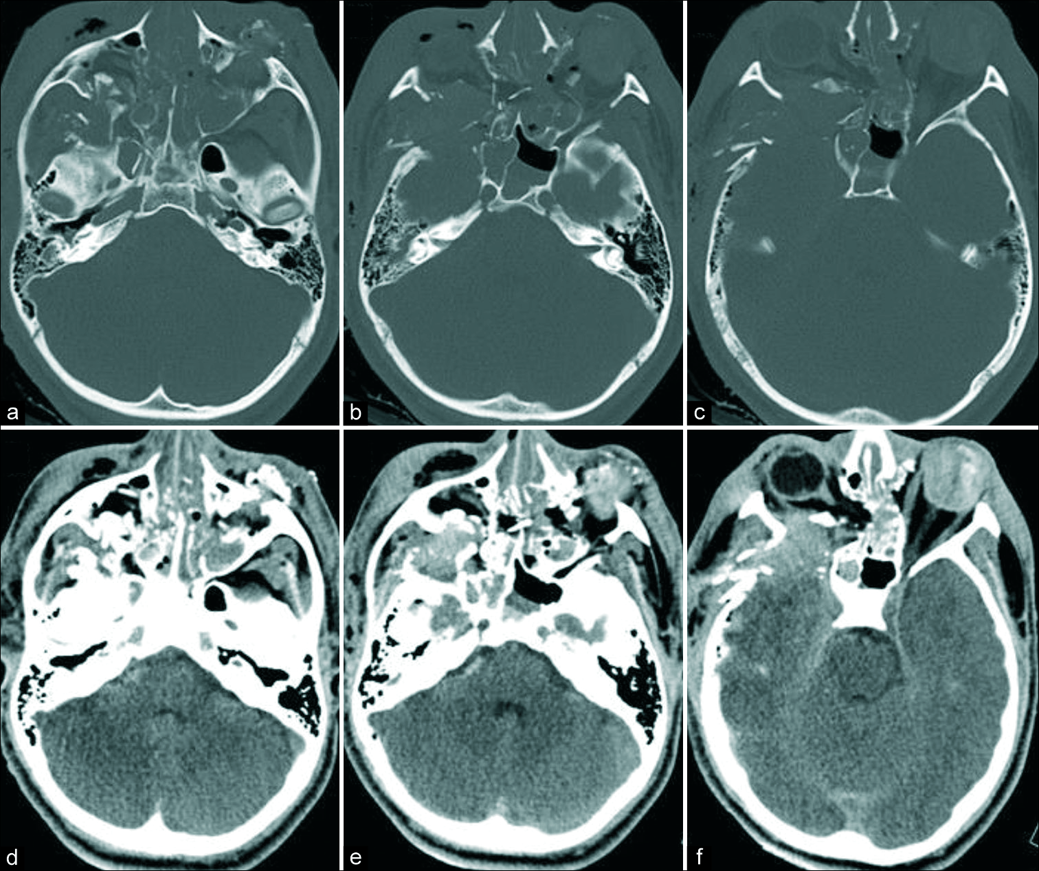
A 15-year-old male was brought to the emergency department after sustaining a gunshot wound to the head. Non-contrast computed tomography (CT) scan of the head demonstrated a trajectory entering the right inferior temporal bone, entering the anterior portion of the middle cranial fossa, traversing the anterior right temporal lobe, entering the right orbital cavity, and exiting through the ethmoid sinus and ultimately the left orbital cavity. There was a right temporal tip contusion along the ballistics path, with associated extra-axial and subarachnoid hemorrhage (SAH). The Sylvian fissures were not involved in the ballistic trajectory [
Figure 1:
Non-contrasted head computed tomography obtained on admission. (a-c) Bone window shows the trajectory of the missile entering the right inferior temporal bone, right middle cranial fossa, the right orbit, through the sinonasal cavity, the left maxillary sinus, and exiting through the left orbit. (d-f) Brain window shows the contusion with the associated extra-axial and subarachnoid hemorrhage in the right temporal tip.
The patient was taken emergently to the operating room for a right-sided decompressive craniectomy and debridement of the right temporal region. The patient started to follow commands, was moving in all extremities, and was extubated by hospital day 3. He underwent repair of his ruptured left globe and persisted with complete vision loss in the right eye.
Examination
Initial CT angiogram (CTA) within 24 h of his admission showed no vascular injury; however, a repeat CTA was performed 5 days later for vascular surveillance for TICA, which showed interval development of a right middle cerebral artery (MCA) bifurcation aneurysm. The same scanner produced both CTAs with 420 slices at 0.6 mm thickness, including three dimensional reconstructions. A subsequent diagnostic cerebral angiogram (DCA) confirmed the presence of a 4 × 3 mm right MCA bifurcation aneurysm, and also identified a 2 mm right internal carotid artery (ICA) terminus aneurysm, as well as severe vasospasm of the proximal ICA and MCA vessels [
Figure 2:
(a) Computed tomography angiogram (CTA) of the head, obtained on hospital day 1, did not show a vascular abnormality. Specifically, no abnormality is seen at the right middle cerebral artery (MCA) bifurcation. (b) Follow-up CTA of the head, obtained on hospital day 5, demonstrates the interval development of a traumatic aneurysm at the right MCA bifurcation. (c) Diagnostic cerebral angiogram (DCA) performed on hospital day 6 demonstrates previously identified 4 × 3 mm right middle cerebral artery bifurcation aneurysm. DCA also identified a 2 mm right internal carotid artery (ICA) terminus aneurysm as well as severe vasospasm of the right distal ICA and right MCA which were not previously appreciated on the CTA from January 1, 2019.
Operation
The patient was taken to surgery for clipping of the right MCA and right ICA terminus TICAs. Direct clipping of the lacerations in the vessels was successful, although bypass was prepared for, depending on the intraoperative findings [
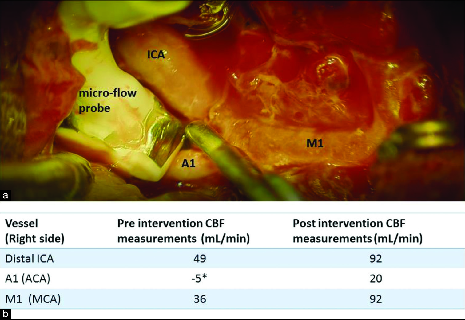
The end-tidal CO2 was kept constant and cerebral blood flow (CBF) measurements were then obtained before and after performing the therapeutic maneuvers using the Charbel Micro-Flowprobe® (Transonic). Measurements were taken at the distal ICA just before the ICA terminus as well as the A1 and M1 segment arteries just after the ICA terminus. This instrument relies on transit-time ultrasound volume flowmetry to obtain blood flow measurements in mL/min. The measurements demonstrated improvement in flow after the therapeutic maneuvers [
Figure 4:
(a) Placement of Charbel Micro-Flowprobe® (Transonic) on the right A1 segment of the anterior cerebral artery. Placement of the two clips on the previously described traumatic intracranial aneurysms at the right internal carotid artery terminus and the middle cerebral artery bifurcation can be seen. (b) Cerebral blood flow measurements pre and post intervention with papaverine and soft tissue dissection are listed. *The negative value depicted for the right A1 preintervention measurements indicates reverse flow.
Post-operative course
The patient returned to the intensive care unit. Repeat catheter angiogram was performed on hospital day 10 and confirmed that the two previously treated aneurysms were excluded from the circulation. Previously-identified vasospasm of the right supraclinoid ICA, M1, and A1 segments was improved. Two-month follow-up DCA demonstrated substantial improvement in the distal ICA caliber and improvement in the M1 irregularity. Several distal dilated vessels in the anterior temporal region were identified as suspicious for pseudoaneurysms, but have largely resolved on the 2-month catheter angiogram. Two weeks after the initial presentation, the patient was neurologically intact, with the exception of bilateral blindness related to the ballistics path, and was discharged to inpatient rehabilitation center.
DISCUSSION
While there is no algorithm for the timing of vascular imaging for vascular complications of PBI, there should be a high clinical suspicion of a ruptured TICA in a patient with new delayed onset neurologic deficit and/or hemorrhage after initial injury from a PBI. In their review, Larson et al. reported that it took an average of 21 days from injury to rupture of a TICA after blunt head trauma with a 50% mortality rate.[
In this case report, the location of the TICAs was the ICA terminus and MCA bifurcation, which is not the typical peripheral branches of the MCA and/or anterior cerebral artery (ACA) seen in closed head injuries or in the direct pathway of the missile, making direct vessel injury a less likely mechanism of injury. It is our suspicion that the location of both TICAs at large bifurcation points, ICA terminus and MCA bifurcation, must be due to a stretch injury from rapid rotational acceleration of the patient’s head after impact from the bullet itself.
Unlike congenital aneurysms, TICAs are usually pseudoaneurysms formed by disruption of all layers of the vessel wall with a contained hematoma covered by a thin layer of connective tissue as displayed in
In addition to TICAs as a vascular complication of PBI, Kordestani et al. reported a higher rate of vasospasm in PBI as compared to aneurysmal SAH.[
In regards to decision making for this case, conservative management seemed inappropriate considering the rapid progress of the TICAs. The location of his two TICAs, right ICA terminus and right MCA bifurcation, ruled out an endovascular treatment with parent vessel sacrifice. Stent- assisted embolization or flow diversion across the distal ICA and right M1 was worrisome due to the degree of vasospasm along the distal right ICA and right M1 segment, calling into question the integrity of those vessels. Furthermore, placement of a stent would require an antiplatelet regimen that would make timely cranioplasty difficult, without risking a thrombotic event due to suspension of his antiplatelet therapy. A similar argument was made for any stent coiling option. Ultimately, we opted for a surgical treatment to protect the outflow vessels with bypass if needed, and explore the integrity of the remaining proximal vessels relative to the vasospasm.
After clipping the two TICAs, papaverine was injected into the intracisternal space over the affected vessels and soft tissue dissection was performed to remove any strictures that may have been compromising blood flow. Pre and post CBF measurements with the Micro-Flowprobe showed improvement in all vessels [
CONCLUSION
While the literature has shifted towards endovascular treatment for TICAs, surgery still offers a safe and efficacious treatment strategy especially when TICAs present at large vessel bifurcation points where parent vessel sacrifice and stent assisted coiling are less favorable strategies. Severe flow limiting vasospasm can be seen in post-traumatic setting specifically PBI. Vasospasm can be treated during open surgery with intracisternal papaverine and arterial soft dissection as confirmed in this case report with intraoperative micro-flow probe measurements.
Declaration of patient consent
Patient’s consent not required as patients identity is not disclosed or compromised.
Financial support and sponsorship
Nil.
Conflicts of interest
There are no conflicts of interest.
References
1. Aarabi B. Traumatic aneurysms of brain due to high velocity missile head wounds. Neurosurgery. 1988. 22: 1056-63
2. Amin-Hanjani S, Meglio G, Gatto R, Bauer A, Charbel FT. The utility of intraoperative blood flow measurement during aneurysm surgery using an ultrasonic perivascular flow probe. Neurosurgery. 2006. 58: ONS-305-12
3. Britz GW ND, West GA, Winn RH, Winn RH.editors. Traumatic cerebral aneurysms secondary to penetrating intracranial injuries. Youmans Neurological Surgery. Philadelphia, PA: Saunders; 2004. 2: 2131-5
4. Chohan MO, Carlson AP, Murray-Krezan C, Taylor CL, Yonas H. Microsurgical vascular manipulation in aneurysm surgery and delayed ischemic injury. Can J Neurol Sci. 2017. 44: 410-4
5. Cohen JE, Gomori JM, Segal R, Spivak A, Margolin E, Sviri G. Results of endovascular treatment of traumatic intracranial aneurysms. Neurosurgery. 2008. 63: 476-85
6. Dalbasti T, Karabiyikoglu M, Ozdamar N, Oktar N, Cagli S. Efficacy of controlled-release papaverine pellets in preventing symptomatic cerebral vasospasm. J Neurosurg. 2001. 95: 44-50
7. Dubey A, Sung WS, Chen YY, Amato D, Mujic A, Waites P. Traumatic intracranial aneurysm: A brief review. J Clin Neurosci. 2008. 15: 609-12
8. Jung SH, Kim SH, Kim TS, Joo SP. Surgical Treatment of Traumatic Intracranial Aneurysms: Experiences at a Single Center over 30 Years. World Neurosurg. 2017. 98: 243-50
9. Kieck CF. Intracranial aneurysm with subarachnoid haemorrhage. Surgical results and long-term outcome in 100 cases. S Afr Med J. 1984. 65: 722-4
10. Kordestani RK, Counelis GJ, McBride DQ, Martin NA. Cerebral arterial spasm after penetrating craniocerebral gunshot wounds: Transcranial Doppler and cerebral blood flow findings. Neurosurgery. 1997. 41: 351-9
11. Larson PS, Reisner A, Morassutti DJ, Abdulhadi B, Harpring JE. Traumatic intracranial aneurysms. Neurosurg Focus. 2000. 8: e4-
12. Lundell A, Bergqvist D, Mattsson E, Nilsson B. Volume blood flow measurements with a transit time flowmeter: An in vivo and in vitro variability and validation study. Clin Physiol. 1993. 13: 547-57
13. Medel R, Crowley RW, Hamilton DK, Dumont AS. Endovascular obliteration of an intracranial pseudoaneurysm: The utility of Onyx. J Neurosurg Pediatr. 2009. 4: 445-8
14. Pruitt BA. Vascular complications of penetrating brain injury. J Trauma. 2001. 51: S26-8
15. Sui M, Mei Q, Sun K. Surgical treatment achieves better outcome in severe traumatic pericallosal aneurysm: Case report and literature review. Int J Clin Exp Med. 2015. 8: 1598-603


