

- Department of Neurosurgery, Mohammed VI University Hospital Center of Marrakesh, Cadi Ayyad University, Faculty of Medicine, Marrakesh, Morocco,
- Department of Pediatric Neurosurgery, Institute for Brain Protection Sciences, Johns Hopkins All Children’s Hospital, St. Petersburg, Florida, United States.
Correspondence Address:
Malak El Marrakchi, Department of Neurosurgery, Mohammed VI University Hospital Center of Marrakesh, Cadi Ayyad University, Faculty of Medicine, Marrakesh, Morocco.
DOI:10.25259/SNI_165_2024
Copyright: © 2024 Surgical Neurology International This is an open-access article distributed under the terms of the Creative Commons Attribution-Non Commercial-Share Alike 4.0 License, which allows others to remix, transform, and build upon the work non-commercially, as long as the author is credited and the new creations are licensed under the identical terms.How to cite this article: Malak El Marrakchi1, Nahla Zian1, Farouk Hajhouji1, Mehdi Laghmari1, Houssine Ghannane1, George Jallo2, Said Ait Benali1. Association of limited dorsal myeloschizis and corpus callosum lipoma: A case report and literature review. 03-May-2024;15:151
How to cite this URL: Malak El Marrakchi1, Nahla Zian1, Farouk Hajhouji1, Mehdi Laghmari1, Houssine Ghannane1, George Jallo2, Said Ait Benali1. Association of limited dorsal myeloschizis and corpus callosum lipoma: A case report and literature review. 03-May-2024;15:151. Available from: https://surgicalneurologyint.com/?post_type=surgicalint_articles&p=12879
Date of Submission
06-Mar-2024
Date of Acceptance
08-Apr-2024
Date of Web Publication
03-May-2024
Abstract
Background: Intracranial lipomas are a rare clinical entity. These lesions are frequently asymptomatic and originate in the pericallosal area. As they are fat-containing lesions which are intimately attached to the surrounding structures, surgery is not recommended. In some individual reports, subtotal resection is recommended to lessen complications. There have been no previous reports of corpus callosum lipoma (CCL) associated with limited dorsal myeloschizis (LDM).
Case Description: We describe the case of a combination of CCL and bilateral choroid plexus lipoma discovered incidentally during the investigation of LDM in a 3-month-old male child. Given the asymptomatic behavior of the lipoma and the vascular elements of the pericallosal area, it was decided to monitor it regularly. Thus, the patient underwent surgery only for LDM. Histological examination confirmed the diagnosis, and postoperative follow-up 1 year after showed good evolution. To the best of our knowledge, this association has never been described in the literature.
Conclusion: This case suggests a possible developmental relationship between CCL and spinal dysraphism.
Keywords: Corpus callosum, Dysraphism, Intracranial, Limited dorsal myeloschizis, Lipoma
INTRODUCTION
Intracranial lipomas (ICLs) are rare fat containing lesions, generally considered a congenital malformation. The first reported case was documented by Rokitansky in 1858.[
CASE REPORT
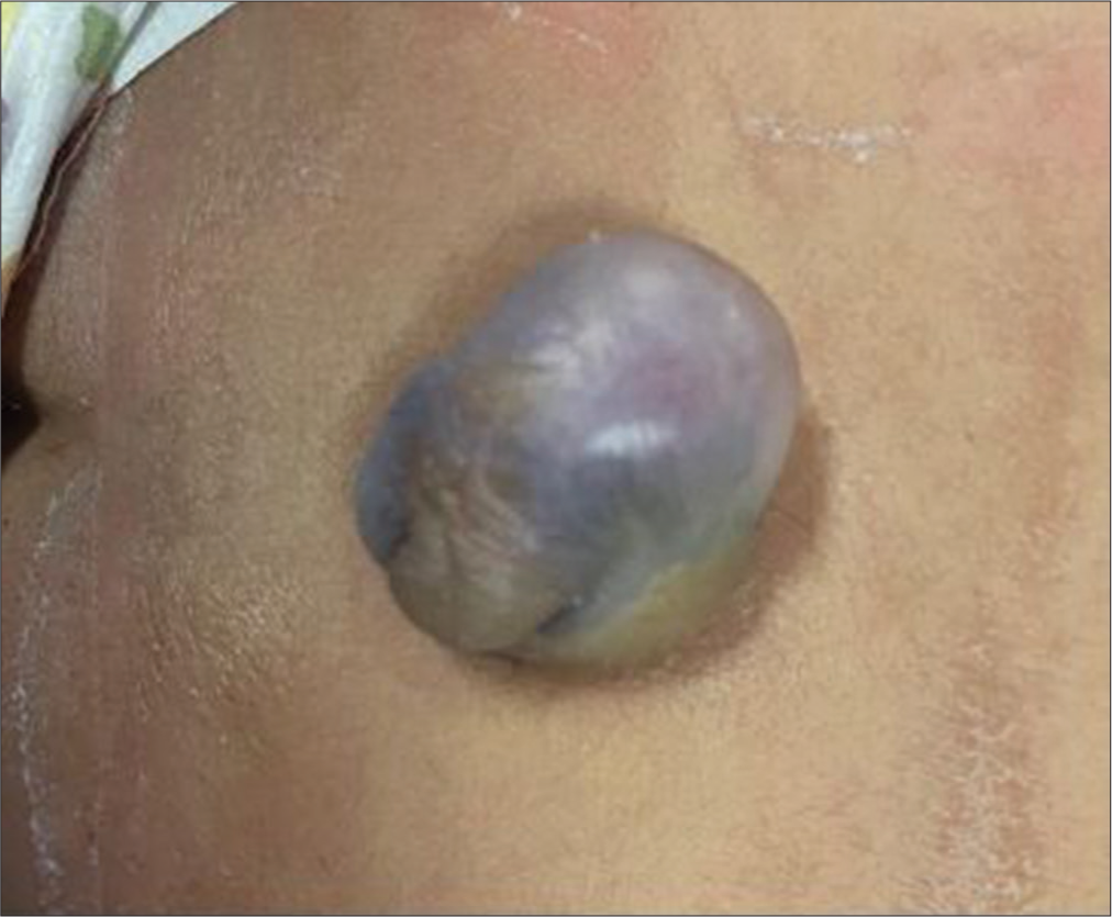
A 3-month male child, born term, and youngest of three siblings, fully vaccinated, and of nonconsanguineous parents presented with a lumbosacral mass. This was a well-monitored single fetal pregnancy carried to term without any incident, admitted for a bluish and rounded lumbosacral mass, painless, and of regular contours, measuring approximately 4 cm [


DISCUSSION
ICLs are very rare congenital abnormalities representing <0.5% of cerebral lesions. They are often found in the pericallosal area (40%). However, other locations have been reported in the literature, such as the Sylvian fissure, prepontine cistern, Galen vein system, craniocervical junction, and cerebral convexity.[
As most ICLs are asymptomatic, they are incidentally discovered when assessing for a traumatic brain injury. Otherwise, clinical presentation is mostly related to location. CCL might cause headaches, seizures, psychomotor disorders, or interhemispheric disconnection, resulting in dyspraxia.
Two types of CCL are described in the literature: curvilinear and tubulonodular. While curvilinear lipomas are thin, slender shaped, and seem to be posterior along the splenium, tubulonodular lesions are thick (more than 2 cm), tubular shaped, and localize more often anteriorly. They are also frequently associated with cerebral abnormalities, mainly hypogenesis or agenesis of the corpus callosum (CC). Yilmaz et al. noticed four cases of CC hypoplasia within ten patients presenting CCL. Three of them had curvilinear lesions, which challenged this idea.[
On CT scans, CCL have a fat density and appears homogeneous with no enhancement when injecting the contrast product. When they are calcified, they can be misdiagnosed and taken for a dermoid cyst or teratoma. On the MRI, these lesions are hyperintense on T1 sequences and FLAIR, hypointense on T2, and fat suppression images.[
Most CCL is unique asymptomatic slow-growing benign lesions. However, they usually include neurovascular components justifying why surgery is generally avoided except in symptomatic patients. In these cases, subtotal resection is recommended for less morbimortality. On the other hand, the planning of the approach depends essentially on the location and dimensions of the lipoma.[
CCL can occur as a part of complex malformations such as Pai syndrome or encephalocraniocutaneous lipomatosis. Our patient does not meet major criteria, making these diagnoses less probable. Another pathology that could have been considered is familial multiple lipomatosis, also called symmetrical multiple lipomatosis or Madlung’s disease. However, it preferentially occurs in men in the 3rd decade and has never been described in children. In addition to that, it frequently has a metabolic component resulting in systematic signs and other criteria missing in our patient. Considering polymicrogyria associated with hypogenesis of the CC, Aicardi syndrome was also a possible diagnosis. Nevertheless, it primarily affects females. Moreover, the classic triad, as well as the modified criteria necessary to make the diagnosis, are missing [

The association of CCL and spinal dysraphism has been described in the literature. Aggarwal et al. described the only case of lipomeningocele associated with interhemispheric lipoma. This suggests a possible continuum in the etiopathogenesis of ICL and spinal dysraphism.[
In fact, the pathogenesis of CCL is not fully elucidated. A lot of theories have been advanced. The most accepted one is that they originate from abnormal differentiation of mesenchymal cells of meninx primitive. Other authors evoke a defective fusion of the ectoderm when recovering the mesoderm during neural development. Considering LDM is the result of incomplete disjunction between cutaneous and neural ectoderms, we believe that this case supports this theory and suggests a possible developmental relationship between CCL and LDM.[
On the other hand, during the 9th week of gestation, the cells from lamina reunians (LRs) migrate rapidly to the sulcus medianus telencephalic medium (SMTM), allowing the CC to grow anteroposteriorly, with the constitution of the genu first followed by the body and the splenium. CCL interferes with the fusion of LR and SMTM, resulting in hypogenesis or agenesis of the CC.[
CONCLUSION
CCL could be part of complex malformations. It is usually discovered incidentally. Association with spinal dysraphism is very rare. A good understanding of the pathophysiology of each entity is the key to comprehend the developmental relationship leading to this association. Subtotal resection of CCL is recommended in symptomatic patients for less morbimortality.[
Ethical approval
The Institutional Review Board approval is not required.
Declaration of patient consent
The authors certify that they have obtained all appropriate patient consent.
Financial support and sponsorship
Nil.
Conflicts of interest
There are no conflicts of interest.
Use of artificial intelligence (AI)-assisted technology for manuscript preparation
The authors confirm that there was no use of artificial intelligence (AI)-assisted technology for assisting in the writing or editing of the manuscript and no images were manipulated using AI.
Disclaimer
The views and opinions expressed in this article are those of the authors and do not necessarily reflect the official policy or position of the Journal or its management. The information contained in this article should not be considered to be medical advice; patients should consult their own physicians for advice as to their specific medical needs.
References
1. Aggarwal N, Gehlot KB, Kumar SD, Khan NK. Frontal subcutaneous lipoma associated with interhemispheric lipoma, lipomeningocele, and corpus callosal dysgenesis in a young adult: CT and MRI findings. Indian J Radiol Imaging. 2018. 28: 22-6
2. Ben Elhend S, Belfquih H, Hammoune N, Athmane EM, Mouhsine A. Lipoma with agenesis of corpus callosum: 2 case reports and literature review. World Neurosurg. 2019. 125: 123-5
3. Britt PM, Bindal AK, Balko MG, Yeh HS. Lipoma of the cerebral cortex: Case report. Acta Neurochir (Wien). 1993. 121: 88-92
4. Clarici G, Heppner F. The operative approach to lipomas of the corpus callosum. Neurochirurgia (Stuttg). 1979. 22: 77-81
5. Douira-Khomsi W, Jhaouet I, Lahmar L, Louati H, Ben Hassine L, Ammar I. A rare cause of seizures in infants: sylvian fissure lipoma. Arch Pédiatr. 2016. 23: 867-8
6. Duman O, Goksu E, Akyuz M, Haspolat S, Senol U. Brain stem lipoma presenting as paroxysmal headache with autonomic features. Headache. 2004. 44: 105
7. Erbay MF, Tecellioglu M. A pericallosal lipoma case with abnormal vasculature mimicking arteriovenous malformation. Neurol India. 2019. 67: 1331
8. Fandiño J, Bermúdez J, Arán E. Quadrigeminal cistern and calcarine fissure lipoma: Case report and review of the literature. Neurocir Astur Spain. 2005. 16: 173-6
9. Fu Y, Fang X, Li Y, Zhao X. Teaching neuroimages: Pericallosal curvilinear lipoma. Neurology. 2019. 93: e212-3
10. Georgy BA, Hesselink JR, Jernigan TL. MR imaging of the corpus callosum. AJR Am J Roentgenol. 1993. 160: 949-55
11. Jerbi Omezzine S, Fkih Hassen S, Ben Rhouma K, Younes S, Hamza HA. Imaging of central nervous system lipoma. J Neuroradiol. 2014. 41: 41
12. Jerbi S, Chouchane N, Salem AB, Farik S, Zayeni O, Hamza HA. Rare locations of head and neck lipoma. J Neuroradiol. 2017. 44: 114
13. Jiménez Caballero PE. Interhemispheric lipoma associated with agenesis of the corpus callosum. Neurol Barc Spain. 2012. 27: 515-7
14. Kannuki S, Okajima K, Sato K, Kusaka K, Matsumoto K. Intracranial lipoma of the temporal lobe--report of a case and review of the literature. No Shinkei Geka. 1986. 14: 379-84
15. Kim TH, Joh JH, Kim MY, Kim YM, Han KS. Fetal pericallosal lipoma: US and MR findings. Korean J Radiol. 2002. 3: 140-3
16. Nunes JC, Martins RF, Bastos A, Claudino LS, Guarnieri R, Lima DC. Brain lipoma, corpus callosum hypoplasia and polymicrogyria in familial multiple lipomatosis. Clin Neurol Neurosurg. 2013. 115: 1157-9
17. Olivero F, Foiadelli T, Luzzi S, Marseglia GL, Savasta S. Pai syndrome: A review. Childs Nerv Syst. 2020. 36: 2635-40
18. Pang D, Zovickian J, Oviedo A, Moes GS. Limited dorsal myeloschisis: A distinctive clinicopathological entity. Neurosurgery. 2010. 67: 1555-79 discussion 1579-80
19. Pashaj S, Merz E. Detection of fetal corpus callosum abnormalities by means of 3D ultrasound. Ultraschall Med. 2016. 37: 185-94
20. Popa RT, Feier D, Fufezan O, Blaga L. Interhemispheric lipoma associated with agenesis of corpus callosum in an infant: Case report. Med Ultrason. 2010. 12: 249-52
21. Seidl Z, Vaneckova M, Vitak T. Intracranial lipomas: A retrospective study. Neuroradiol J. 2007. 20: 30-6
22. Sherer DM, Onyeije CI. Prenatal ultrasonographic diagnosis of fetal intracranial tumors: A review. Am J Perinatol. 1998. 15: 319-28
23. Shinar S, Lerman-Sagie T, Telleria ME, Viñals F, García R, Quiroga H. Fetal pericallosal lipomas-clues to diagnosis in the second trimester. Eur J Paediatr Neurol. 2018. 22: 929-34
24. Sms Q, Iqbal Km H. Incidental detection of intra-cranial lipoma in patient with quadriparesis. J Clin Diagn Res. 2014. 8: MD06-7
25. Szeto C, Tewfik TL, Jewer D, Rideout A. Pai syndrome (median cleft palate, cutaneous nasal polyp, and midline lipoma of the corpus callosum): A case report and literature review. Int J Pediatr Otorhinolaryngol. 2005. 69: 1247-52
26. Taydas O, Ogul H, Kantarci M. The clinical and radiological features of cisternal and pericallosal lipomas. Acta Neurol Belg. 2020. 120: 65-70
27. Ventura E, Ormitti F, Crisi G, Sanna M, Bacciu A. Bilateral cerebellopontine angle lipomas. Auris Nasus Larynx. 2012. 39: 103-6
28. Yildiz H, Hakyemez B, Koroglu M, Yesildag A, Baykal B. Intracranial lipomas: Importance of localization. Neuroradiology. 2006. 48: 1-7
29. Yilmaz MB, Genc A, Egemen E, Yilmaz S, Tekiner A. Pericallosal lipomas: A series of 10 cases with clinical and radiological features. Turk Neurosurg. 2016. 26: 364-8

