

- Department of Neurosurgery, University of Kansas Medical Center, Kansas City, Kansas, USA
Correspondence Address:
Kyle A. Smith
Department of Neurosurgery, University of Kansas Medical Center, Kansas City, Kansas, USA
DOI:10.4103/2152-7806.194520
Copyright: © 2016 Surgical Neurology International This is an open access article distributed under the terms of the Creative Commons Attribution-NonCommercial-ShareAlike 3.0 License, which allows others to remix, tweak, and build upon the work non-commercially, as long as the author is credited and the new creations are licensed under the identical terms.How to cite this article: Kyle A. Smith, Samuel K. Asante, John Clough. Intradural-extramedullary isolated compressive sarcoid lesion. 21-Nov-2016;7:
How to cite this URL: Kyle A. Smith, Samuel K. Asante, John Clough. Intradural-extramedullary isolated compressive sarcoid lesion. 21-Nov-2016;7:. Available from: http://surgicalneurologyint.com/surgicalint_articles/intradural%e2%80%91extramedullary-isolated-compressive-sarcoid-lesion/
Date of Submission
13-Mar-2016
Date of Acceptance
24-May-2016
Date of Web Publication
21-Nov-2016
Abstract
Background:Sarcoid involvement of the central nervous system is a rare occurrence, with involvement in approximately 5–10% of all cases. Isolated spinal involvement is an even rarer encounter, only 0.3–1% of all cases. These lesions can form compressive nodules leading to myelopathy. In the presented case of cervical sarcoid, the patient required a decompressive procedure to address cord compression.
Case Description:This is the case of a 39-year-old male presenting with cervical myelopathy caused by a compressive sarcoid nodule who underwent a successful posterior decompressive procedure. The pathology demonstrated a non-caseating granuloma, consistent with sarcoid. Postoperatively, the patient's myelopathic symptoms improved.
Conclusions:Sarcoid is rarely associated with an isolated compressive cervical lesion. Although sarcoid management typically involves immune suppression, in cases of active cord compression surgical intervention is warranted.
Keywords: Cervical mass, cervical myelopathy, neurosarcoid, sarcoid, spinal sarcoid
INTRODUCTION
Sarcoidosis manifests itself in the central nervous system in approximately 5–10% of the cases.[
CASE REPORT
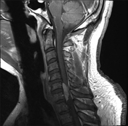
A 39 year-old male presented with a 3-month history of progressive cervical myelopathy. The magnetic resonance imaging of the cervical spine demonstrated a homogenously enhancing right-sided ventrolateral, intradural-extramedullary mass compressing the cord from C6–T1 levels [
DISCUSSION
An extramedullary spinal sarcoid lesion is extremely rare; there are approximately 16 such cases reported in the literature.[
Financial support and sponsorship
Nil.
Conflicts of interest
There are no conflicts of interest.
References
1. Bose B. Extramedullary sarcoid lesion mimicking intraspinal tumor. Spine J. 2002. 2: 381-5
2. Sohn M, Culver DA, Judson MA, Scott TF, Tavee J, Nozaki K. Spinal cord neurosarcoidosis. Am J Med Sci. 2014. 347: 195-8

