

- Department of Neurosurgery, Specialties Hospital, La Raza National Medical Center, Mexico City, Mexico
- Department of Neurosurgery, Western General Hospital, Zapopan, Mexico.
- Department of Neurosurgery, University of Guadalajara, Guadalajara, Mexico.
Correspondence Address:
Héctor Alonso Tirado-Ornelas, Department of Neurosurgery, Specialties Hospital, La Raza National Medical Center, Mexico City, Mexico.
DOI:10.25259/SNI_227_2024
Copyright: © 2024 Surgical Neurology International This is an open-access article distributed under the terms of the Creative Commons Attribution-Non Commercial-Share Alike 4.0 License, which allows others to remix, transform, and build upon the work non-commercially, as long as the author is credited and the new creations are licensed under the identical terms.How to cite this article: Héctor Alonso Tirado-Ornelas1, Jesús Ricardo Cazadero-Márquez1, Carlos Antonio Cruz-Argüelles1, José Enrique Kleemann-Jaramillo2, Miguel Ángel Meza-Bautista3, Jorge Arturo Santos-Franco1. Morphometric analysis of the foramen ovale in the Mexican population using computed tomography scan. 03-May-2024;15:148
How to cite this URL: Héctor Alonso Tirado-Ornelas1, Jesús Ricardo Cazadero-Márquez1, Carlos Antonio Cruz-Argüelles1, José Enrique Kleemann-Jaramillo2, Miguel Ángel Meza-Bautista3, Jorge Arturo Santos-Franco1. Morphometric analysis of the foramen ovale in the Mexican population using computed tomography scan. 03-May-2024;15:148. Available from: https://surgicalneurologyint.com/surgicalint-articles/12882/
Date of Submission
27-Mar-2024
Date of Acceptance
10-Apr-2024
Date of Web Publication
03-May-2024
Abstract
Background: The assessment of cranial foramina is an important part of the objective diagnostic and therapeutic study relevant to pathologies involving structures of the skull base. The study of the foramen ovale not only holds significance for anatomical development but also bears profound surgical importance, such as in trigeminal neuralgia, and diagnostic importance in tumors and various types of epilepsy. It becomes relevant in fine-needle aspiration techniques in perineural tumor procedures, for electroencephalographic analysis in seizures, and therapeutic procedures such as percutaneous trigeminal rhizotomy for trigeminal neuralgia.
Methods: A cross-sectional study at the Department of Neurosurgery, Specialties Hospital, La Raza National Medical Center, Mexico City, involved 70 patients aged >18 years who underwent a single skull computed tomography scan between July 2023 and March 2024. Patients with sufficient scan quality and optimal visualization of skull base foramina were included in the study. Measurements of tomographic images were taken using Inobitec’s DICOM file viewer. Data analysis in Microsoft Excel yielded mean, standard deviation, and 95% confidence interval (CI) for morphometric parameters of the foramen ovale.
Results: Analysis of tomographies from 70 patients revealed a total of 140 foramen ovale, evenly split between 25 males (35.7%) and 45 females (64.3%). The measurements for the maximum anteroposterior diameter, transverse diameter, and surface area of all foramina were as follows: 6.61 ± 0.25 mm (95% CI), 3.97 ± 0.21 mm (95% CI), and 20.84 ± 1.58 mm2 (95% CI), respectively. Specific measurements for the right and left sides were obtained: for the right side, 6.59 ± 0.26 mm (95% CI) and 3.89 ± 0.21 mm (95% CI) for the maximum anteroposterior and transverse diameters, respectively, and 20.38 ± 1.62 mm2 (95% CI) for the surface area. For the left side, the measurements were 6.63 ± 0.24 mm (95% CI), 4.05 ± 0.21 mm (95% CI), and 21.31 ± 1.55 mm2 (95% CI) for the maximum anteroposterior diameter, transverse diameter, and surface area, respectively. The maximum and minimum dimensions for anteroposterior and transverse diameters were 10.67 mm, 4.41 mm, 7.09 mm and 2.36 mm, respectively, with a corresponding range for the surface area of 10.16 mm2–44.13 mm2. The average minimum distance between the foramen ovale and the foramen spinosum was 2.32 ± 0.24 mm (95% CI). In males, the average size of the foramen ovale was 23.66 ± 1.61, which was 22% larger than the average size in females (19.28 ± 1.45) (P = 0.0001).
Conclusion: The foramen ovale is one of the main anatomical structures of the skull base, and besides that, it is complex and not directly accessible for clinical evaluation, useful information can be obtained through morphometric analysis. The present study provides specific anatomical data with morphological patterns to increase the understanding of the characteristics of the foramen ovale in the Mexican population. These are intended to be helpful in the pursuit of acknowledging the morphometrics and thus being able to plan neurosurgical procedures in the middle cranial fossa.
Keywords: Foramen ovale, Morphometrics, Neuroanatomy, Neurosurgery
INTRODUCTION
The evaluation of cranial foramina is an important part of the objective diagnostic and therapeutic study, particularly relevant to pathologies involving structures of the skull base.
The topographic anatomy of the sphenoid bone is then of great clinical importance due to the presence of several cranial foramina. Rotundum foramen, for transmission of the maxillary nerve, located in the anterior part and medial part of the sphenoid bone, and the foramen ovale, for mandibular nerve transmission, accessory meningeal artery, venous plexus, and sometimes the lesser petrosal nerve.[
The foramen ovale is an opening located in the base of the skull, situated in the greater wing of the sphenoid bone, between the foramen rotundum and the foramen spinosum. It typically has a maximum diameter of 7.48 and a minimum of 4.17, through which the mandibular branch of the trigeminal nerve, the accessory meningeal artery, and occasionally the lesser petrosal nerve and emissary veins pass through the skull base.[
This is one of the important foramina situated in the transition zone between intracranial and extracranial structures, opening into the infratemporal fossa and through its other opening on the lateral surface of the greater wing.[
Several studies have measured the dimension of the foramen ovale, including reports with measurements in cadaver skulls and morphometric studies carried out with the assistance of simple skull computed tomography.
Although variations in the shape of this foramen may be present, a careful evaluation of these holes will facilitate the diagnosis of lesions present in the middle cranial fossa and nasopharynx.
The study of this foramen not only holds importance for anatomical development but also bears profound surgical significance, such as in trigeminal neuralgia, and diagnostic importance in tumors and various types of epilepsy. It becomes relevant in fine-needle aspiration techniques in perineural tumor procedures, for electroencephalographic analysis in certain types of epilepsy, and in therapeutic procedures such as percutaneous trigeminal rhizotomy for trigeminal neuralgia.[
Lesions involving the medial parts of the middle cranial base can be accessed through an intracranial or subcranial route or a combination of the two routes; therefore, knowledge of its anatomy is of vital importance to carry out minimally invasive diagnostic and therapeutic procedures.[
Some studies indicate that the morphology of the foramen ovale is not entirely precise, as it may sometimes be covered by ossified ligaments extending between the lateral pterygoid process and the sphenoidal spine, or its venous portion may be compartmentalized by a bony spur located anteromedially, resulting in a double foramen ovale.[
Given these precedents, it is important to highlight that this study presents the morphometry of the foramen ovale in the Mexican population.
MATERIALS AND METHODS
A cross-sectional study was conducted at the Department of Neurosurgery, Specialties Hospital, La Raza National Medical Center in Mexico City, Mexico. Seventy patients aged >18 years who underwent a single skull computed tomography scan for any medical indication with at least 150 slices and optimal visualization of the skull base foramina between July 2023 and March 2024 were included in the study. Patients with a history of cranial surgery, brain or cranial tumors, or insufficient quality computed tomography scans for proper visualization of the foramen ovale were excluded from the study.
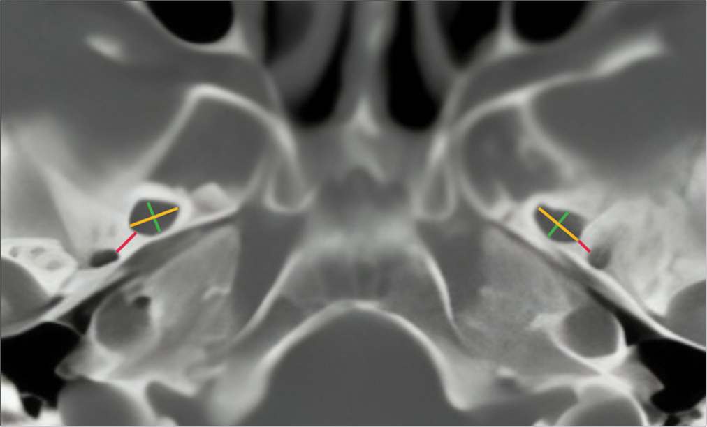
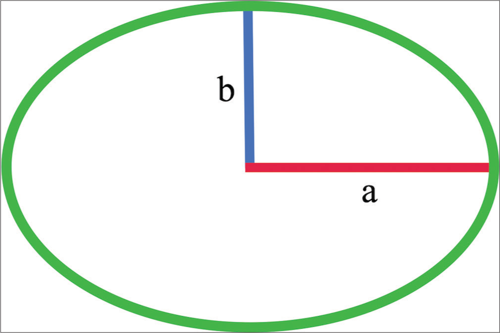
Measurements of the tomographic images were performed using the DICOM file viewer Inobitec. The inion-nasion line was used to define the axial plane, as well as the septum pellucidum to define the sagittal and coronal planes. Subsequently, using the bone window, the foramen ovale was identified, and its major anteroposterior diameter, the diameter perpendicular to this, and the shortest distance to the foramen spinosum were measured [
Data were collected in Microsoft Excel for Mac version 16.78.3, and statistical analysis was performed to obtain the mean standard deviation with a 95% confidence interval (CI).
RESULTS
The tomographies of the 70 patients were analyzed, comprising 25 males (35.7%) and 45 females (64.3%), totaling 140 foramen ovale, with 70 on the right side and 70 on the left side. The average of the maximum anteroposterior diameter, transverse diameter, and surface area of all analyzed foramina (n = 140) was calculated, resulting in 6.61 ± 0.25 mm (95% CI), 3.97 ± 0.21 mm (95% CI), and 20.84 ± 1.58 mm2 (95% CI), respectively [

The maximum anteroposterior diameter, transverse diameter, and surface area of the foramina on the right side resulted in 6.59 ± 0.26 mm (95% CI), 3.89 ± 0.21 mm (95% CI), and 20.38 ± 1.62 mm2 (95% CI), respectively [

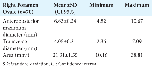
The maximum anteroposterior diameter, transverse diameter, and surface area of the foramina on the left side resulted in 6.63 ± 0.24 mm (95% CI), 4.05 ± 0.21 mm (95% CI), and 21.31 ± 1.55 mm2 (95% CI), respectively [
The maximum anteroposterior diameter among all analyzed foramen ovale was 10.67 mm, the minimum was 4.41 mm, the maximum transverse diameter was 7.09 mm, and the minimum was 2.36 mm. Regarding the surface area of the foramen ovale, including all analyzed foramina, the maximum was 44.13 mm2, and the minimum was 10.16 mm2.
The average minimum distance from the foramen ovale to the foramen spinosum was 2.32 ± 0.24 (95% CI) [

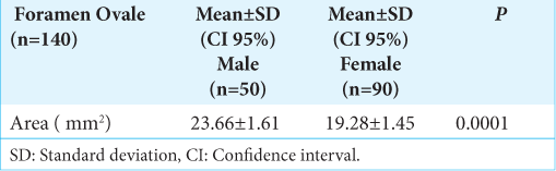
When comparing all measured foramen ovale between males and females, we found that in males (23.66 ± 1.61), the foramen ovale is 22% larger than in females (19.28 ± 1.45) (P = 0.0001) [
DISCUSSION
While there are existing morphometric studies analyzing various foramens of the human skull base, the objective of this study is to obtain, through tomographic images, an analysis that allows us to understand the morphometry of the foramen ovale in the Mexican population. In addition, we aim to determine the distance between the foramen ovale and the nearest foramen in the skull base, the foramen spinosum. This will provide a clearer understanding of this important foramen in neurosurgery, through which diagnostic and therapeutic procedures can be performed.
CONCLUSION
The foramen ovale is one of the main anatomical structures of the skull base, and besides that, it is complex and not directly accessible for clinical evaluation; useful information can be obtained through morphometric analysis. The present study provides specific anatomical data with morphological patterns to increase the understanding of the characteristics of the foramen ovale in the Mexican population. These are intended to be helpful in the pursuit of acknowledging the morphometrics and thus being able to plan neurosurgical procedures in the middle cranial fossa.
Ethical approval
The Institutional Review Board approval is not required.
Declaration of patient consent
Patients’ consent not required as patients’ identities were not disclosed or compromised.
Financial support and sponsorship
Nil.
Conflicts of interest
There are no conflicts of interest.
Use of artificial intelligence (AI)-assisted technology for manuscript preparation
The authors confirm that there was no use of artificial intelligence (AI)-assisted technology for assisting in the writing or editing of the manuscript and no images were manipulated using AI.
Disclaimer
The views and opinions expressed in this article are those of the authors and do not necessarily reflect the official policy or position of the Journal or its management. The information contained in this article should not be considered to be medical advice; patients should consult their own physicians for advice as to their specific medical needs.
References
1. Açikgöz AK, Babacan S, Tuncel Çini N, Bozkir MG. Anatomical dimensions and variances of the foramen ovale in adult human skulls: Morphometry of the foramen ovale. J Surg Med. 2022. 6: 839-43
2. Bhattarai R, Panthi S, Yadav GK, Bhandari S, Acharya R, Sharma A. Morphometric analysis of foramen ovale, foramen spinosum, and foramen rotundum of human skull using computed tomography scan: A cross-sectional study. Ann Med Surg. 2012. 85: 1731-6
3. Khairnar KB, Bhusari PA. An anatomical study on the foramen ovale and the foramen spinosum. J Clin Diagn Res. 2013. 7: 427-9
4. Raguž M, Dumić-Čule I, Almahariq F, Romić D, Gajski D, Blažević A. Foramen ovale and foramen rotundum: Characterization of postnatal development. Acta Clin Croat. 2022. 60: 415-22
5. Ray B, Gupta N, Ghose S. Anatomic variations of foramen ovale. Kathmandu Univ Med J (KUMJ). 2005. 3: 64-8
6. Rhoton AL. The anterior and middle cranial base. Neurosurgery. 2002. 51: S273-302
7. Somesh MS, Sridevi HB, Prabhu LV, Swamy MS, Krishnamurthy A, Murlimanju BV. A morphometric study of foramen ovale. Turk Neurosurg. 2011. 21: 378-83
8. Yanagi S. Developmental studies on the foramen rotundum, foramen ovale and foramen spinosum of the human sphenoid bone. Hokkaido Igaku Zasshi [Hokkaido J Med Sci]. 1987. 62: 485-96

