

- Ghaly Neurosurgical Associates, Aurora, Illinois, USA
Correspondence Address:
R. F. Ghaly
Ghaly Neurosurgical Associates, Aurora, Illinois, USA
DOI:10.4103/sni.sni_433_16
Copyright: © 2017 Surgical Neurology International This is an open access article distributed under the terms of the Creative Commons Attribution-NonCommercial-ShareAlike 3.0 License, which allows others to remix, tweak, and build upon the work non-commercially, as long as the author is credited and the new creations are licensed under the identical terms.How to cite this article: R. F. Ghaly, A. Lissounov. A modified transcondylar screw to accommodate anatomical skull base variations. 05-Jun-2017;8:98
How to cite this URL: R. F. Ghaly, A. Lissounov. A modified transcondylar screw to accommodate anatomical skull base variations. 05-Jun-2017;8:98. Available from: http://surgicalneurologyint.com/surgicalint-articles/a-modified-transcondylar-screw-to-accommodate-anatomical-skull-base-variations/
Date of Submission
31-Oct-2016
Date of Acceptance
19-Mar-2017
Date of Web Publication
05-Jun-2017
Abstract
Background:Occipitocervical instability may be attributed to congenital, bony/ligamentous abnormalities, trauma, neoplasm, degenerative bone disease, and failed atlantoaxial fixation. Indications for occipitocervical fixation include the prevention of disabling pain, cranial nerve dysfunction, paralysis, or even sudden death.
Methods:The screw trajectory for the modified transcondylar screw (mTCS) is optimally planned utilizing a three-dimensional skull reconstructed image.
Results:The modified mTCS technique is helpful where there is a loss of bone, such as after prior suboccipital craniotomy and/or an inadequate occipital condyle. The new proposed technique is similar to the classical transcondylar screw placement but follows a deeper course along the bony lip of foramen magnum toward clivus from a dorsolateral approach.
Conclusion:The modified mTCS technique allows for direct visualization and, therefore, helps to avoid damage to the hypoglossal nerve and lateral aspect of brain stem.
Keywords: Clivus, occipital condyle, occipitocervical fixation, transcondylar screw
INTRODUCTION
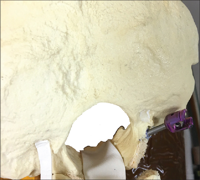
The modified transcondylar screw (mTCS) is helpful in cases lacking bone following suboccipital craniotomy and/or an inadequate/incompletely formed occipital condyle (OC). This technique is similar to the classical transcondylar screw placement for occipitocervical fusion but involves deeper placement of the screw utilizing the dorsolateral approach along the bony lip of the foramen toward the clivus [
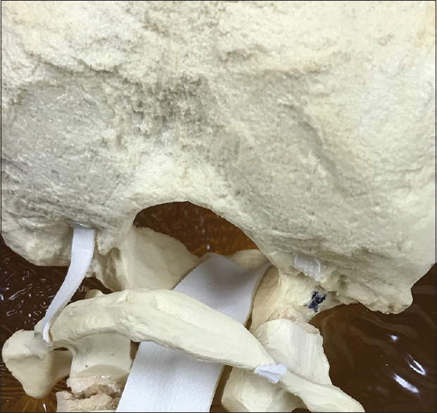
Figure 1
Posterior–inferior view of occipital bone. Posterior–lateral approach for a modified transcondylar screw is marked with black marker in the absence of occipital condyle. Entry of the screw is similar steps as transcondylar screw under guidance of intraoperative navigation and preoperative CT scan
MATERIALS AND METHODS
Utilizing a posterior approach through the occipital condyle toward the posterior–inferior region of clivus,[
RESULTS
This alternative application of modified transcondylar screw (mTCS) to the occipital condyle proved to be injury-free. There were no hypoglossal nerve, jugular bulb, and carotid or vertebral artery injuries.[
DISCUSSION
Classical techniques, such as occipital condyle screw[
The mTCS was helpful in cases where adequate bone volume was lacking, either following prior suboccipital craniotomy or inadequate OC. Utilizing this mTCS technique with the perioperative 3D-CT skull image for appropriate screw trajectory planning, the authors avoided hypoglossal nerve damage, inadvertent brain stem injury, and nerve tissue irritation.
Financial support and sponsorship
Nil.
Conflicts of interest
There are no conflicts of interest.
References
1. Grob D, Dvorak J, Panjabi M, Froehlich M, Hayek J. Posterior occipitocervical fusion. A preliminary report of a new technique. Spine. 1991. 16: 17-24
2. Hong JT, Takigawa T, Sugisaki K, Espinoza Orías AA, Inoue N. Biomechanical and morphometric evaluation of occipital condyle for occipitocervical segmental fixation. Neurol Med Chir (Tokyo). 2011. 51: 701-6
3. Ji W, Wang XY, Xu HZ, Yang XD, Chi YL, Yang JS. The anatomic study of clival screw fixation for the craniovertebral region. Eur Spine J. 2012. 21: 1483-91
4. Leggon R, Lindsey RW, Doherty BJ, Alexander J, Noble P. The holding strength of cannulated screws compared with solid core screws in cortical and cancellous bone. J Orthop Trauma. 1993. 7: 450-7
5. Menezes AH. Occipitocervical fixation. World Neurosurg. 2010. 73: 635-7
6. Uribe JS, Ramos E, Vale F. Feasibility of occipital condyle screw placement for occipitocervical fixation: A cadaveric study and description of a novel technique. J Spinal Disord Tech. 2008. 21: 540-6
7. Uribe JS, Ramos E, Youssef AS, Levine N, Turner AW, Johnson WM. Craniocervical fixation with occipital condyle screws: Biomechanical analysis of a novel technique. Spine. 2010. 35: 931-8

