- Department of Neurosurgery, Maastricht University Medical Center, Debyelaan, The Netherlands
Correspondence Address:
Yasin Temel
Department of Neurosurgery, Maastricht University Medical Center, Debyelaan, The Netherlands
DOI:10.4103/2152-7806.194494
Copyright: © 2016 Surgical Neurology International This is an open access article distributed under the terms of the Creative Commons Attribution-NonCommercial-ShareAlike 3.0 License, which allows others to remix, tweak, and build upon the work non-commercially, as long as the author is credited and the new creations are licensed under the identical terms.How to cite this article: Saeed Banaama, Jacobus van Overbeeke, Yasin Temel. An unusual case of repeated intracranial hemorrhage in vestibular schwannoma. 21-Nov-2016;7:
How to cite this URL: Saeed Banaama, Jacobus van Overbeeke, Yasin Temel. An unusual case of repeated intracranial hemorrhage in vestibular schwannoma. 21-Nov-2016;7:. Available from: http://surgicalneurologyint.com/surgicalint_articles/an-unusual-case-of-repeated-intracranial-hemorrhage-in-vestibular-schwannoma/
Abstract
Background:Symptomatic intratumoral hemorrhage (ITH) in vestibular schwannoma (VS) is rare. A repeated hemorrhage is, therefore, even more exceptional. Repeated ITH has been reported in four cases thus far in English literature. Here, we describe a patient with a Koos grade D VS who presented to our Skull Base team with repeated ITH and an unexpected disease course.
Case Description:A 76-year-old woman presented with hearing loss due to polycystic VS on the left side. Five years later, the patient was presented with facial palsy caused by hemorrhage in the VS. The patient had an eventful medical history that necessitated the use of anti-coagulants. The patient suffered from three subsequent hemorrhages preoperatively and one hemorrhage 36 h postoperatively.
Conclusion:We have experienced multiple repeated hemorrhages in a patient with a polycystic VS, and despite surgical intervention, the outcome was unfavorable.
Keywords: Acoustic neuroma, repeated intratumoral hemorrhages, vestibular schwannoma
INTRODUCTION
Vestibular schwannomas (VS) are benign masses of the 8th cranial nerve (CN VIII). Approximately 8–10% of all intracranial tumors are VS. They are considered to be the most common tumor in the cerebellopontine angle (CPA), as they constitute 75% of the tumors found in this area.[
CASE DESCRIPTION
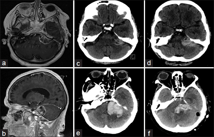
A 76-year-old female patient was diagnosed with a left polycystic VS Koos grade D in the cerebellopontine angle in 2011 [Figure
Figure 1
(a) An axial T1 weighted magnetic resonance imaging (MRI) of the polycystic vestibular schwannoma with a mass effect. (b) A T1 weighted MRI sagittal view of the vestibular schwannoma. (c) Computerized tomography imaging of the first intratumoral hemorrhage in vestibular schwannoma; (d) Second hemorrhage; (e) Third hemorrhage; (f): fourth hemorrhage 36 h after intervention
The medical history of the patient included hypertension, hypercholesterolemia, thrombosis of the right carotid artery, acute myocardial infarction, total knee replacement, thoracic and lumbar fractures, diabetic retinopathy, aortic valve replacement, coronary artery bypass surgery, and postoperative arterial fibrillation. The patient used an extensive list of medications, which included anti-coagulants.
The VS remained stable on follow-up. In May 2016, the patient presented with a severe headache and a facial palsy House and Brackmann (HB) grade 5. Computerized tomography (CT) scan showed an ITH [
Histopathological examination confirmed the diagnosis of VS. Furthermore, it showed an extensive hemorrhage with small focal fragments of fusiform proliferation and thick-walled dilated blood vessels. Immunological examination showed a positive S100 and a low mitotic activity (MIB1) in tumor areas. Glial fibrillary acidic protein (GFAP) staining was negative. The tumor area showed inflammation and contained eosinophil cytoplasm with elongated nuclei.
DISCUSSION
ITH in VS is uncommon.[
Cystic vestibular schwannoma (CVS) has been described to have a more rapid expansion rate than the solid ones, faster involvement of nerves, and expression of symptoms as well as erratic behavior. It is also postulated that the formation of the cyst is due ITH.[
A notable similarity between our case and that of Mandl[
Financial support and sponsorship
Nil.
Conflicts of interest
There are no conflicts of interest.
References
1. Kim SH, Youm JY, Song SH, Kim Y, Song KS. Vestibular schwannoma with repeated intratumoral hemorrhage. Clin Neurol Neurosurg. 1998. 100: 68-74
2. Kurata A, Saito T, Akatsuka M, Kann S, Takagi H. Acoustic neurinoma presenting with repeated intratumoral hemorrhage. Case report. Neurol Med Chir. 1989. 29: 328-32
3. Mandl ES, Vandertop WP, Meijer OW, Peerdeman SM. Imaging-documented repeated intratumoral hemorrhage in vestibular schwannoma: A case report. Acta Neurochir. 2009. 151: 1325-7
4. Niknafs YS, Wang AC, Than KD, Etame AB, Thompson BG, Sullivan SE. Hemorrhagic vestibular schwannoma: Review of the literature. World Neurosurg. 2014. 82: 751-6
5. Piccirillo E, Wiet MR, Flanagan S, Dispenza F, Giannuzzi A, Mancini F. Cystic vestibular schwannoma: Classification, management, and facial nerve outcomes. Otol Neurotol. 2009. 30: 826-34
6. Sughrue ME, Kaur R, Kane AJ, Rutkowski MJ, Yang I, Pitts LH. Intratumoral hemorrhage and fibrosis in vestibular schwannoma: A possible mechanism for hearing loss: Clinical article. J Neurosurg. 2011. 114: 386-93
7. Takeuchi S, Nawashiro H, Otani N, Sakakibara F, Nagatani K, Wada K. Vestibular schwannoma with repeated intratumoral hemorrhage. J Clin Neurosci. 2012. 19: 1305-7


