- Department of Neurosurgery, University of Sao Paulo, São Paulo,
- Department of Neurosurgery, Atenas University Center, Paracatu, Minas Gerais, Brazil.
Correspondence Address:
Eberval Gadelha Figueiredo
Department of Neurosurgery, University of Sao Paulo, São Paulo,
DOI:10.25259/SNI_515_2019
Copyright: © 2020 Surgical Neurology International This is an open-access article distributed under the terms of the Creative Commons Attribution-Non Commercial-Share Alike 4.0 License, which allows others to remix, tweak, and build upon the work non-commercially, as long as the author is credited and the new creations are licensed under the identical terms.How to cite this article: Francisco Matos Ureña, Jose Gregorio Matos Ureña, Saul Almeida, Nícollas Nunes Rabelo, Julian Reis da Silva, Mauricio Mandel, Manoel Jacobsen Teixeira, Eberval Gadelha Figueiredo. Anterior communicating artery duplication associated with a triplication of anterior cerebral artery – A rare anatomical variation. 06-Mar-2020;11:36
How to cite this URL: Francisco Matos Ureña, Jose Gregorio Matos Ureña, Saul Almeida, Nícollas Nunes Rabelo, Julian Reis da Silva, Mauricio Mandel, Manoel Jacobsen Teixeira, Eberval Gadelha Figueiredo. Anterior communicating artery duplication associated with a triplication of anterior cerebral artery – A rare anatomical variation. 06-Mar-2020;11:36. Available from: https://surgicalneurologyint.com/surgicalint-articles/9951/
Abstract
Background: The anterior communicating artery complex may presente several anatomical variations, and many abnormalities have been reported in radiologiacal and cadaveric studies.
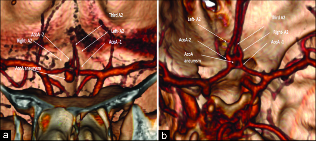
Case Description: The authors present a case of a 44-year-old Caucasian female, with a prior history of smoking and arterial systemic hypertension, admitted in the emergency department complaining of a sudden headache, nausea, and vomiting followed by tonic-clonic seizures. Computerized tomography (CT) and angiography (angio- CT) were carried out and showed Fisher Grade IV subarachnoid hemorrhage. Angio-CT revealed an anterior communicating artery (AComA) aneurysm. Minimally invasive craniotomy and microsurgical clipping were performed uneventfully. An unusual anatomical variation of the AComA complex characterized by duplication of the AComA associated with a triplication of anterior cerebral artery (ACA) was observed. The patient was discharged with no neurological deficits.
Concluision: This unique anatomical variation of the AComA-ACA complex constitute risck factors for development and rupture of aneurysms.
Keywords: Anatomy, Anterior cerebral artery, Anterior communicating artery
INTRODUCTION
The circle of Willis was originally described in 1664.[
Several authors have studied these anatomical variations and proposed many common anatomical variations. Understanding the anatomy of the anterior communicating artery complex and its possible variations is fundamental for the treatment of aneurysm in this topography, considering therapeutic planning decision-making process (endovascular or microsurgical clipping) and to prevent complications related to the procedure.
In this paper, we present a case with an unusual association of duplication of the AComA associated with a triplication of ACA. To the best of our knowledge, this is the second description of this kind of variation in the literature, the first that was depicted in the radiological study.
CASE REPORT
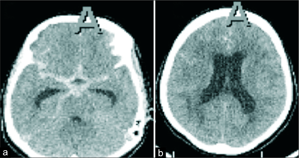
A 44-year-old Caucasian female, with a previous history of smoking and arterial systemic hypertension, was admitted to our emergency department complaining of a sudden headache, nausea, and vomiting. At her initial presentation, she was awake and aware, without any neurological deficits. Six hours after, she presented generalized tonic-clonic seizures. The patient received an attack dose of phenytoin and improved her level of consciousness after the postictal period. Head computerized tomography (CT) and angiography (angio-CT) were performed. Subarachnoid hemorrhage Fisher Grade IV was diagnosed [
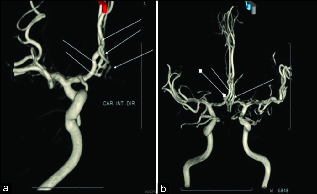
Figure 3:
(a) Frontal and (b) coronal view. Postoperative 3D digital subtraction angiography illustrating the described malformation with no residual neck of the ACoA an aneurysm, after surgery, shown in arrows: left, right, and third A2, ACoA 1 and 2, and ACoA aneurysm. Narrows show the anatomy variation.
DISCUSSION
It is well known that some anatomical variations of the A1 artery (diameter asymmetry) are associated with more risk of intracranial aneurysm development and rupture.[
CONCLUSION
We reported the first radiological description of a very rare duplication of the AComA associated with triplication of the ACA and a ruptured intracranial aneurysm. Anatomical variations in the ACA-AComA complex constitute risk factors for the development and rupture of aneurysms. The perfect understanding of the regional anatomy and its variants is fundamental for successful endovascular or surgical treatment of aneurysms located at this site.
Declaration of patient consent
The authors certify that they have obtained all appropriate patient consent.
Financial support and sponsorship
Nil.
Conflicts of interest
There are no conflicts of interest.
References
1. Cui Y, Xu T, Chen J, Tian H, Cao H. Anatomic variations in the anterior circulation of the circle of Willis in cadaveric human brains. Int J Clin Exp Med. 2015. 8: 15005-10
2. Dimmick SJ, Faulder KC. Normal variants of the cerebral circulation at multidetector CT angiography. Radiographics. 2009. 29: 1027-43
3. He J, Liu H, Huang B, Chi C. Investigation of morphology and anatomic variations of circle of Willis and measurement of diameter of cerebral arteries by 3D-TOF angiography. Sheng Wu Yi Xue Gong Cheng Xue Za Zhi. 2007. 24: 39-44
4. Krayenbuhl HA, Yasargil MG.editors. Cerebral Angiography. New York: Thieme Medical Publishers Inc.; 1982. p. 91-3
5. Sun C, Xv ZD, Yuan ZG, Wang XM, Wang LJ, Liu C. MSCT diagnosis of aneurysms associated with an unusual variant: Atypical triplication anterior cerebral artery. Surg Radiol Anat. 2012. 34: 777-80
6. Tarulli E, Fox AJ. Potent risk factor for aneurysm formation: Termination aneurysms of the anterior communicating artery and detection of A1 vessel asymmetry by flow dilution. AJNR Am J Neuroradiol. 2010. 31: 1186-91
7. Weir B, Macdonald LR, Wilkins RH, Rengachari S.editors. Aneurysms and subarachnoid hemorrhage. Neurosurgery. New York: Mc Graw Hil; 1996. p. 2191-213
8. Wolpert SM. The circle of Willis. AJNR Am J Neuroradiol. 1997. 18: 1033-4
9. Zunon-Kipré Y, Peltier J, Haïdara A, Havet E, Kakou M, Le Gars D. Microsurgical anatomy of distal medial striate artery (recurrent artery of Heubner). Surg Radiol Anat. 2012. 34: 15-20


