

- College of Medicine, King Saud bin Abdulaziz University for Health Sciences, Riyadh, Saudi Arabia.
- King Abdullah International Medical Research Center, Riyadh, Saudi Arabia.
- Division of Neurosurgery, Department of Surgery, King Abdulaziz Medical City, Ministry of National Guard - Health Affairs, Riyadh, Saudi Arabia.
Correspondence Address:
Ali Alkhaibary, College of Medicine, King Saud bin Abdulaziz University for Health Sciences, Riyadh, Saudi Arabia.
DOI:10.25259/SNI_874_2023
Copyright: © 2024 Surgical Neurology International This is an open-access article distributed under the terms of the Creative Commons Attribution-Non Commercial-Share Alike 4.0 License, which allows others to remix, transform, and build upon the work non-commercially, as long as the author is credited and the new creations are licensed under the identical terms.How to cite this article: Ali Alkhaibary1,2,3, Ahoud Alharbi1,2,3, Sami Khairy1,2,3. Calvarial multiple myeloma: Raindrop skull. 09-Feb-2024;15:34
How to cite this URL: Ali Alkhaibary1,2,3, Ahoud Alharbi1,2,3, Sami Khairy1,2,3. Calvarial multiple myeloma: Raindrop skull. 09-Feb-2024;15:34. Available from: https://surgicalneurologyint.com/surgicalint-articles/12741/
Date of Submission
27-Oct-2023
Date of Acceptance
12-Dec-2023
Date of Web Publication
09-Feb-2024
Abstract
Background: The “Raindrop skull” appearance represents the multiple punched-out and lytic lesions hitting a surface and creating a scattered splash pattern.
Case Description: A 73-year-old female presented with multiple painless lumps over the forehead and head. The patient reported unintentional weight loss, fatigability, loss of appetite, fever, night sweats, and back pain for seven months (B symptoms). The examination revealed multiple, nonmobile, calvarial lesions with defined borders, measuring approximately 1 × 1 cm. Laboratory investigations of serum-free light chains showed a free kappa level of 12.91 mg/L, a lambda level of 4549.28 mg/L, and a free kappa/lambda ratio of 0.00. Radiological imaging of the skull and brain showed a “raindrop skull” appearance and multiple calvarial osteolytic lesions. The patient underwent a right superior iliac crest bone marrow aspirate and trephine biopsy. The laboratory and histopathological sections were compatible with multiple myeloma. A diagnosis of multiple myeloma (free light chain lambda) was rendered.
Conclusion: Calvarial multiple myeloma is rare and requires a high index of suspicion to diagnose. “Raindrop skull” appearance is pathognomonic of calvarial multiple myeloma.
Keywords: Kappa, Lambda, Osteolytic, Plasma cell, Skull
CASE DESCRIPTION
A 73-year-old female presented with multiple painless lumps over the forehead and head. The patient reported unintentional weight loss, fatigability, loss of appetite, fever, night sweats, and back pain for seven months (B symptoms). The examination revealed multiple, nonmobile, calvarial lesions with defined borders, measuring approximately 1 × 1 cm. No communicating sinuses, skin breakthroughs, or discharges were noted.
Laboratory investigations of serum free light chains showed free kappa level of 12.91 mg/L (reference range: 3.30–19.4 mg/L), lambda level of 4549.28 mg/L (reference range: 5.71–26.30 mg/L), and free kappa/lambda ratio of 0.00 (reference range: 0.26–1.65). Urine electrophoresis was notable for the Bence-Jones protein. A skeletal survey radiograph showed multiple compression fractures of the thoracolumbar spine and bilateral femoral lytic lesions. Radiological imaging of the skull and brain showed a “raindrop skull” appearance and multiple calvarial osteolytic lesions [
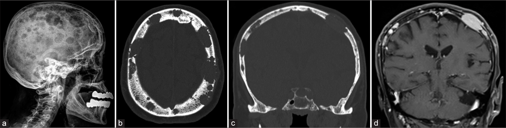
Figure 1:
(a) Lateral skull radiograph demonstrating the classic “raindrop skull” appearance. (b and c) Axial and coronal brain computed tomography, “bone window,” shows variable-sized, well-defined osteolytic lesions of the calvarial vault; the largest lesion measures 2.4 cm at the left frontal bone. (d) Coronal brain magnetic resonance imaging with contrast showing subgaleal and epidural soft tissue component enhancement. An incidental left cerebellar arachnoid cyst is noted. The brain parenchyma is unremarkable.
The patient underwent a right superior iliac crest bone marrow aspirate and trephine biopsy. The histopathological sections were compatible with multiple myeloma. Peripheral blood smear was notable for rouleaux formation of red blood cells. The immune profile was suggestive of plasma cell neoplasm (80% of total bone marrow cellularity). A diagnosis of multiple myeloma (free light chain lambda) was rendered. The patient was commenced on immunotherapy (DARA-RD protocol; daratumumab, lenalidomide, and dexamethasone). She was discharged and followed up for one year and is still being followed up clinicoradiologically.
DISCUSSION
The appearance of a “Raindrop skull” represents the multiple punched-out and lytic lesions hitting a surface and creating a scattered splash pattern.[
CONCLUSION
The present article illustrates the clinical and radiological appearance of a patient with a classic “raindrop skull” appearance of calvarial multiple myeloma. Neurosurgeons should keep in mind the radiological appearance of such a rare finding in patients with multiple myeloma.
Ethical approval
Ethical approval was obtained from King Abdullah International Medical Research Center. The assigned protocol number was IRB/2107/23.
Declaration of patient consent
Patient’s consent not required as patient’s identity is not disclosed or compromised.
Financial support and sponsorship
Nil.
Conflicts of interest
There are no conflicts of interest.
Use of artificial intelligence (AI)-assisted technology for manuscript preparation
The authors confirm that there was no use of artificial intelligence (AI)-assisted technology for assisting in the writing or editing of the manuscript and no images were manipulated using AI.
Disclaimer
The views and opinions expressed in this article are those of the authors and do not necessarily reflect the official policy or position of the Journal or its management. The information contained in this article should not be considered to be medical advice; patients should consult their own physicians for advice as to their specific medical needs.
References
1. Gomez CK, Schiffman SR, Bhatt AA. Radiological review of skull lesions. Insights Imaging. 2018. 9: 857-82
2. Simsek AT, Calis F, Dursun FE, Simsek BC, Akdemir H, Alyanak D. Giant cranial plasmacytoma: Case report and discussion of a potential relationship with sex hormones. Neurol Neurochir Pol. 2023. 57: 314-6
3. Solis F, Gonzalez C. Raindrop skull. N Engl J Med. 2018. 378: 1930

