

- Institute of Neurosurgery and Pathology, Catholic University, Rome, Italy
Correspondence Address:
Nicola Montano
Institute of Neurosurgery and Pathology, Catholic University, Rome, Italy
DOI:10.4103/2152-7806.162551
Copyright: © 2015 Pignotti F. This is an open-access article distributed under the terms of the Creative Commons Attribution License, which permits unrestricted use, distribution, and reproduction in any medium, provided the original author and source are credited.How to cite this article: Pignotti F, Coli A, Fernandez E, Montano N. Capillary hemangioma of the cauda equina. Surg Neurol Int 10-Aug-2015;6:133
How to cite this URL: Pignotti F, Coli A, Fernandez E, Montano N. Capillary hemangioma of the cauda equina. Surg Neurol Int 10-Aug-2015;6:133. Available from: http://surgicalneurologyint.com/surgicalint_articles/capillary-hemangioma-of-the-cauda-equina/
Sir,
We read with great interest, the recent review about capillary hemangiomas of cauda equina published by Liu et al.[
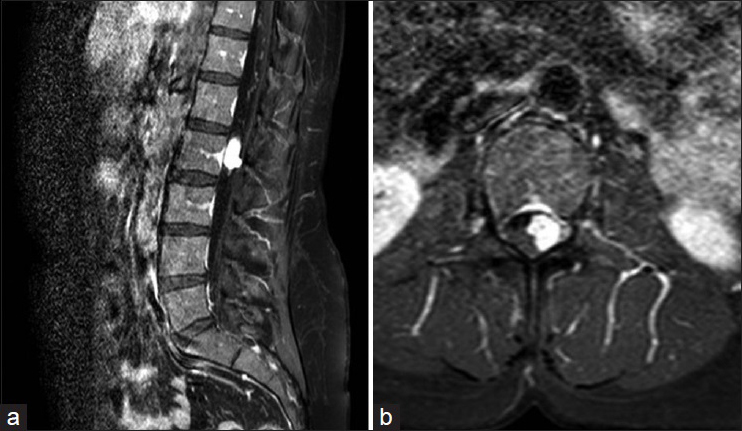
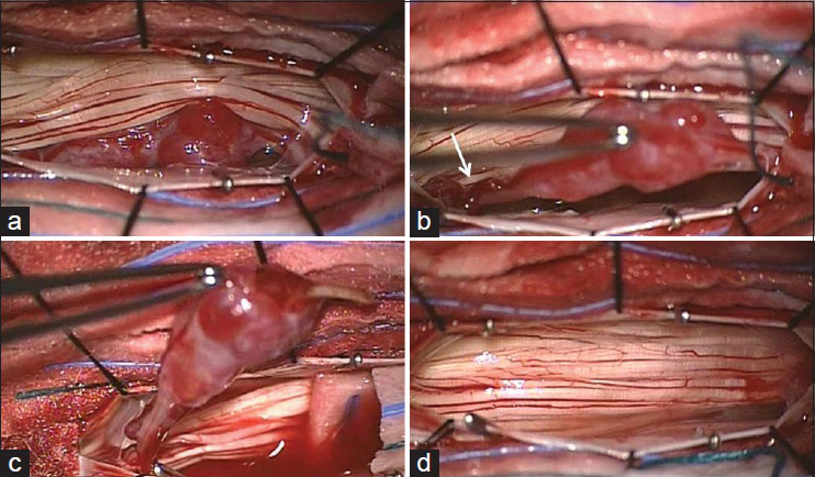
Figure 2
Intraoperative view of lesion. The lesion appeared as a white reddish elliptical-nodular lesion intimately involved with the nerve root displacing the cauda equina (a) Enlarged vessels were also evident along the nerve root (b, white arrow). After dissecting the lesion from cauda equina and cutting the originating nerve root distally (c) the lesion was totally removed (d)
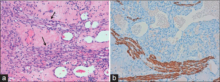
Figure 3
Histological picture (a) of the surgical specimen shows many, variably sized, thin-walled vascular channels lined by endothelial cells with interposed nerve fibers (black arrows) (H and E, original magnification ×20). Immunohistochemical staining (b) for neurofilament protein highlights the nerve fibers interspersed among the vascular spaces. The lesion was also positive at S-100 staining (not showed) (original magnification ×20)
To our knowledge, we report the first intraoperative picture of a capillary hemangioma of cauda equina. Only a few cases of capillary hemangiomas have been reported at cauda equina.[
References
1. Ganapathy S, Kleiner LI, Mirkin LD, Hall L. Intradural capillary hemangioma of the cauda equina. Pediatr Radiol. 2008. 38: 1235-8
2. Liu JJ, Lee DJ, Jin LW, Kim KD. Intradural extramedullary capillary hemangioma of the cauda equina: Case report and literature review. Surg Neurol Int. 2015. 6: S127-31
3. Mastronardi L, Guiducci A, Frondizi D, Carletti S, Spera C, Maira G. Intraneural capillary hemangioma of the cauda equina. Eur Spine J. 1997. 6: 278-80
4. Nowak DA, Gumprecht H, Stölzle A, Lumenta CB. Intraneural growth of a capillary haemangioma of the cauda equina. Acta Neurochir (Wien). 2000. 142: 463-7
5. Nowak DA, Widenka DC. Spinal intradural capillary haemangioma: A review. Eur Spine J. 2001. 10: 464-72

