

- Department of Neurosurgery, Kobe City Medical Center General Hospital, Kobe, Japan.
- Department of Neurosurgery, Hikone Chuo Hospital, Hikone, Japan.
- Department of Neurosurgery, Japanese Red Cross Otsu Hospital, Otsu, Japan.
Correspondence Address:
Yoshinori Maki, Department of Neurosurgery, Hikone Chuo Hospital, Hikone, Japan.
DOI:10.25259/SNI_613_2023
Copyright: © 2023 Surgical Neurology International This is an open-access article distributed under the terms of the Creative Commons Attribution-Non Commercial-Share Alike 4.0 License, which allows others to remix, transform, and build upon the work non-commercially, as long as the author is credited and the new creations are licensed under the identical terms.How to cite this article: Kota Nakajima1, Yoshinori Maki2, Toshinari Kawasaki3, Motohiro Takayama3. Cervical dumbbell-type tumor spontaneously shrinking following an ischemic stroke. 25-Aug-2023;14:301
How to cite this URL: Kota Nakajima1, Yoshinori Maki2, Toshinari Kawasaki3, Motohiro Takayama3. Cervical dumbbell-type tumor spontaneously shrinking following an ischemic stroke. 25-Aug-2023;14:301. Available from: https://surgicalneurologyint.com/surgicalint-articles/12512/
Date of Submission
21-Jul-2023
Date of Acceptance
04-Aug-2023
Date of Web Publication
25-Aug-2023
Abstract
Background: Asymptomatic cervical dumbbell-type tumors can be incidentally diagnosed. Notably, the chronological changes in the size of these tumors have not been satisfactorily described.
Case Description: A 57-year-old man was clinically followed for an asymptomatic cervical dumbbell-type tumor that had the appearance of a schwannoma on magnetic resonance (MR) images obtained over a 7-year period. Notably, the tumor compressed both the spinal cord and the right vertebral artery. At the end of the 7-year period, the patient sustained a cerebral infarction due to atherosclerosis of the right vertebral artery; the angiogram revealed both atherosclerosis and the tumor compressing the right vertebral artery. After the stroke/ischemic event, the tumor progressively shrunk on MR images obtained for the following 4 years, and the spinal cord compression was similarly relieved.
Conclusion: Here, we report on a 57-year-old man with cervical MR images revealing that a cervical dumbbell schwannoma was progressively compressing both the spinal cord and the right vertebral artery. However, following a cerebral infarction, the tumor underwent spontaneous shrinkage over the next 4 years, thus relieving the compression.
Keywords: Cervical dumbbell-type tumor, Ischemic stroke, Natural course, Spontaneous shrinkage, Vertebral artery stenosis
INTRODUCTION
In some cases, conservative treatment may be appropriate for the management of asymptomatic dumbbell-type tumors.[
CASE PRESENTATION
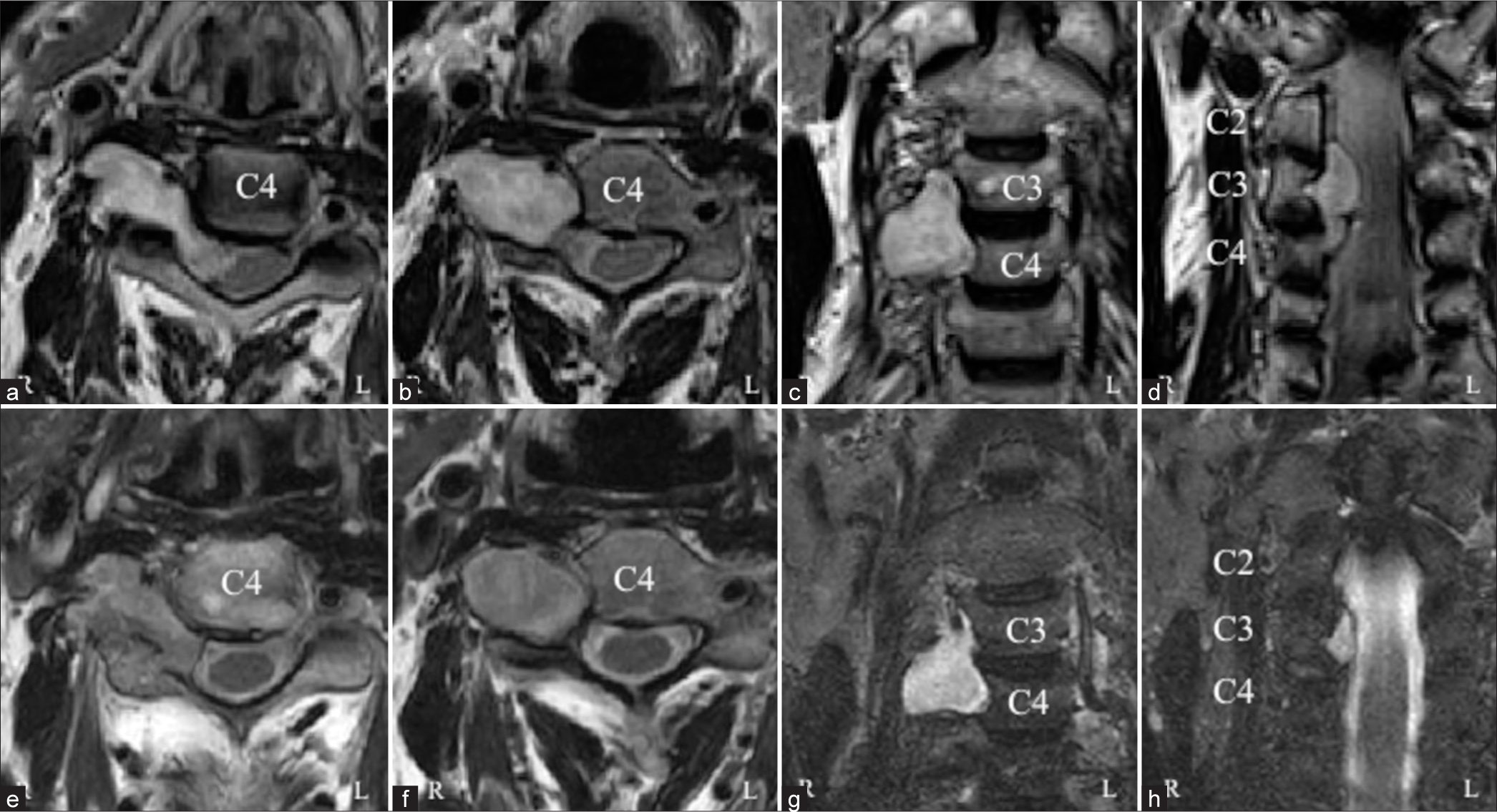
For 7 years, a 57-year-old man underwent repeated magnetic resonance (MR) scans that documented progressive spinal cord and right vertebral artery compression attributed to a dumbbell schwannoma with a likely origin in the C4 root. The lesion was isointense on T1-weighted and hyperintense on T2-weighted MR images, and it was heterogeneously enhanced with gadolinium [
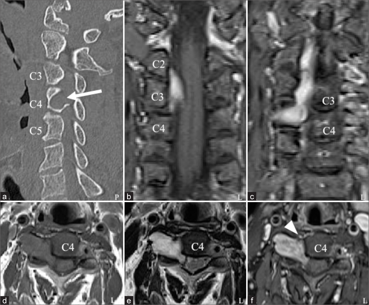
Figure 1:
Initial radiological characteristics. Sagittal computed tomography images reveal the enlargement of the right C3/4 intervertebral foramen (arrow) and no calcification (a). A cervical dumbbell-type tumor exhibits isointensity on an axial T1-weighted magnetic resonance image (d) and heterogeneous hyperintensity on an axial T2-weighted magnetic resonance image (e). The tumor is heterogeneously enhanced with a gadolinium contrast agent (f). The tumor is located at the C2–4 level (b and c: coronal T1-weighted images with a gadolinium contrast agent). The cervical spinal cord and right vertebral artery (f, arrowhead) are compressed by the tumor.
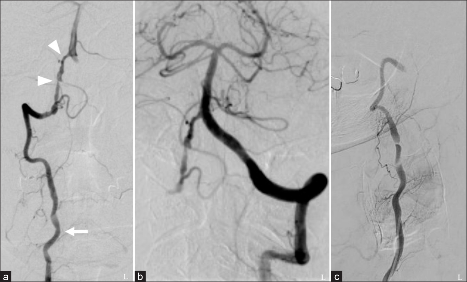
Figure 2:
Angiographical characteristics. Right vertebral angiography reveals atherosclerotic stenosis of the V4 segment (arrowheads), and the V2 segment of the right vertebral artery is medially shifted (arrow) (a). Blood flow to the right V4 segment from the left vertebral artery is observed through left vertebral angiography (b). The blood flow is interrupted upon right vertebral angiography performed during right rotation of the neck (c).
DISCUSSION
Concerning spontaneous shrinkage of spinal schwannomas, both a vascular theory (necrosis and bleeding due to thrombi in hyalinized and dilated vessels) and a mechanical theory (laceration of intratumoral arteries due to vertebral motion) of shrinkage have been proposed [
CONCLUSION
A cervical dumbbell-type tumor (likely a schwannoma) may spontaneously shrink following a cerebral infarction.
Declaration of patient consent
The authors certify that they have obtained all appropriate patient consents.
Financial support and sponsorship
Nil.
Conflicts of interest
There are no conflicts of interest.
Use of artificial intelligence (AI)-assisted technology for manuscript preparation
The authors confirm that there was no use of Artificial Intelligence (AI)-Assisted Technology for assisting in the writing or editing of the manuscript and no images were manipulated using the AI.
Disclaimer
The views and opinions expressed in this article are those of the authors and do not necessarily reflect the official policy or position of the Journal or its management. The information contained in this article should not be considered to be medical advice; patients should consult their own physicians for advice as to their specific medical needs.
References
1. De Divitiis E, Maiuri F, Corriero G, Donzelli R. Subarachnoid hemorrhage due to a spinal neurinoma. Surg Neurol. 1985. 24: 187-90
2. Luxon LM, Harrison MJ. Subarachnoid hemorrhage and papilledema due to a cervical neurilemmoma. Case report. J Neurosurg. 1978. 48: 1015-8
3. Mills B, Marks PV, Nixon JM. Spinal subarachnoid haemorrhage from an “ancient” schwannoma of the cervical spine. Br J Neurosurg. 1993. 7: 557-9
4. Suzuki M, Aizawa T, Hashimoto K, Kanno H, Onoda Y, Itoi E. Spontaneous regression of a thoracic dumbbell tumor: A case report. J East Jpn Orthop Traumatol. 2020. 32: 198-201


JOSE LOMELI
Posted September 15, 2023, 10:20 am
It is a very interesting paper, however I have 1 question, Why you didn´t operate the patient, earlier? Why wait for complications?