

- Department of Neurosurgery, “Riuniti” Hospital,
- Department of Neurosurgery, University of Foggia,
- Department of Neurosurgery, Faculty of Medicine and Surgery, University of Foggia, Foggia,
- Department of Neurosurgery, Giovanni XXIII Hospital, Bari, Italy,
- Department of Neurosurgery, Städtisches Klinikum Karlsruhe, Karlsruhe, Germany.
Correspondence Address:
Augusto Leone, Department of Neurosurgery, Städtisches Klinikum Karlsruhe, Karlsruhe, Germany.
DOI:10.25259/SNI_814_2022
Copyright: © 2022 Surgical Neurology International This is an open-access article distributed under the terms of the Creative Commons Attribution-Non Commercial-Share Alike 4.0 License, which allows others to remix, transform, and build upon the work non-commercially, as long as the author is credited and the new creations are licensed under the identical terms.How to cite this article: Antonio Colamaria1, Nicola Pio Fochi2, Yasser Andres Dallos Laguado3, Maria Blagia4, Augusto Leone5, Francesco Carbone2. Cervical intra and extramedullary hemangioblastoma with associated syringomyelia: A case report and review of the literature. 30-Sep-2022;13:448
How to cite this URL: Antonio Colamaria1, Nicola Pio Fochi2, Yasser Andres Dallos Laguado3, Maria Blagia4, Augusto Leone5, Francesco Carbone2. Cervical intra and extramedullary hemangioblastoma with associated syringomyelia: A case report and review of the literature. 30-Sep-2022;13:448. Available from: https://surgicalneurologyint.com/surgicalint-articles/11902/
Date of Submission
04-Sep-2022
Date of Acceptance
15-Sep-2022
Date of Web Publication
30-Sep-2022
Abstract
Background: Spinal hemangioblastoma (HB) is a highly vascularized tumor commonly presenting in the lower thoracic and lumbar segments. It typically causes spinal compression, extensive bleeding, and/or syringomyelia.
Case Description: A 32-year-old female presented with persistent headaches with a cervical MRI showing an intradural and extradural mass extending from the obex to C2. Following surgical tumor resection, the patient’s symptoms resolved.
Conclusion: Resection of spinal HB requires direct removal of the tumor mass as the accompanying cystic components typically spontaneously regress.
Keywords: Cervical hemangioblastoma, Fenestration, Hemangioblastoma surgery, Intra and extradural, Syringomyelia
INTRODUCTION
Spinal hemangioblastomas (HBs) are slow-growing and highly vascularized tumors, accounting for 2–15% of primary spinal cord malignancies. They can arise as an isolated lesion or as multiple tumors spread throughout the central nervous system in association with syringomyelia, intramedullary hemorrhages, or Von Hippel-Lindau (VHL) syndrome (in 20–30% of the cases).[
CASE PRESENTATION
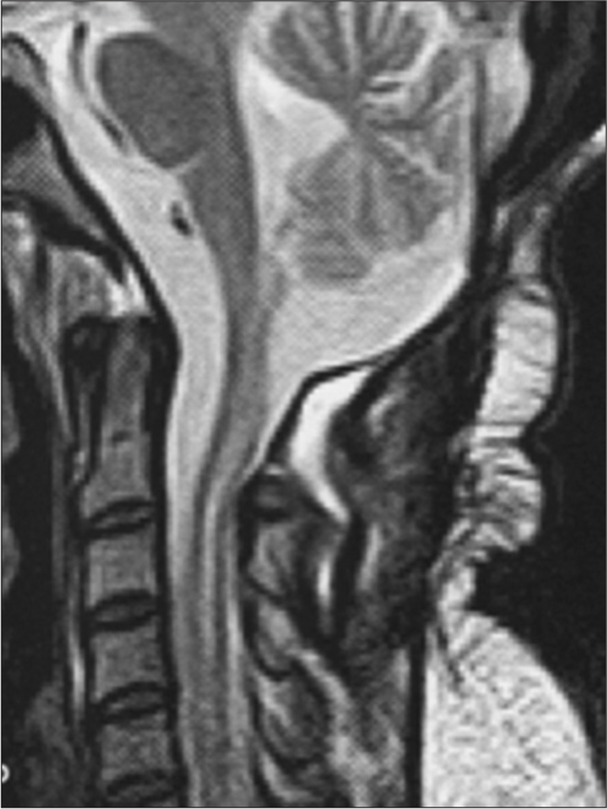
A 32-year-old female presented with occipital headaches and sudden onset of numbness in the upper extremities with gait instability without focal neurological deficits (Mc Cormick grade II). The cervical MRI with gadolinium showed an oval mass in the intradural space at the obex extending to the superior margin of C2 markedly enhancing following the administration of contrast and associated with syringomyelia extending from the obex to C6 [

Surgery
The patient underwent suboccipital craniectomy and total C1 laminectomy for complete removal of the intra and extramedullary mass under continuous spinal cord monitoring. Intraoperatively, the lesion was red and elastic in consistency, richly vascularized, and extended to the central canal of the spinal cord [

Postoperative course
Postoperatively, the gait was slightly impaired, while the headaches subsided, but the hypoesthesia in the upper extremities remained unchanged. The postoperative MRI demonstrated total tumor removal with a reduction of the syrinx. The patient was discharged 7 days later without focal deficits (Mc Cormick Grade I). At 6 months of follow-up, the patient had no residual complaints or deficits. Further, the MRI confirmed no tumor recurrence and a significant reduction of the accompanying syringomyelia [
Pathology
The pathological examination confirmed the diagnosis of a capillary HBL. It showed groups of large polygonal, lipid-laden stromal cells, interspersed with thin-walled, and closely packed blood-filled channels or vessels. Immunohistochemical staining revealed the presence of stromal cells positive for inhibin A and NSE while immunonegative for CD10 and EMA [

DISCUSSION
Sporadic spinal HBs usually present as a single lesion,[

Management options
The most effective and definitive treatment for spinal HB are represented by microsurgical resection, which should only be considered if neurological deficits are present. In VHL patients with multiple small lesions, gamma-knife radiosurgery has shown promising results although further evidence is foreseen to recommend this treatment.[
Prognosis
Gross-total resection (i.e., minimally invasive cytoreductive surgery under neurophysiological monitoring) can be achieved in over 90% of the cases and can result in full functional recovery in 96% of cases.[
Recurrence of HB
Moreover, recurrence is frequently seen after partial resections[
CONCLUSION
Spinal cervical HBs are rare malignancies and are best managed with gross total resection without the need for complete syrinx excision.
Declaration of patient consent
Patient’s consent not required as patient’s identity is not disclosed or compromised.
Financial support and sponsorship
Nil.
Conflicts of interest
There are no conflicts of interest.
References
1. Ampie L, Choy W, Khanna R, Smith ZA, Dahdaleh NS, Parsa AT. Role of preoperative embolization for intradural spinal hemangioblastomas. J Clin Neurosci. 2016. 24: 83-7
2. Barrey C, Kalamarides M, Polivka M, George B. Cervical dumbbell intra-extradural hemangioblastoma: Total removal through the lateral approach: Technical case report. Neurosurgery. 2005. 56: E625
3. Chang H, Li J, Wang P, Lu X, Li B. Microsurgical treatment of cervical spinal hemangioblastoma. Neurochirurgie. 2020. 66: 56-60
4. Colamaria A, Sacco M, Iodice S, D’Oria Parbonetti G, Carbone F. Intradural extramedullary cavernous hemangioma of the cervicothoracic junction: A case report and review of the literature. Surg Neurol Int. 2022. 13: 53
5. D’Oria S, Giraldi D, Fanelli V, D’Angelo V.editors. Sporadic hemangioblastoma of cauda equina: A case-report and brief literature review. Neurocirugía. 2022. p.
6. Gluf WM, Dailey AT. Hemorrhagic intramedullary hemangioblastoma of the cervical spinal cord presenting with acute-onset quadriparesis: Case report and review of the literature. J Spinal Cord Med. 2014. 37: 791-4
7. Kim JH, Joo SM, Cho YE, Ha SW, Suh SH. Percutaneous onyx embolization of recurrent cervical nerve root hemangioblastoma: A case report and review of the literature. Clin Neuroradiol. 2021. 31: 1209-13
8. Li D, Choe S, Borys E, Serrone JC, Germanwala AV. Primary intradural extramedullary sporadic spinal hemangioblastomas: Case report and systematic review. World Neurosurg. 2021. 152: 84-94
9. Sun Hİ, Özduman K, Usseli Mİ, Özgen S, Pamir MN. Sporadic spinal hemangioblastomas can be effectively treated by microsurgery alone. World Neurosurg. 2014. 82: 836-47
10. Takai K, Taniguchi M, Takahashi H, Usui M, Saito N. Comparative analysis of spinal hemangioblastomas in sporadic disease and Von Hippel-Lindau syndrome. Neurol Med Chir (Tokyo). 2010. 50: 560-7

