

- Department of Neurosurgery, University of Rochester Medical Center, Rochester, New York, USA
- Department of Neurosurgery, Rochester Regional Health System, Rochester, New York, USA
Correspondence Address:
Hanna Algattas
Department of Neurosurgery, Rochester Regional Health System, Rochester, New York, USA
DOI:10.4103/2152-7806.174887
Copyright: © 2016 Surgical Neurology International This is an open access article distributed under the terms of the Creative Commons Attribution-NonCommercial-ShareAlike 3.0 License, which allows others to remix, tweak, and build upon the work non-commercially, as long as the author is credited and the new creations are licensed under the identical terms.How to cite this article: Algattas H, Kimmell KT, Petraglia AL, Maurer PK. Conservative management of a cervical ligamentum flavum hematoma: Case report. Surg Neurol Int 25-Jan-2016;7:
How to cite this URL: Algattas H, Kimmell KT, Petraglia AL, Maurer PK. Conservative management of a cervical ligamentum flavum hematoma: Case report. Surg Neurol Int 25-Jan-2016;7:. Available from: http://surgicalneurologyint.com/surgicalint_articles/conservative-management-of-a-cervical-ligamentum-flavum-hematoma-case-report/
Abstract
Background:Spontaneous epidural hematoma arising from the ligamentum flavum is a rare cause of acute spinal cord compression. There are only four reports in the cervical spine literature, and all were managed with surgery. Here, we describe an acute case of a spontaneous epidural hematoma arising from the ligamentum flavum in the cervical spine successfully managed without surgery.
Case Description:A 69-year-old woman with a cervical spine epidural hematoma contained within the ligamentum flavum presented with paroxysmal neck pain and stiffness without a history of trauma. The magnetic resonance imaging (MRI) revealed a posterolateral epidural hematoma contained within the ligamentum flavum. As the patient was intact, she was managed conservatively with cervical orthosis. Three months later, she was symptom-free, and the hematoma resolved on the follow-up MRI study.
Conclusion:Spontaneous epidural hematoma arising from ligamentum flavum is a rare cause of spinal cord compression. Previous reports have described success with surgical decompression. However, initial observation and conservative management may be successful as illustrated in this case.
Keywords: Cervical spine, epidural hematoma, ligamentum flavum
INTRODUCTION
Epidural hematoma in the cervical spine contained within the ligamentum flavum is rare but can be a devastating, life-threatening condition. Patients may present with the new, sudden onset of spontaneous neck pain (e.g., without trauma), along with variable neurological deficits. Hematoma rupture into the ligamentum flavum is classically described in the lumbar spine in cases of stenosis caused by ligamental hypertrophy. However, it has only been reported in four cases in the cervical region, and all were treated surgically.[
CASE REPORT
A 69-year-old female presented with the sudden, atraumatic onset, of severe right-sided neck pain without focal symptoms or neurological deficit. Both the computed tomography and magnetic resonance imaging (MRI) of the cervical spine identified an epidural, hyperdense right-sided posterolateral mass compressing the spinal cord between the C2 and C5 vertebrae [Figures
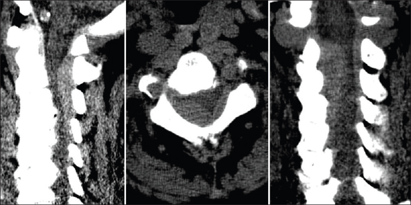
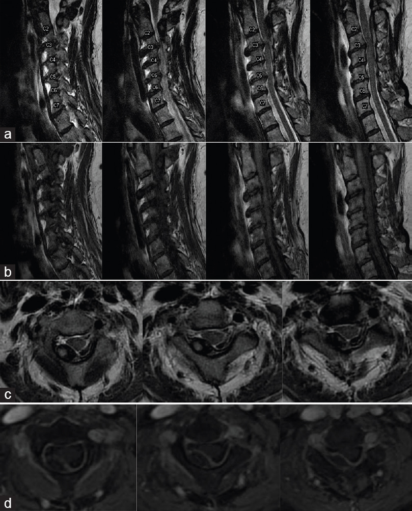
Figure 2
MRI Cervical Spine with Contrast: MRI cervical spine revealing right posterolateral epidural mass contained within ligamentum flavum and extending from C2-C5 vertebral levels. Mass appears as isointense lesion on T1-weighted images and heterogeneous isointense to hyperintense lesion on T2-weighted images. The addition of Gd-DTPA contrast enhanced the outer border of the ligament on T1-weighted images. (a) Sagittal T2- Weighted MRI, (b) Sagittal T1-Weighted MRI, (c) Axial T2-Weighted MRI, (d) Axial T1-Weighted MRI with contrast
Since the patient was neurologically intact, she was treated conservatively in a collar and discharged 2 days later. Three months later, the MRI of the cervical spine showed complete resolution of the hematoma [
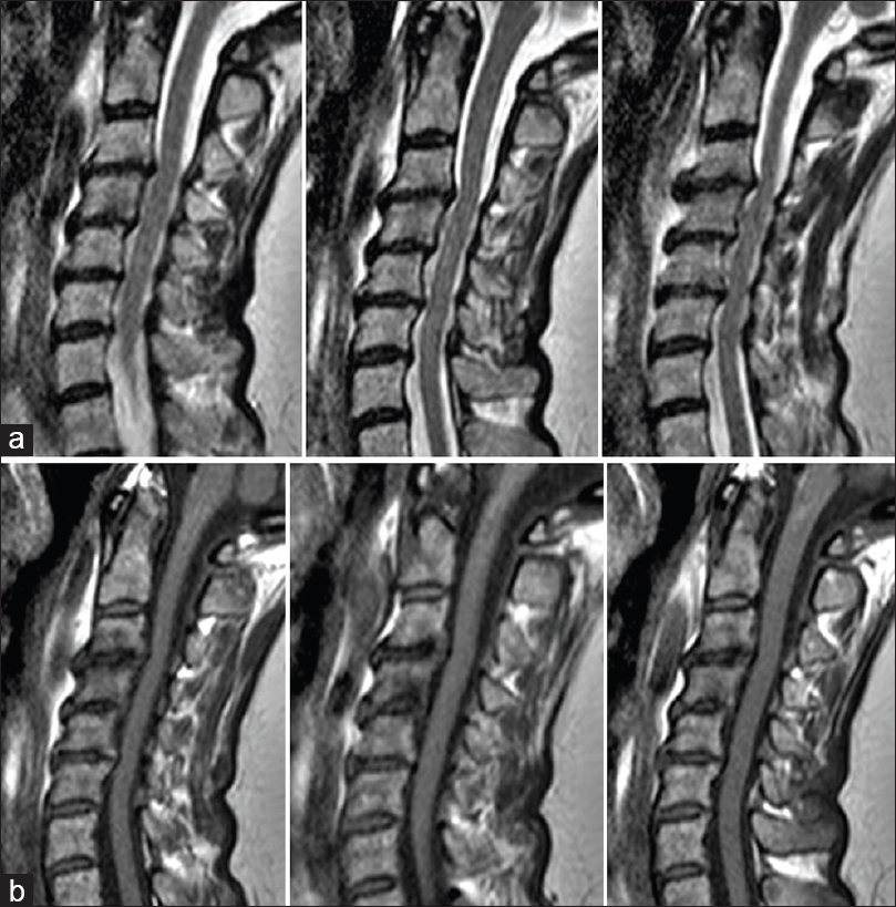
Figure 3
Follow-up MRI Cervical Spine with Contrast: Three month follow-up MRI (T1 and T2-weighted) of cervical spine demonstrating complete resolution of hematoma with moderate stenosis from pre-existing osteophyte complexes emanating from the vertebral bodies of C2-C4. (a) Follow-up Sagittal T2-Weighted MRI, (b) Follow-up Sagittal T1-Weighted MRI
DISCUSSION
Spontaneous epidural hematoma arising from the ligamentum flavum is a rare occurrence. There are rare reports of this entity in the cervical, thoracic, and lumbar spine. To date, there have been only four reported cases in the cervical spine.[
MRI is helpful in diagnosing ligamentum flavum hematomas, but there may be considerable variation in the intensity of T1, T2, and contrast enhanced images.[
Histological degenerative changes within the ligamentum flavum increased angiogenesis in regions of degeneration, and loss of elastic fibers likely constitute the pathogenic mechanism for ligamentum flavum hematomas.[
Yayama et al. have corroborated the spatial association between angiogenesis and fiber degeneration in histological and immunohistochemical analyses.[
Previous cases have been managed operatively with hemilaminectomy or laminectomy and occasionally facetectomy or interlaminotomy. In this case, the lack of neurologic compromise despite an acute presentation warranted watchful waiting and observation for hematoma progression. Cervical orthosis was placed, and the patient instructed to remain in the collar for 4 weeks. At follow-up, the patient was symptom-free with complete resolution of the hematoma.
CONCLUSIONS
The present case is the first to report on the successful conservative management of a patient with a spontaneous epidural hematoma arising from the ligamentum flavum. While surgical decompression has proven immediately beneficial in previous cases with acute neurologic decline, stable patient condition and examination may warrant initial observation and conservative management.
Financial support and sponsorship
Nil.
Conflicts of interest
There are no conflicts of interest.
References
1. Chen HC, Hsu PW, Lin CY, Tzaan WC. Symptomatic hematoma of cervical ligamentum flavum: Case report. Spine (Phila Pa 1976). 2005. 30: E489-91
2. Kotani Y, Sudo H, Abumi K, Ito M, Matsubara S, Minami A. Spinal cord compression by hematoma in the cervical ligamentum flavum: A case report. Spine J. 2012. 12: e18-21
3. Miyakoshi N, Shimada Y, Kasukawa Y, Ando S. Ligamentum flavum hematoma in the cervical spine – Case report. Neurol Med Chir (Tokyo). 2006. 46: 556-8
4. Tamura T, Sakai T, Sairyo K, Takao S, Kagawa S, Katoh S. Hematoma in the cervical ligamentum flavum. Report of a case and review of the literature. Skeletal Radiol. 2010. 39: 289-93
5. Yayama T, Kobayashi S, Sato R, Uchida K, Kokubo Y, Nakajima H. Calcium pyrophosphate crystal deposition in the ligamentum flavum of degenerated lumbar spine: Histopathological and immunohistological findings. Clin Rheumatol. 2008. 27: 597-604

