Tools

Nancy Epstein
  1. Clinical Professor of Neurosurgery, School of Medicine, State University of New York at Stony Brook.

DOI:10.25259/SNI_294_2020

Copyright: © 2020 Surgical Neurology International This is an open-access article distributed under the terms of the Creative Commons Attribution-Non Commercial-Share Alike 4.0 License, which allows others to remix, tweak, and build upon the work non-commercially, as long as the author is credited and the new creations are licensed under the identical terms.

How to cite this article: Nancy Epstein. Diagnosis, and Treatment of Cervical Epidural Abscess and/or Cervical Vertebral Osteomyelitis with or without Retropharyngeal Abscess; A Review. 20-Jun-2020;11:160

How to cite this URL: Nancy Epstein. Diagnosis, and Treatment of Cervical Epidural Abscess and/or Cervical Vertebral Osteomyelitis with or without Retropharyngeal Abscess; A Review. 20-Jun-2020;11:160. Available from: https://surgicalneurologyint.com/surgicalint-articles/10095/

Date of Submission
20-May-2020

Date of Acceptance
20-May-2020

Date of Web Publication
20-Jun-2020

Abstract

Background: Every year approximately 19.6 patients/100,000 per year are admitted to hospitals with spinal epidural abscesses (CSEA), 7.4/100,000 have vertebral osteomyelitis (VO)/100,000/year, while 4.1/100.000 children/year have cervical retropharyngeal abscesses (RPA) (i.e., data insufficient for adults).

Methods: Here we evaluated 11 individual case studies, 6 multiple patient series, and looked at 9 general review articles focusing on CSEA, and/or VO, with/without RPA.

Results: Of the 11 case studies involving 15 patients, 14 had cervical spinal epidural abscesses (CSEA: 10 CSEA/ VO/RPA, 2 CSEA/VO, 1 CSEA/TSEA, 1 CSEA/ TSEA/LSEA), 13 had cervical osteomyelitis (VO: 11 VO/CSEA, 2 VO/RPA), and 12 had cervical retropharyngeal abscesses (RPA: 10 RPA/CSEA/VO, 2 RPA/VO alone). When patients were treated surgically, they required 12 anterior, and 2 posterior approaches; 1 patient required no surgery. In the 6 larger cervical series involving 355 patients, 4 series involved CSEA (3 CSEA, 1 CSEA/VO), and 2 seires had cervical VO. Primary surgery was performed in 298 patients, while 57 were initially managed medically; 24 of these latter patients failed non-surgical therapy, and required delayed cervical surgery. Notably, all 17 clinical studies advocated early surgery where clinically appropriate for varying combinations of CSEA and/or VO with or without RPA. The 8 final articles reviewed all-levels of SEA and or VO, while also providing additional unique information regarding RPA.

Conclusion: We analyzed 11 case studies and 6 multiple case series regarding the diagnosis and treatment of combinations of cervical CSEA, and/or VO with or without RPA. We also reviewed 8 articles on the evaluation/ management of all-level SEAs and/or VOs, along with the unique features of RPAs.

Keywords: Cervical spine epidural abscesses, How to recognize failure of medical management, Retropharyngeal abscesses, Success of early surgery where appropriate for CSEA and/or VO with/without RPA, Vertebral osteomyelitis

INTRODUCTION

Every year approximately 19.6 patients/100,000 are admitted to hospitals with spinal epidural abscesses (CSEA), 7.4/100,000 have vertebral osteomyelitis (VO) /100,000, and 4.1/100,000 children have cervical retropharyngeal abscesses (RPA) (i.e. data insufficient for adults).[ 1 , 2 , 9 , 10 , 13 , 16 , 18 , 20 , 22 ] Our focus was on how to best recognize, diagnose, treat (surgically vs. non- surgically), and analyze outcomes for varying combinations of cervical CSEA, and/or VO with/ without RPA in 11 case studies and 6 multiple-patient series. We also reviewed 8 articles addressing all-level SEA, VO, and some additional background on RPAs.[ 1 - 25 ] Most articles emphasized the need for the early diagnosis of CSEA, VO, and RPAs, and to recognize when surgery is primarily or secondarily indicated (i.e. when medical management has failed).[ 1 , 2 , 9 , 10 , 16 , 18 , 20 , 22 ]

11 CASE STUDIES INVOLVING 15 PATIENTS WITH CERVICAL EPIDURAL ABSCESSES (CSEA) AND/OR CERVICAL VERTEBRAL OSTEOMYELITIS (VO) WITH OR WITHOUT CERVICAL RETROPHARYNGEAL ABSCESS (RPA)

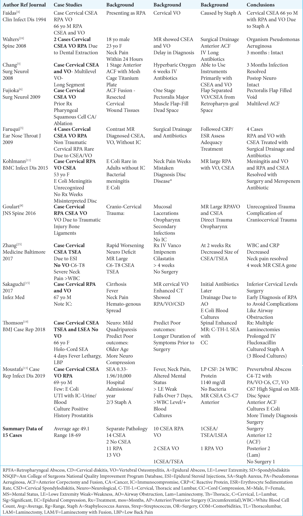
In 11 case studies, there were 15 patients who presented with varying frequencies of cervical epidural abscesses (CSEA: 14 patients), and/or vertebral osteomyelitis (VO: 13 patients) with or without retropharyngeal abscesses (RPA 11 patients) [ Table 1 ].[ 3 - 6 , 8 , 11 , 13 , 15 , 19 , 24 , 25 ] Patients averaged 49.1 years of age in 6 studies (Range 18–69).[ 4 , 11 , 13 , 15 , 19 , 24 ] Note that in all but 2 studies, patients had multiple overlapping pathologies. In the 14 patients with CSEA, 10 had accompanying VO/RPA, 2 had VO, while 2 had SEA alone (i.e. 1 CSEA/TSEA, and 1 holocord abscess CSEA/TSEA/LSEA).[ 3 - 6 , 8 , 11 , 13 , 19 , 24 , 25 ] For the 13 patients with VO, 10 additionally had CSEA/RPA, 2 had CSEA and 1 had an RPA.[ 3 - 6 , 8 , 11 , 13 , 15 , 24 ] Of the 11 patients with RPA, 10 had accompanying CSEA/VO, while one had additional VO alone.[ 4 , 5 , 8 , 11 , 13 , 15 , 24 ]


Table 1:

11 Case Studies of Cervical Spinal Epidural Spinal Abscess (CSEA) and/or Vertebral Osteomyelitis (VO), with or without Retropharyngeal Abscess (RPA).


Sources and Organisms Associated with CSEA and/or Cervical VO with/without RPA

Distinct sources of infection/organisms for cervical CSEA and/or cervical VO, with/without RPA included: 2 dental extractions (Pseudomonas aeruginosa),[ 24 ] 1 ablation of pharyngeal cancer (Staphylococcus aureus),[ 6 ] 1 meningitis (E. coli),[ 11 ] 1 cranio-cervical trauma (Staphylococcus aureus),[ 8 ] 1 cervical epidural steroid spinal injection (ESI: Staph A),[ 25 ], 1 immunocompromised patient with cirrhosis (E. coli),[ 15 ] and 1 urinary tract infection (E. coli);[ 13 ] the remainder of cases were attributed to the hematogenous spread of Staphylococcus aureus [ Table 1 ].[ 3 - 5 , 19 ]

Surgical Options for CSEA, and/or Cervical VO with/ without RPA

Surgical intervention, warranted in 14 ouf of 15 cases, included 12 anterior procedures (i.e. typically multilevel anterior corpectomy with instrumented fusions), and 2 posterior procedures (i.e. including multilevel laminectomies).[ 3 - 6 , 8 , 11 , 13 , 15 , 19 , 24 ] Where the patient had undergone a cervical epidural steroid injection (ESI) resulting in a C6-T8 CSEA/TSEA, antibiotics alone effectively resolved the infection.[ 25 ]

6 LARGER SERIES INVOLVING 355 PATIENTS WITH CERVICAL EPIDURAL ABSCESS (CSEA) AND/OR CERVICAL VERTEBRAL OSTEOMYELITIS (VO) WITH OR WITHOUT CERVICAL RETROPHARYNGEAL ABSCESS (RPA)

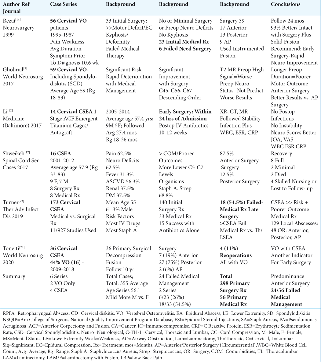
There were 6 larger cervical series involving 355 patients included in this analysis; 4 series specifically addressed CSEA (i.e. 1 also including VO), and 2 series involved cervical VO alone [ Table 2 ].[ 2 , 7 , 14 , 17 , 21 , 23 ] Surgery was primarily performed in 298 patients, while 57 underwent initial medical management. Of the latter 57 patients, 24 from two series failed medical therapy, and required delayed cervical surgery; medical therapy failed in 6 of 23 (26%) patients in one series, and 18 of 33 (54.5%) patients in the other study.[ 14 , 23 ] Data from 3 of the overall 6 series showed that these patients averaged 56.1 years of age, some with a mild male preponderance.[ 7 , 12 , 17 ] Common conclusions from these 6 series focusing on CSEA, and/or VO with/without RPA included; (1) early diagnosis with MR examinations is critical to maximize the quality of outcomes, (2) early recognition of failed medical management and/or the need for primary surgery (where appropriate) is critical, (3) primary application of spinal instrumentation in the presence of infection was safe/effective, (4) and that anterior, posterior, or circumferential surgical procedures had to adequately address the specific pathology as documented in each case [ Table 2 ].[ 2 , 7 , 14 , 17 , 21 , 23 ]


Table 2:

6 Series of Cervical Spinal Epidural Spinal Abscess (CSE) and/or Vertebral Osteomyelitis (VO).


2 Series Focusing on Cervical Vertebral Osteomyelitis Alone (VO)

Two series focused on the non-surgical and/or surgical management of cervical vertebral osteomyelitis [ Table 2 ].[ 7 , 14 ] One study (1999) pointed out that for 56 patients with VO, there was an average 10.6 week delay before correctly establishing the diagnosis; they emphasized that this was unacceptable, and how it led to major increased permanent morbidity and/or mortality for patients with VO.[ 14 ] Primary surgery was chosen for 33 of the 56 patients who met the following criteria; a significant presenting motor deficit, substantial MR- documented epidural spinal cord compression, and/or the presence of kyphosis. The other 23 patients without initial neurological deficits and/or kyphosis underwent primary non-surgical management; 6 failed medical therapy, and required delayed surgery. The total surgical procedures for the 39 patients in this series included; 17 anterior, 13 posterior, and 9 circumferential operations.[ 14 ] In another study by Ghobrial et al. (2017) involving 59 patients with cervical VO, patients averaged 59 years of age, and underwent initial anterior C4-C5, C5-C6, and/or C6-C7 surgical procedures (descending order).[ 7 ] Interestingly, better postoperative results were observed for those undergoing anterior surgery alone vs. circumferential procedures. Further, a preoperative intramedullary hyperintense T2 weighted MR cord signal correlated with the severity of the patients’ preoperative deficits, but did not correlate or predict their postoperative outcomes. Notably, authors from both studies strongly advocated for earlier recognition of when medical therapy failed, and when surgery was warranted. They further emphasized that primary surgery was indicated in the presence of significant preoperative neurological deficits to avoid permanent postoperative sequelae and/or death.

4 Series of Cervical Spine Epidural Abscess (CSEA)

From 2017-2020, there were 4 series focusing on CSEA, one of which included VO [ Table 2 ].[ 12 , 17 , 21 , 23 ] In Li et al. (2017), within 24 hours of admission, 14 patients (average age 57.4) with CSEA underwent anterior cervical corpectomy/fusion (ACF) utilizing titanium cages/autograft.[ 12 ] Postoperative follow-up utilized successive X-rays, CT, and MR studies to confirm the resolution of CSEA infections, regression of spinal cord compression, and identify progressive fusion without increased instability/deformity. These patients were also closely and repeatedly followed with inflammatory markers (white blood cell count (WBC), Erythrocyte Sedimentation Rate (ESR), C-Reactive Protein (CRP), and blood cultures) to further confirm the failure or efficacy of antibiotic/surgical treatment in resolving infections. They also noted that placing instrumentation during the index procedure was both safe and effective as there were no recurrent infections, and no reoperations for residual/recurrent infections and/or instability/deformity. In 2017, Shweikeh et al. looked at 16 CSEA (average age 57.9) involving the C5-C7 levels; 8 had initial surgery (7 anterior, 1 posterior), while 8 were treated medically.[ 17 ] Outcomes included; 8 full recoveries, 2 with minimal improvement, 2 deaths, and 4 patients referred to nursing homes or lost to follow-up. For the 173 patients in Turner et al. (2019) evaluation of 11 studies (culled out of 927 articles) involving CSEA, 140 patients had primary surgery, while 33 were initially medically managed.[ 23 ]Notably, 18 (54.5%) of the latter failed medical therapy; the uniquely high failure rate prompted the authors to conclude that CSEA resulted in a higher rate of failed medical management vs. thoracic (TSEA) and/or lumbar (LSEA) epidural abscesses. For Tonetti et al. (2020) series involving 36 CSEA, 44% of whom also had VO, all patients underwent initial surgical decompression with fusion (e.g. 7 anterior, 27 posterior, and 2 circumferential procedures).[ 21 ] Over a 10 year period, they observed an 11% (4 patients) reoperation rate; interestingly, all occurred in patients with CSEA accompanied by VO.

REVIEW OF ARTICLES CONCERNING ALL- LEVEL SEA, VO, AND ADDED REVIEW OF RPA

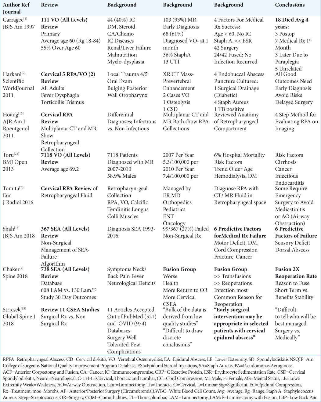
A review of an additional 8 articles provided a general overview of the diagnosis and treatment of all-level spinal epidural abscesses (SEA), vertebral osteomyelitis (VO), and also discussed more background information regarding RPA [ Tables 1 - 3 ].[ 1 , 2 , 9 , 10 , 16 , 18 , 20 , 22 ]


Table 3:

General Review of Cervical Epidural Abscess and/or Vertebral Osteomyelitis and/or Retropharyngeal Abscess (RPA).


Frequency and Definition of Cervical Spine Vertebral Osteomyelitis

Vertebral osteomyelitis is seen in hospitals at the rate of approximately 7.4 cases/100,000 patients/per year [ Tables 2 and 3 ].[ 22 ] Carragee et al. (1997) and Toru et al. (2013) respectively evaluated 111 and 7118 patients with vertebral osteomyelitis (VO) involving all spinal levels.[ 1 , 22 ] Patients respectively averaged 60 and 69.2 years of age, and exhibited the following significant comorbidities; older age, diabetes, steroid use, cancer, chemotherapy, renal/liver disease, renal failure requiring dialysis, endocarditis, malnutrition, myelodysplasia, and others [ Tables 2 and 3 ]. Those with spinal VO often presented with pain, fever, elevated clinical markers (i.e. WBC, ESR, CRP, positive blood cultures), and motor/sensory neurological deficits (i.e. VO deformity with resultant cord compression, and/or accompanying SEA).[ 1 , 14 , 22 ] Enhanced MR scans best documented VO (i.e. positive in over 90% of cases), and were typically positive for VO in as few as 2–4 weeks.[ 1 ] Further, whenever initial MRs studies were “negative”, but patients remained symptomatic, repeated MRs often subsequently readily document VO. Authors generally advocated early diagnosis and early surgery to achieve the best clinical outcomes, and largely supported utilizing instrumentation at the index procedure to avoid deformity/reoperations.[ 1 , 2 , 7 , 14 , 17 , 22 ] The in-hospital mortality rate in 2013 for all-level VOs was 6%.[ 22 ]

Frequency and Definition of Spinal Epidural Abscesses (SEA)

Spinal epidural abscesses (SEA) occur in approximately 19.6 patients/100,000 per year, and their frequency has tripled over the last 2 decades [ Tables 1 - 3 ].[ 13 , 21 ] Typical etiologies for SEA include; intravenous drug abuse, diabetes, older age, cancer, chemotherapy, immunological compromise, renal failure, and cirrhosis.[ 2 , 16 , 18 , 23 ] Chaker et al. (2018) found the classical triad for diagnosing 738 SEA; neck/back pain, fever, and a neurological deficit (e.g. weakness).[ 2 ] SEA also frequently resulted in elevated WBC levels (no elevation in some cases where the peripheral WBC and cerebropsinal fluid WBC may remain normal), but classically ESR and CRP levels are increased. Further, Procalcitonin levels (normal 0.05 ng/ mL) may be elevated to >2 ng/mL or >10 ng/mL indicating respectively infection, and severe sepsis. Blood cultures are often positive. In most cases the organism is Staphyloccosus aureus, but there may be other pathogens (e.g. Streptococcus, Pseudomonas aeruginosa E. coli, and others). [ 4 , 11 , 13 , 23 ] In some cases (e.g. immunosuppression, drug addiction), multiple organisms may appear simultaneously. With appropriate medical/surgical treatment, inflammatory markers should decrease; if they remain high and/or increase, treatment is likely failing.[ 13 , 17 , 23 , 24 ] Enhanced MRs remain the study of choice for diagnosing SEA (e.g. typically positive within 2–4 weeks), while CT abnormalities are often more delayed (i.e. positive at 6–10 weeks).[ 2 , 5 , 8 , 11 , 16 , 18 , 19 , 24 , 25 ] If initial non-surgical management is utilized (i.e. antibiotics alone), it is critical to recognize when this strategy has failed [ Tables 2 - 3 ].[ 2 , 14 , 16 , 18 ] Repeat MR studies should be utilzied to follow/recognize progression of SEA/VO with attendant increased neurological compromise/increased MR cord compression/worsening CT- documented deformity. If studies are interpreted as negative but the patient remains clinically symptomatic, obtaining additional MR studies (under sedation if needed to eliminate motion artifact) and second, third, or more radiological/ neuroradiological opinions may be warranted. In Shah’s study (2018), there was a 27% (99/367 patients) failure rate for the non-surgical treatment of SEA, largely attributed to 6 independent risk factors (algorithm): an initial motor deficit, a pathological compression fracture, cancer, diabetes, a sensory deficit, and a dorsally located abscess.[ 16 ] SEA many be managed utilizing anterior,[ 3 - 6 , 8 , 11 , 14 , 17 , 21 , 23 , 24 ] posterior,[ 12 - 14 , 17 , 19 , 21 , 23 , 25 ] or circumferential procedures[ 7 , 23 ] depending on the location of pathology. Performing early surgery, if patients meet the appropriate criteria, is critical to achieve the best results, and avoid permanent neurological sequelae.[ 2 , 3 , 7 , 12 , 14 , 16 , 18 ] Interestingly, Chaker et al. study was one of the few that documented that the additonal performance of instrumented fusions doubled the reoperation rate for SEA (e.g. series of 130 patients undergoing laminectomies/fusion vs. 608 laminectomies alone for SEA).[ 2 ]

Review of Retropharyngeal Abscesses (RPA)

Cervical retropharyngeal abscesses (RPA) occur in 4.10/100,000 children/year.[ 9 , 10 ] They predominate in the pediatric age group due to their proliferation of retropharyngeal lymph nodes [ Tables 1 and 3 ]. In adults, symptoms of RPA may include; fever, dysphagia, respiratory compromise, torticollis, and the onset of new neurological deficits (e.g. particularly in conjunction with SEA/VO).[ 9 , 10 , 20 ] These patients are often immunocompromised (e.g. diabetes, cancer, older age), have sustained spinal trauma, and/or local trauma to the oropharynx (i.e. foreign bodies in the retropharyngeal space). The retropharyngeal compartment is defined by the buccopharyngeal fascia anteriorly, prevertebral fascia posteriorly, and bilaterally, the carotid sheaths. Tomita et al. and Hoang et al. both noted how multiplanar CT and MR studies readily documented prevertebral enhancement plus retropharyngeal “fluid/other” collections, indicative of RPA often in conjuntion with CSEA and/or cervical VO.[ 10 , 20 ] Differential diagnoses for RPA included; cervical osteomyelitis, CSEA, calcific tendinitis of the longus colli muscle, jugular venous thrombosis, necrotizing fasciitis, sepsis, mediastinitis, and erosion into the carotid artery.[ 9 , 10 , 20 ]

Early diagnosis (e.g. with MR and/or CT) and early surgical treatment were typically critical to avoid respiratory collapse. Most pathogens involved a Staphylococcus aureus species, but others included; Beta-hemolytic Streptococci, anaerobic, and/or Gram-negative organisms. In Harkani et al. (2001) study of 5 patients with RPA, 4 were treated with endobuccal abscess puncture/culture and antibiotics, while 1 required surgical drainage (diabetic); 4 cultured positive for Staph Aureus, while one was positive for Tuberculosis.[ 9 ] For the 11 patients with RPA from the 11 case series involving a total of 15 patients, 10 also had CSEA/VO, while one additional patient had RPA/VO; note all 11 patients required anterior surgical procedures [ Table 1 ].[ 3 - 5 , 8 , 11 , 13 , 15 , 24 ]

CONCLUSION

Patients presenting with CSEA and/or cervical VO, with or without RPA typically exhibit fever with elevations of WBC, ESR, CRP, and Procalcitonin, have positive blood cultures, and abnormal early diagnostic MR studies (i.e. within 2–4 weeks). With persistent or increasingly elevated laboratory studies, repeatedly positive blood cultures, and progressive pathological findings on MR studies correlating with worsening neurological deficits, early surgery must be considered to maximize recovery, and limit permanent neurological sequelae along with other attendant morbidity, and mortality.

Declaration of patient consent

Patient’s consent not required as there are no patients in this study.

Financial support and sponsorship

Nil.

Conflicts of interest

There are no conflicts of interest.

References

1. Carragee EJ. Pyogenic vertebral osteomyelitis. J Bone Joint Surg Am. 1997. 79: 874-80

2. Chaker AN, Bhimani AD, Esfahani DR, Rosinski CL, Geever BW, Patel AS. Epidural Abscess: A Propensity Analysis of Surgical Treatment Strategies. Spine. 2018. 43: E1479-E1485

3. Chang WC, Tsou HK, Kao TH, Yang MY, Shen CC. Successful treatment of extended epidural abscess and long segment osteomyelitis: a case report and review of the literature. Surg Neurol. 2008. 69: 117-20

4. Faidas A, Ferguson JV Jr, Nelson JE, Baddour LM. Cervical vertebral osteomyelitis presenting as a retropharyngeal abscess. Clin Infect Dis. 1994. 18: 992-4

5. Faruqui S, Palacios E, Friedlander P, Melgar M, Alvernia J, Parry PV. Nontraumatic retropharyngeal abscess complicated by cervical osteomyelitis and epidural abscess in post-Katrina New Orleans: four cases. Ear Nose Throat J. 2009. 88: E14-

6. Fujioka M, Oka K, Kitamura R, Yakabe A. Cervical osteomyelitis and epidural abscess treated with a pectoralis major muscle flap. Surg Neurol. 2009. 72: 761-4

7. Ghobrial GM, Franco D, Theofanis T, Margiotta PJ, Andrews E, Wilson JR. Cervical Spondylodiscitis: Presentation Timing and Surgical Management in 59 Patients. World Neurosurg. 2017. 103: 664-670

8. Goulart CR, Mattei TA, Fiore ME, Thoman WJ, Mendel E. Retropharyngeal abscess with secondary osteomyelitis and epidural abscess: proposed pathophysiological mechanism of an underrecognized complication of unstable craniocervical injuries: case report. J Neurosurg Spine. 2016. 24: 197-205

9. Harakani A, Hassani R, Ziad L, Nouri H, Rochdi Y, Raji A. Retropharyngeal Abscess in Adults: Five Case Reports and Review of the Literature. Scientific WorldJournal. 2011. 11: 1623-1629

10. Hoang JK, Branstetter BF, Eastwood JD, Glastonbury CM. Multiplanar CT and MRI of collections in the retropharyngeal space: is it an abscess?. AJR Am J Roentgenol. 2011. 196: W426-32

11. Kohlmann R, Nefedev A, Kaase M, Gatermann SG. Community-acquired adult Escherichia coli meningitis leading to diagnosis of unrecognized retropharyngeal abscess and cervical spondylodiscitis: a case report. BMC Infect Dis. 2015. 15: 567-

12. Li H, Chen Z, Yong Z, Li X, Huang Y, Wu D. Emergency 1-stage anterior approach for cervical spine infection complicated by epidural abscess. Medicine (Baltimore). 2017. 96: e7301-

13. Moustafa A, Kheireldine R, Khan Z, Alim H, Khan MS, Alsamman MA.editors. Cervical Spinal Osteomyelitis with Epidural Abscess following an Escherichia coli Urinary Tract Infection in an Immunocompetent Host. Case Rep Infect Dis. 2019. 2019: 5286726-

14. Rezai AR, Woo HH, Errico TJ, Cooper PR. Contemporary management of spinal osteomyelitis. Neurosurgery. 1999. 44: 1018-25

15. Sakaguchi A, Ishimaru N, Ohnishi H, Kawamoto M, Takagi A, Yoshimura S. Escherichia coli in a patient with liver cirrhosis. Infez Med. 2017. 25: 169-173

16. Shah AA, Ogink PT, Nelson SB, Harris MB, Schwab JH. Nonoperative Management of Spinal Epidural Abscess: Development of a Predictive Algorithm for Failure. J Bone Joint Surg Am. 2018. 100: 546-555

17. Shweikeh F, Hussain M, Sangtani A, Issa H, Bashir A, Johnson JP. Cervical spine epidural abscess: a single center analytical comparison to the literature. Spinal Cord Ser Cases. 2017. 3: 17036-

18. Stricsek G, Iorio J, Mosley Y, Prasad S, Heller J, Jallo J. Etiology and Surgical Management of Cervical Spinal Epidural Abscess (SEA): A Systematic Review. Global Spine J. 2018. 8: 59S-67S

19. Thomson C. Spinal cord compression secondary to epidural abscess: the importance of prompt diagnosis and management. BMJ Case Rep. 2018. 2018:

20. Tomita H, Yamashiro T, Ikeda H, Fujikawa A, Kurihara Y, Nakajima Y. Fluid collection in the retropharyngeal space: A wide spectrum of various emergency diseases. Eur J Radiol. 2016. 85: 1247-56

21. Tonetti DA, Eichar B, Ares WJ, Kanter AS, Hamilton DK. Should the Presence of Spondylodiscitis Alter the Surgical Treatment of Patients with Symptomatic Ventral Cervical Epidural Abscesses? An Institutional Analysis. World Neurosurg. 2020. p.

22. Toru Akiyama, Hirotaka Chikuda, Hideo Yasunaga, Hiromasa Horiguchi Kivohide Fushimi, Kazuo Saita. Incidence and risk factors for mortality of vertebral osteomyelitis: a retrospective analysis using the Japanese diagnosis procedure combination database. BMJ Open. 2013. 3: e002412-

23. Turner A, Zhao L, Gauthier P, Chen S, Roffey DM, Wai EK. Management of cervical spine epidural abscess: a systematic review. Ther Adv Infect Dis. 2019. 6: 2049936119863940-

24. Walters HL, Measley R. Two cases of Pseudomonas aeruginosa epidural abscesses and cervical osteomyelitis after dental extractions. Spine. 2008. 33: E293-6

25. Zhang JH, Wang ZL, Wan L. Cervical epidural analgesia complicated by epidural abscess: A case report and literature review. Medicine (Baltimore). 2017. 96: e7789-

Leave a Reply

Your email address will not be published. Required fields are marked *