Tools

Yasuhiko Nariai, Tomoji Takigawa, Akio Hyodo, Kensuke Suzuki
  1. Department of Neurosurgery, Dokkyo Medical University Saitama Medical Center, Saitama, Japan.

Correspondence Address:
Yasuhiko Nariai, Department of Neurosurgery, Dokkyo Medical University Saitama Medical Center, Saitama, Japan.

DOI:10.25259/SNI_1279_2021

Copyright: © 2022 Surgical Neurology International This is an open-access article distributed under the terms of the Creative Commons Attribution-Non Commercial-Share Alike 4.0 License, which allows others to remix, transform, and build upon the work non-commercially, as long as the author is credited and the new creations are licensed under the identical terms.

How to cite this article: Yasuhiko Nariai, Tomoji Takigawa, Akio Hyodo, Kensuke Suzuki. Distal migration of the flow-redirection endoluminal device immediately after treatment: A case report and literature review. 04-Mar-2022;13:81

How to cite this URL: Yasuhiko Nariai, Tomoji Takigawa, Akio Hyodo, Kensuke Suzuki. Distal migration of the flow-redirection endoluminal device immediately after treatment: A case report and literature review. 04-Mar-2022;13:81. Available from: https://surgicalneurologyint.com/surgicalint-articles/11419/

Date of Submission
31-Dec-2021

Date of Acceptance
16-Feb-2022

Date of Web Publication
04-Mar-2022

Abstract

Background: A flow diverter (FD) has been a promising endovascular therapeutic modality for challenging intracranial aneurysms. However, stent migration has been an unusual complication. Until recently, among some types of FDs, the migration of the flow-redirection endoluminal device (FRED; MicroVention Inc., Aliso Viejo, CA, USA) has almost never been reported. Herein, we report a case of acute distal migration of a single FRED secondary to in-stent thrombi with symptomatic ischemic stroke and review the literature on the distal migration of FDs.

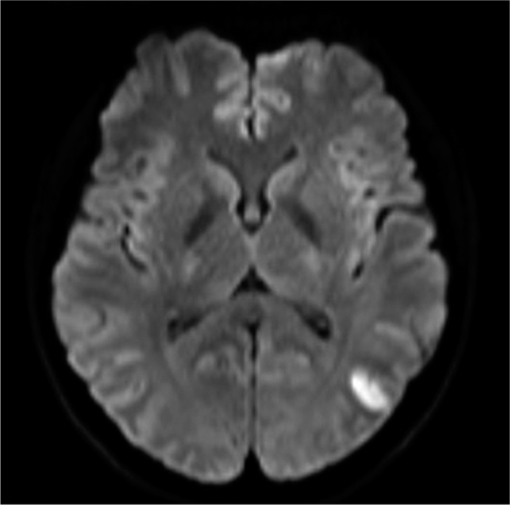
Case Description: A 35-year-old woman was diagnosed with a left unruptured internal carotid-ophthalmic artery aneurysm. A 3.5 mm diameter and 17 mm long FRED was adequately deployed. The patient awoke from general anesthesia without neurological deficits. However, shortly after the procedure, the patient presented with conjugate deviation toward the left side, right severe hemiparesis, and total aphasia. Although the symptoms gradually improved, angiography was performed. Angiography revealed some in-stent thrombi and distal migration of the FRED, and initially, one of the left M2 inferior trunk branches was occluded by an embolic thrombus. However, the thrombus spontaneously migrated distally without any specific treatment. Finally, despite leaving the migrated stent in situ, the flow almost completely improved, and the patient’s neurologic deficits disappeared. Magnetic resonance imaging following treatment revealed only a small cerebral infarction in the left temporo-occipital area.

Conclusion: Distal migration of an FD in an acute setting, including the FRED, may occur even following appropriate placement. In-stent thrombosis can cause distal stent migration and thromboembolic stroke.

Keywords: Flow diverter, Flow-redirection endoluminal device, Stent migration

INTRODUCTION

Endoluminal vessel reconstruction treatment with a flow diverter (FD) stent has been a promising endovascular therapeutic modality for challenging intracranial aneurysms at various locations. The FD is a low porosity metal stent that can decrease the velocity and pressure of intra-aneurysmal blood flow and lead to delayed thrombosis with reconstruction of the parent artery. Moreover, they provide a scaffold for endothelialization across an aneurysm neck by diverting blood flow away from the aneurysmal sac, resulting in clotting of the aneurysm.[ 11 ] The flow-redirection endoluminal device (FRED; MicroVention Inc., Aliso Viejo, CA, USA) is one of the FDs with a paired, integrated dual-layer self-expanding braided design, which was released for clinical use in Europe in 2012. It has a high occlusion rate (complete and near-complete occlusion rate of 97.2% at a median follow-up of 10.8 months) and relatively low rates of complications, such as hemorrhagic (0.8%) and thromboembolic complications (5.3%).[ 8 ] Apart from the two aforementioned complications, FD migration has been an unusual complication. It may occur in both acute and delayed phases following treatment, and sometimes lead to symptomatic complications, such as aneurysm rupture and thromboembolic events.[ 2 , 3 , 5 , 16 ] To date, the types of FDs with the reported complication of stent migration are the pipeline embolization device (PED; Covidien/ev3, Irvine, CA, USA) and the SILK FD (Balt Extrusion, Montmorency, France).[ 2 - 5 , 10 , 13 , 16 ] Considering migration of the FRED, Gawlitza et al. first reported a distal migration case of the FRED. However, they bridged with two telescoping FDs using a p64 (Phenox, Bochum, Germany) (distal side) and a FRED (proximal side). Six days after stenting, they incidentally identified distal migration of the FRED with its proximal part floating in the aneurysm without evident clinical symptoms.[ 6 ] We report a case of acute distal migration of a single FRED secondary to in-stent thrombi with symptomatic ischemic stroke and review the literature on the distal migration of FDs.

CASE REPORT

A 35-year-old woman was incidentally diagnosed with a left unruptured internal carotid-ophthalmic artery aneurysm with a maximum size of 6.1 mm and neck size of 3.6 mm [ Figures 1a and b ]. Endovascular treatment with FRED was scheduled. Two weeks before treatment, the patient was premedicated with 100 mg aspirin and 75 mg clopidogrel daily. The day before the procedure, using the VerifyNow point-of-care platelet assay (Accumetrics, San Diego, CA, USA), the aspirin reaction unit and P2Y12 reaction unit (PRU) were measured at 493 and 192, respectively. Both values were considered to be within adequate platelet inhibition function, and no preoperative enhanced antiplatelet therapy was needed.[ 1 ]


Figure 1:

A 35-year-old woman with an unruptured internal carotid-ophthalmic artery aneurysm with a maximum size of 6.1 mm and a neck size of 3.6 mm. (a and b) Initial three-dimensional reconstruction of digital subtraction angiography and normal angiogram, respectively.


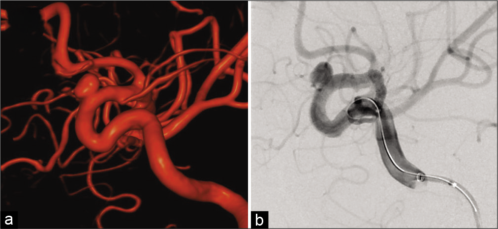
Under general anesthesia, a 7-French shuttle sheath (Cook Medical, Bloomington, IN, USA) was placed with a coaxial system at the cervical portion of the left internal carotid artery (ICA) through the right femoral site. A Headway27 microcatheter (MicroVention Inc.) was navigated distal to the aneurysm using a microguidewire (ASAHI CHIKAI 14; Asahi Intecc Co., Ltd., Aichi, Japan). Moreover, the SOFIA SELECT EX (MicroVention Inc.) was used as a distal access catheter. The size of the FRED was determined according to the diameter of the ICA vessel proximal (3.3 mm) and distal (3.2 mm) to the aneurysm. A 3.5 mm diameter and 17 mm long FRED was adequately deployed through the microcatheter across the neck of the aneurysm under appropriate therapeutic heparinization [ Figure 2a ]. Post deployment angiography revealed intra-aneurysm contrast stagnation after FRED deployment [ Figure 2b ]. Full expansion of the FRED was confirmed using cone-beam computed tomography angiography. The patient awoke without any neurological symptoms.


Figure 2:

A flow-redirection endoluminal device (FRED) was adequately deployed with good coverage of the neck (a). Postdeployment angiography revealing intra-aneurysm contrast stagnation (b). The orange circles indicate each flair end of the FRED. The blue arrow shows the aneurysm.


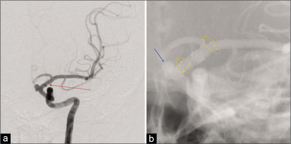
However, only 1 h following the procedure, the patient presented with conjugate deviation toward the left side, right severe hemiparesis, and total aphasia. Diffusion-weighted magnetic resonance imaging (MRI) revealed no evident acute cerebral infarction. However, magnetic resonance angiography revealed the aneurysm, which meant that the FD was no longer covering the aneurysm. Furthermore, a signal defect of the left ICA terminus, which meant that the FD was located there, and decline of the flow of the left peripheral middle cerebral artery (MCA) [
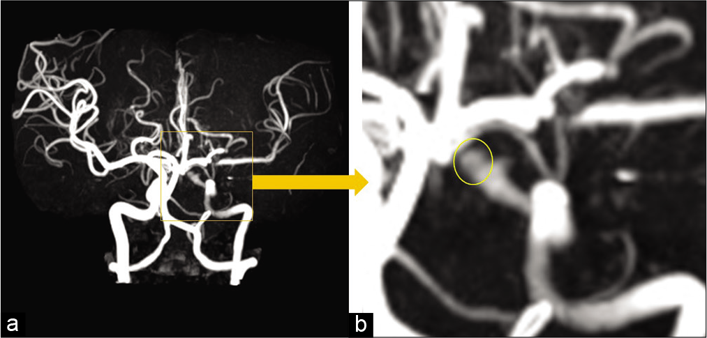
 Figure 3 ] were also observed by magnetic resonance angiography. Immediately, after the MRI, angiography was promptly performed, although the patient’s symptoms gradually improved. An angiogram revealed some in-stent thrombi with distal migration of the FRED [ Figures 4a and b ]. Initially, one of the left M2 inferior trunk branches was occluded by an embolic thrombus, and the left temporo-occipital area was avascular [ Figure 5a ]. However, during the preparation of recanalization, the thrombus spontaneously migrated distally, and the flow gradually improved without any specific treatment. The final angiogram revealed almost complete recanalization, except for the delay of the flow in the left MCA temporo-occipital area [ Figures 5b and c ]. At that time, the patient’s symptoms completely improved. Thus, we administered a bolus of 20 mg prasugrel per oral and intravenous drip of argatroban, leaving the migrated stent in situ. The MRI on the day after the treatment revealed only a small cerebral infarction in the left temporo-occipital area without any evident neurological symptoms [ Figure 6 ].


Figure 3:

Magnetic resonance angiography shortly after the flow-redirection endoluminal device deployment and occurrence of the neurological deficits (a) revealed the aneurysm (yellow circle) and signal defect of the left internal carotid artery terminus with decline of the left peripheral middle cerebral artery (b).


Figure 4:

The left internal carotid angiography revealing some in-stent thrombi (red arrow) (a) and distal migration of the flow-redirection endoluminal device (FRED) (b). The orange circles indicate each flair end of the distally migrated FRED. The blue arrow indicates the aneurysm.


Figure 5:

One of the left M2 inferior trunk branches was occluded by an embolic thrombus (red arrow) (a). The thrombus spontaneously migrated distally (red curved arrow). Final angiogram showed almost complete recanalization except for the left temporo-occipital area (red circle) (b and c).


Figure 6:

Diffusion-weighted magnetic resonance imaging on the day after the treatment revealing only small cerebral infarction in the left temporo-occipital area without any evident neurological symptoms.


DISCUSSION

This case report describes the acute distal migration of a single FRED resulting from in-stent thrombi causing ischemic stroke. Among FD migration cases, FRED migration has almost never been reported. Furthermore, the clinical appearance of the neurologic symptoms enabled extremely early confirmation of the stent migration shortly after treatment.

Spontaneous migrations of the FD have been reported both early and late, that is, from immediately after treatment to 14 months following treatment.[ 3 , 5 , 6 , 10 , 16 ] Proximal FD migration during treatment or shortly after treatment is a frequent intraoperative complication observed in up to 12% of cases, and it rarely generates neurological symptoms.[ 2 ] Proximal FD migration has also been considered a delayed complication, frequently presenting with aneurysm recurrence due to incomplete coverage of the aneurysm neck.[ 2 , 3 , 10 , 15 , 16 ] A chief cause of proximal FD migration is elongation of the device during deployment, which may lead to spontaneous foreshortening.[ 5 , 10 ] Moreover, as causes of both proximal and distal migrations, the significant mismatch (>1 mm) between the inflow and outflow vessels has been considered important.[ 2 ] In that case, the smaller vessel end of the stent cannot be fully flared to enhance the apposition of the stent. Consequently, a constant squeezing force can occur at the end of the stent.[ 3 ] Large aneurysms with long neck or fusiform aneurysms may also decrease the surface area of the stent landing zone and increase the risk of stent migration.[ 16 ] Some studies have reported the following preventive measures for migration: using longer FDs, appropriate device diameter sizing (0.25–0.5 mm larger than the distal parent vessel), avoiding excessive stretching of the device, obtaining complete expansion of the stent, and using adjunctive coiling to prevent the FD from prolapsing into large aneurysms.[ 2 , 3 , 16 ]

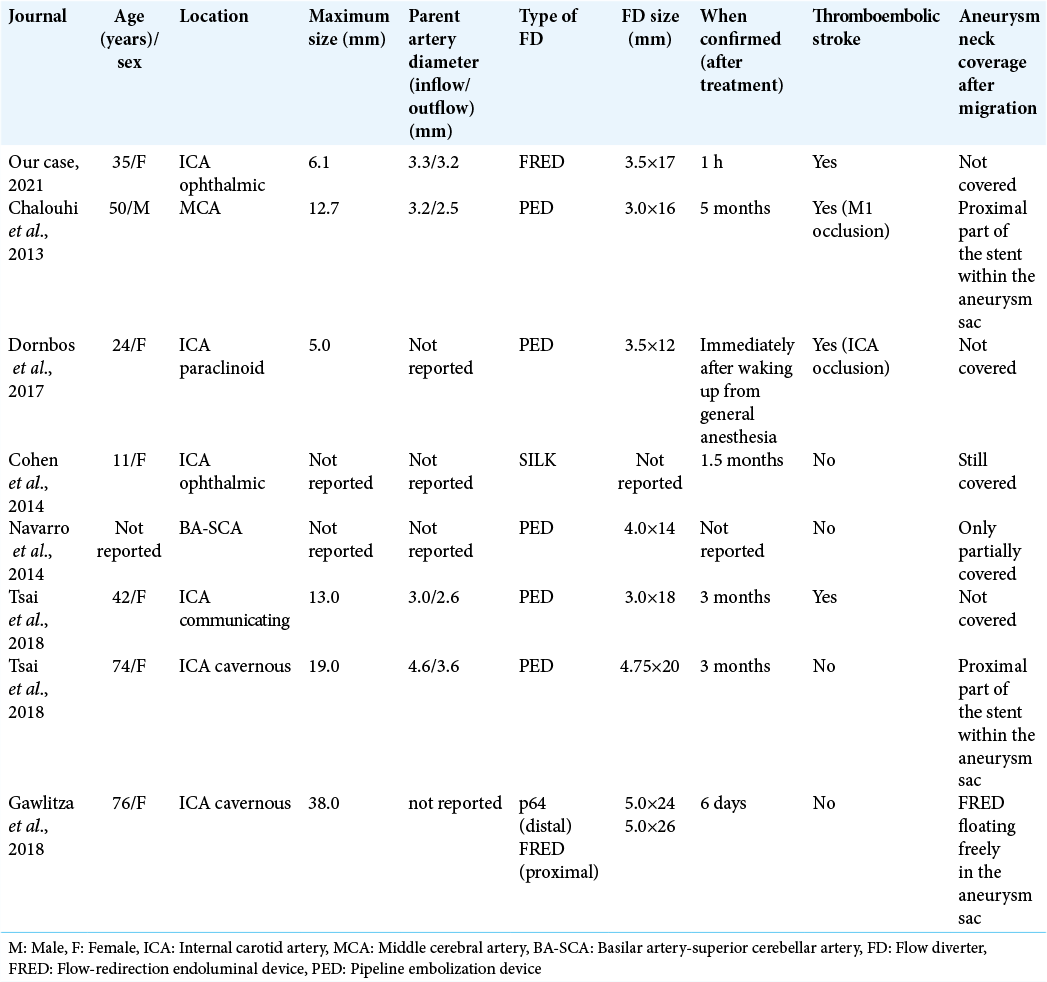
Compared with proximal migration, distal migration of an FD is rare. Apart from our case, seven cases of distal FD migration have been identified in the literature [ Table 1 ].[ 3 , 4 , 6 , 13 , 16 ] Limited to an acute period of FD migration, proximal migration may occur more frequently because of the foreshortening mechanism. There are only two reported cases of distal migration of the FD in an acute setting (within 24 h), including our case.[ 5 ] In both cases, the acute distal migrations were recognized by the patients’ symptoms of thromboembolic stroke shortly following stent placement. Considering the factors of distal migration, in addition to the aforementioned factors, instent thrombus formation has also been reported.[ 4 , 5 , 16 ] Tsai et al. reported that microthrombus formation on the FD may increase the dragging force on the stent and ultimately lead to distal migration.[ 16 ] In our case, the aneurysm was not large and there was no significant difference between the distal and proximal vessel diameters. Therefore, migration could be affected by an in-stent thrombus as an underlying thrombotic pathology. In fact, our case presented with some in-stent thrombi and MCA branch occlusion by an embolic thrombus, which must have occurred within the FRED. Given our patient’s initial severe symptoms and spontaneous improvement, ICA or M1 occlusion by a larger thrombus could have occurred. Fortunately, the thrombus might have spontaneously migrated distally, which resulted in the gradual improvement of the patient’s symptoms. Regarding initial images on the clinical appearance of neurologic deficits following distal migration of an FD, in the present case, MRI images were checked first. After checking the MRI images, angiography was promptly performed. A policy where computed tomography is performed first to check for an intracranial hemorrhage may also be good because the time required to obtain MRI images is generally longer. In the absence of an hemorrhage, prompt angiography to evaluate for the presence of a thromboembolic stroke and correct positioning of the stent should be performed.


Table 1:

Cases of distal migration of the flow diverter in the literature.


Among all types of FDs, the FRED is the only dual-layered type nitinol device consisting of a low porosity inner mesh with higher pore attenuation (48 nitinol wires) and an outer stent with high porosity (16 nitinol wires). An interwoven double helix of radiopaque tantalum strands attaches the inner mesh to the outer stent and improves visibility over its full length of dual-layer coverage.[ 12 ] Considering the thrombogenicity, Hagen et al. compared FRED with PED as a representative of single-layered FDs in an ex vivo shunt model. They demonstrated that FRED showed a trend toward greater platelet accumulation in the absence of clopidogrel, which may indicate the effectiveness of clopidogrel in the use of FRED. They also identified FRED as having more fibrin-driven thrombogenicity than a PED, even under the administration of dual antiplatelet therapy (DAPT; acetylsalicylic acid [ASA] with clopidogrel). Reportedly, the finding resulted from the unique design of the FRED: the separation design between the two layers could potentially lead to localized disturbed flow conditions, which could entrap activated platelets and serve as a nidus for further thrombus growth.[ 9 ] Girdhar et al. also reported the high thrombogenicity of the FRED using an in vitro human blood physiological flow loop model. They confirmed that a large amount of thrombus accumulated in the spaces between the two layers (<100 μm on average) using sectional high-resolution scanning electron microscopy.[ 7 ] Thus, in our case, the high thrombogenicity of the FRED can cause in-stent thrombus formation and finally lead to distal migration of the FRED. In general, the use of FDs requires perioperative systemic heparinization and DAPT before and for at least 3 months following treatment. Unlike ASA, clopidogrel has widely variable effects on platelet activity in response to individual cytochrome P450 CYP2C19 activity, which can potentially reduce the antiplatelet effect.[ 14 ] In the present case, the preoperative PRU value was 193. Considering the high thrombogenicity of the FRED, especially in the case of insufficient administration of clopidogrel,[ 9 ] the preoperative PRU values may need to be managed more strictly to prevent distal migration of the FRED. Further prospective studies are needed to clarify the association between the high thrombogenicity of the FRED and distal migration of the FD.

CONCLUSION

Distal migration of an FD in an acute setting, including the FRED, may occur even following appropriate placement. In-stent thrombosis can cause distal stent migration and thromboembolic stroke.

Declaration of patient consent

The authors certify that they have obtained all appropriate patient consent.

Financial support and sponsorship

Nil.

Conflicts of interest

There are no conflicts of interest.

References

1. Ajadi E, Kabir S, Cook A, Grupke S, Alhajeri A, Fraser JF. Predictive value of platelet reactivity unit (PRU) value for thrombotic and hemorrhagic events during flow diversion procedures: A meta-analysis. J Neurointerv Surg. 2019. 11: 1123-8

2. Chalouhi N, Satti SR, Tjoumakaris S, Dumont AS, Gonzalez LF, Rosenwasser R. Delayed migration of a pipeline embolization device. Neurosurgery. 2013. 72: ons229-34

3. Chalouhi N, Tjoumakaris SI, Gonzalez LF, Hasan D, Pema PJ, Gould G. Spontaneous delayed migration/shortening of the pipeline embolization device: Report of 5 cases. AJNR Am J Neuroradiol. 2013. 34: 2326-30

4. Cohen JE, Gomori JM, Moscovici S, Leker RR, Itshayek E. Delayed complications after flow-diverter stenting: reactive in-stent stenosis and creeping stents. J Clin Neurosci. 2014. 21: 1116-22

5. Dornbos D, Powers CJ. Acute distal migration of a flow diverting stent. J Clin Neurosci. 2017. 44: 223-5

6. Gawlitza M, Soize S, Manceau PF, Pierot L. Delayed intraaneurysmal migration of a flow diverter construct after treatment of a giant aneurysm of the cavernous internal carotid artery. J Neuroradiol. 2020. 47: 233-6

7. Girdhar G, Andersen A, Pangerl E, Jahanbekam R, Ubl S, Nguyen K. Thrombogenicity assessment of Pipeline Flex, Pipeline Shield, and FRED flow diverters in an in vitro human blood physiological flow loop model. J Biomed Mater Res Part A. 2018. 106: 3195-202

8. Griessenauer CJ, Thomas AJ, Enriquez-Marulanda A, Deshmukh A, Jain A, Ogilvy CS. Comparison of pipeline embolization device and flow re-direction endoluminal device flow diverters for internal carotid artery aneurysms: A propensity score-matched cohort study. Neurosurgery. 2019. 85: E249-55

9. Hagen MW, Girdhar G, Wainwright J, Hinds MT. Thrombogenicity of flow diverters in an ex vivo shunt model: Effect of phosphorylcholine surface modification. J Neurointerv Surg. 2017. 9: 1006-11

10. Jabbour P, Atallah E, Chalouhi N, Tjoumakaris S, Rosenwasser RH. A case of pipeline migration in the cervical carotid. J Clin Neurosci. 2019. 59: 344-6

11. Leung GK, Tsang AC, Lui WM. Pipeline embolization device for intracranial aneurysm: A systematic review. Clin Neuroradiol. 2012. 22: 295-303

12. Möhlenbruch MA, Herweh C, Jestaedt L, Stampfl S, Schönenberger S, Ringleb PA. The FRED flow-diverter stent for intracranial aneurysms: clinical study to assess safety and efficacy. AJNR Am J Neuroradiol. 2015. 36: 1155-61

13. Navarro R, Cano EJ, Brasiliense LB, Dabus G, Hanel RA. Fatal outcome after delayed pipeline embolization device migration for the treatment of a giant superior cerebellar artery aneurysm: technical note for complication avoidance. Open J Mod Neurosurg. 2014. 4: 6

14. Sim SC, Ingelman-Sundberg M. Update on allele nomenclature for human cytochromes P450 and the human cytochrome P450 allele (CYP-allele) nomenclature database. Methods Mol Biol. 2013. 987: 251-9

15. Takong W, Kobkitsuksakul C. Delayed proximal flow diverting stent migration in a ruptured intracranial aneurysm: A case report. Neurointervention. 2020. 15: 154-7

16. Tsai YH, Wong HF, Hsu SW. Endovascular management of spontaneous delayed migration of the flow-diverter stent. J Neuroradiol. 2020. 47: 38-45

Leave a Reply

Your email address will not be published. Required fields are marked *