

- Department of Surgery, Federal Medical Center, Owo,
- Department of Surgery, University of Ibadan/University College Hospital, Ibadan, Oyo State, Nigeria.
Correspondence Address:
Toyin Ayofe Oyemolade
Department of Surgery, University of Ibadan/University College Hospital, Ibadan, Oyo State, Nigeria.
DOI:10.25259/SNI_346_2020
Copyright: © 2020 Surgical Neurology International This is an open-access article distributed under the terms of the Creative Commons Attribution-Non Commercial-Share Alike 4.0 License, which allows others to remix, tweak, and build upon the work non-commercially, as long as the author is credited and the new creations are licensed under the identical terms.How to cite this article: Toyin Ayofe Oyemolade, Augustine Abiodun Adeolu. Extensive vertebral scalloping in a thoracolumbar junction spinal schwannoma. 11-Jul-2020;11:183
How to cite this URL: Toyin Ayofe Oyemolade, Augustine Abiodun Adeolu. Extensive vertebral scalloping in a thoracolumbar junction spinal schwannoma. 11-Jul-2020;11:183. Available from: https://surgicalneurologyint.com/surgicalint-articles/10125/
Date of Submission
11-Jun-2020
Date of Acceptance
25-Jun-2020
Date of Web Publication
11-Jul-2020
Abstract
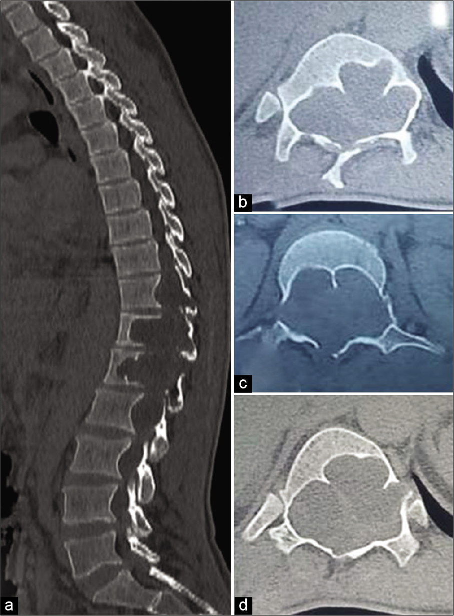
Severe vertebral scalloping in spinal schwannoma is very rare. When present, extensive scalloping of the vertebral bodies possesses significant treatment challenges in patients with spinal tumors. We present the computed tomography scan and magnetic resonance images of spinal schwannoma with marked vertebral scalloping in a 40-year-old Nigerian.
Keywords: Extensive, Schwannoma, Vertebral scalloping
DESCRIPTION
A 40-year-old man presented with back pain and progressive weakness of the lower limbs of 6 years. He had been bed ridden for the 3 years preceding presentation. There was associated paraesthesia and sphincter dysfunction. Neurological examination revealed flaccid paraplegia. The sensory level was at T12. Magnetic resonance imaging showed a heterogeneously contrast enhancing tumor at T11–L2 with marked scalloping of T12 and L1 vertebral bodies [

Spinal schwannomas are benign tumors arising from the Schwann cells of the nerve sheaths.[
Declaration of patient consent
Patient’s consent not required as patients identity is not disclosed or compromised.
Financial support and sponsorship
Nil.
Conflicts of interest
There are no conflicts of interest.
References
1. Borges G, Bonilha L, Proa M, Fernandes YB, Ramina R, Zanardi V. Imaging features and treatment of an intradural lumbar cystic schwannoma. Arq Neuropsiquiatr. 2005. 63: 681-4
2. Corley J, Kasliwal MK, O’Toole JE, Byrne RW. Extensive vertebral scalloping in a case of giant cystic spinal schwannoma: More than just a radiological diagnosis. J Neurooncol. 2014. 120: 219-20
3. Wakely SL. The posterior vertebral scalloping sign. Radiology. 2006. 239: 607-9

