

- Almoosa Specialist Hospital, Al-Ahsa, Saudi Arabia.
- Department of Neurosurgery, College of Medicine, King Faisal University, Al-Ahsa, Saudi Arabia.
Correspondence Address:
Abdulelah S. Almousa, Department of Neurosurgery, College of Medicine, King Faisal University, Al-Ahsa, Saudi Arabia.
DOI:10.25259/SNI_6_2024
Copyright: © 2024 Surgical Neurology International This is an open-access article distributed under the terms of the Creative Commons Attribution-Non Commercial-Share Alike 4.0 License, which allows others to remix, transform, and build upon the work non-commercially, as long as the author is credited and the new creations are licensed under the identical terms.How to cite this article: Ibrahim H. Alahmed1, Abdulelah S. Almousa2, Abdulsalam Aleid2. Fontanellar bone - A rarity in pediatric cranial abnormalities. 01-Mar-2024;15:64
How to cite this URL: Ibrahim H. Alahmed1, Abdulelah S. Almousa2, Abdulsalam Aleid2. Fontanellar bone - A rarity in pediatric cranial abnormalities. 01-Mar-2024;15:64. Available from: https://surgicalneurologyint.com/surgicalint-articles/12780/
Date of Submission
02-Jan-2024
Date of Acceptance
01-Feb-2024
Date of Web Publication
01-Mar-2024
Abstract
Background: Fontanelles, membranous gaps in the infant skull, are integral for accommodating the expansion of the skull by the growing brain postnatally. The anterior fontanelle, situated at the frontal-parietal bone intersection, typically closes gradually within the first two years. Fontanellar bone, an exceedingly rare ossification anomaly of the anterior fontanelle, clinically mimics craniosynostosis.
Case Description: We present the case of a 22-day-old male with an almost closed anterior fontanelle who underwent evaluation. Prenatal and postnatal history were unremarkable. Physical examination revealed a well-nourished infant with a nearly closed fontanelle but no other anomalies. The initial diagnosis was craniosynostosis; however, a head computed tomography scan revealed fontanellar bone. Consequently, a conservative management approach with regular follow-ups was adopted.
Conclusion: This case provides valuable insights into fontanellar bone, emphasizing its consideration in differential diagnoses for almost closed anterior fontanelles. The report aims to enhance awareness and understanding of this rare condition, promoting accurate diagnosis and optimal patient outcomes.
Keywords: Case report, Cranial abnormalities, Craniosynostosis, Fontanellar bone
INTRODUCTION
Fontanelles, membranous gaps in the infant skull, serve as crucial anatomical landmarks, allowing for the expansion of the skull by the growing brain during early postnatal life. The anterior fontanelle, positioned at the intersection of the frontal and parietal bones, typically undergoes a gradual closure process within the first two years of life. Deviations from this norm can signify a spectrum of conditions ranging from benign variations to severe pathological states.[
CASE PRESENTATION
A 22-day-old male infant came to attention after a routine physical examination raised concerns about a nearly closed anterior fontanelle. The baby, delivered full-term through spontaneous vaginal delivery, had an uneventful pregnancy and birth.
A thorough examination revealed an almost closed anterior fontanelle. The rest of the head circumference fell within normal limits, and there were no signs of abnormal head shape. Dysmorphic features or other apparent congenital anomalies were not observed. The primary differential diagnosis considered was craniosynostosis.
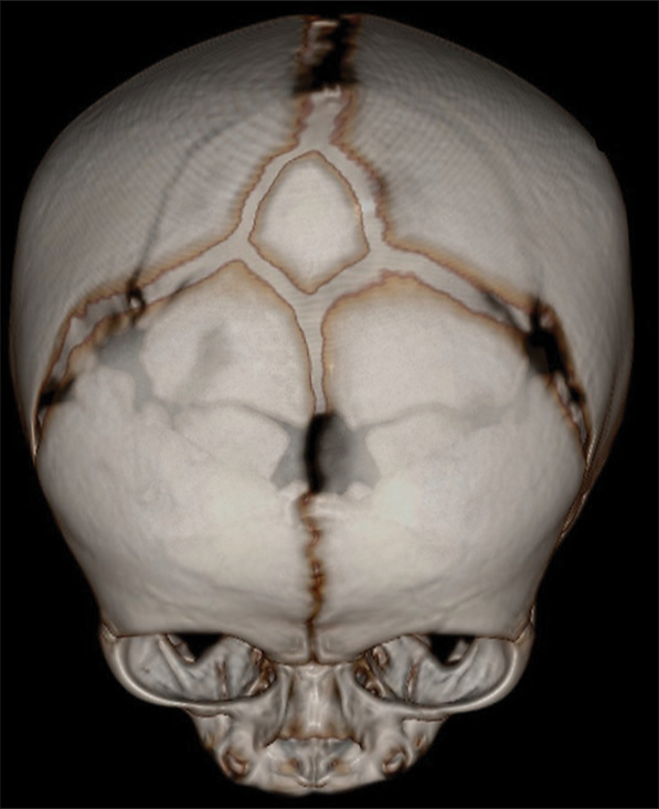
To further evaluate the closure of the fontanelle, a head computed tomography scan was conducted. The scan disclosed the presence of fontanellar bone, an unusual finding characterized by the ossification of the fontanelle, clarifying the almost closed appearance [
Given the absence of associated symptoms or complications, a conservative approach was recommended. Regular clinical follow-ups were scheduled to monitor the infant’s head growth.
DISCUSSION
The identification of fontanellar bone in the presented case sheds light on the diagnostic challenges associated with this rare cranial anomaly. Fontanellar bone represents a unique subset within this diagnostic landscape, characterized by the premature ossification of the anterior fontanelle.[
In our case, the nearly closed anterior fontanelle prompted a comprehensive work-up to distinguish between benign variations and pathological states. The initial consideration of craniosynostosis was a reasonable differential diagnosis, given the clinical presentation. However, it is crucial to acknowledge that the assessment of head shape holds more substantial diagnostic value in the clinical evaluation of craniosynostosis. It should also be emphasized that, considering radiation exposure, ultra-low dose computed tomography or ultrasound should indeed be considered.[
The scarcity of documented cases of fontanellar bone in the existing scientific literature underscores the uniqueness of this diagnostic entity. The limited awareness of this anomaly can potentially lead to misinterpretation, emphasizing the significance of presenting and documenting such cases. The management of fontanellar bone in our case exemplifies the necessity for an individualized approach. Given the absence of associated symptoms or complications, a conservative management strategy was adopted. This decision aligns with the broader principle of avoiding unnecessary interventions, particularly in cases where the condition poses no immediate threat to the patient’s well-being.[
CONCLUSION
Our case report on fontanellar bone adds valuable information to the existing literature, underlining the significance of including this rare anomaly in the considerations for almost closed anterior fontanelles. The collaboration between clinical examination and diagnostic imaging, combined with an individualized management approach, emphasizes the detailed and specific care needed for such cases in a clinical setting.
Ethical approval
Institutional Review Board approval is not required.
Declaration of patient consent
The authors certify that they have obtained all appropriate patient consent.
Financial support and sponsorship
Nil.
Conflicts of interest
There are no conflicts of interest.
Use of artificial intelligence (AI)-assisted technology for manuscript preparation
The authors confirm that there was no use of artificial intelligence (AI)-assisted technology for assisting in the writing or editing of the manuscript and no images were manipulated using AI.
Disclaimer
The views and opinions expressed in this article are those of the authors and do not necessarily reflect the official policy or position of the Journal or its management. The information contained in this article should not be considered to be medical advice; patients should consult their own physicians for advice as to their specific medical needs.
References
1. D’Antoni AV, Donaldson OI, Schmidt C, Macchi V, De Caro R, Oskouian RJ. A comprehensive review of the anterior fontanelle: Embryology, anatomy, and clinical considerations. Childs Nerv Syst. 2017. 33: 909-14
2. Goske MJ, Applegate KE, Boylan J, Butler PF, Callahan MJ, Coley BD. Image gently(sm): A national education and communication campaign in radiology using the science of social marketing. J Am Coll Radiol. 2008. 5: 1200-5
3. Governale LS. Craniosynostosis. Pediatr Neurol. 2015. 53: 394-401
4. Johal J, Iwanaga J, Loukas M, Tubbs RS. Anterior fontanelle wormian bone/fontanellar bone: A review of this rare anomaly with case illustration. Cureus. 2017. 9: e1443
5. Pickett AT, Montes MA. Wormian bone in the anterior fontanelle of an otherwise well neonate. Cureus. 2019. 11: e4741
6. Whittall I, Lambert WA, Moote DJ, Bookland MJ, Martin JE, Hughes CD. Postnatal diagnosis of single-suture craniosynostosis with cranial ultrasound: A systematic review. Childs Nerv Syst. 2021. 37: 3705-14
7. Woods RH, Johnson D. Absence of the anterior fontanelle due to a fontanellar bone. J Craniofac Surg. 2010. 21: 448-9

