- Department of Neurosurgery with Pediatric Neurosurgery, Charité-University Medicine, Campus Virchow, Berlin, Germany
- Department of Neurosurgery, Brigham and Woman's Hospital, Harvard Medical School, Boston, Massachusetts, USA
- Department of Neuroradiology, Beth Israel Deaconess Medical Center, Harvard Medical School, Boston, Massachusetts, USA
- Department of Pathology and Neurology, Beth Israel Deaconess Medical Center, Harvard Medical School, Boston, Massachusetts, USA
- Department of Neurosurgery, Beth Israel Deaconess Medical Center, Harvard Medical School, Boston, Massachusetts, USA
Correspondence Address:
Ekkehard M. Kasper
Department of Neurosurgery, Beth Israel Deaconess Medical Center, Harvard Medical School, Boston, Massachusetts, USA
DOI:10.4103/2152-7806.194506
Copyright: © 2016 Surgical Neurology International This is an open access article distributed under the terms of the Creative Commons Attribution-NonCommercial-ShareAlike 3.0 License, which allows others to remix, tweak, and build upon the work non-commercially, as long as the author is credited and the new creations are licensed under the identical terms.How to cite this article: Laura-Nanna Lohkamp, Christian Strong, Rafael Rojas, Matthew Anderson, Yosef Laviv, Ekkehard M. Kasper. Hypervascular glioblastoma multiforme or arteriovenous malformation associated Glioma? A diagnostic and therapeutic challenge: A case report. 21-Nov-2016;7:
How to cite this URL: Laura-Nanna Lohkamp, Christian Strong, Rafael Rojas, Matthew Anderson, Yosef Laviv, Ekkehard M. Kasper. Hypervascular glioblastoma multiforme or arteriovenous malformation associated Glioma? A diagnostic and therapeutic challenge: A case report. 21-Nov-2016;7:. Available from: http://surgicalneurologyint.com/surgicalint_articles/hypervascular-glioblastoma-multiforme-or-arteriovenous-malformation-associated-glioma-a-diagnostic-and-therapeutic-challenge-a-case-report/
Abstract
Background:Simultaneous presentation of arteriovenous malformation (AVM) and glioblastoma multiforme (GBM) is rarely reported in the literature and needs to be differentiated from “angioglioma”, a highly vascular glioma and other differential diagnosis such as hypervascular glioblastoma. Incorporating critical features of both, malignant glioma and AVM, such lesions lack a standard algorithm for diagnosis and therapy due to their rare incidence as well as their complex radiological and highly individualized clinical presentation.
Case Description:We present a case of a 71-year-old female with newly developing motor deficits and radiographic findings of a heterogeneously contrast enhancing right-sided thalamic lesion with highly prominent vasculature. While computed tomography angiogram and cerebral digital subtraction angiography supported the diagnosis of AVM, contrast-enhancing magnetic resonance imaging (MRI) and MR-spectroscopy was suggestive of malignant glioma. A stereotactic biopsy revealed the diagnosis of a GBM (WHO IV) and the patient was treated accordingly.
Conclusion:The coincidental presentation of vascular lesions such as AVM and malignant glioma is rare and presents a major challenge when establishing a diagnosis. The respective treatment decision is complicated by the fact that available treatment modalities (e.g. radiosurgery and/or open resection) carry disease specific complications for each entity. Finding a suitable solution for such cases requires standardization of early diagnostic and therapeutic management.
Keywords: Angioglioma, arteriovenous malformation, hypervascular glioblastoma multiforme, angiogenesis, case report
BACKGROUND
Arteriovenous malformations (AVMs) associated with gliomas, in particular glioblastoma multiforme (GBM), are uncommon and remain infrequently reported in the literature. Due to the critical features of both entities, such AVM-associated malignant gliomas represent a challenging clinical entity in terms of diagnosis, therapeutic management, and risk assessment.
The pathological entity in these cases differs from other glioma-associated vascular lesions, e.g., the so-called “angiogliomas,” which occur reportedly more frequently. Though potentially misleading during diagnostic evaluation, they are hypervascular low-grade lesions with a favorable prognosis.[
Here, we report a striking case of what was considered an AVM-associated glioma; we evaluate its clinical and radiological presentation and its subsequent management. By illustrating this case in the context of the existing literature, we seek to caution against diagnostic pitfalls and emphasize the need for an accurate histopathological diagnosis.
CASE PRESENTATION
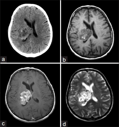
A 71-year-old female presented to an outside hospital with a 1-month history of progressive lower left extremity weakness and a left foot drop with resulting difficult ambulating. Over the days prior to admission, she also developed difficulty in holding objects with her left hand. A cranial computed tomography (CT) showed an approximately 3.5 cm nodular right-sided thalamic lesion with heterogeneous enhancement, a central hypodensity, and mild midline shift [
Figure 1
Initial multimodal imaging after admission. (a) Cranial computed tomography without contrast. The image shows an approximately 3.5 cm measuring lesion thalamic lesion with compression of the right lateral ventricle and consecutive midline-shift of 4 mm. The lesion appears heterogeneous with peripheral enhancement and central hypodensity. No intense perifocal edema was present. (b) Cranial magnetic resonance imaging. Axial T1 sequences without contrast show a poorly demarcated, circumscribed mass in the right thalamic area having a space-consuming effect with compression of the right lateral ventricle. (c) T1 sequence with contrast reveals a heterogeneous pattern of avid enhancement (b, axial T1 with gadolinium) and focal hypertense margins surrounding a hypotense area (c, Axial MPRage). (d) An axial T2 sequence displays dispositions of irregular blood products as well as enlarged vessels draining the lesion at its rostral and caudal margins
She was transferred to our tertiary care center for further evaluation. On admission, the patient showed persistent left hemiparesis. Based on the initial CT results, the differential diagnosis of the lesion included primary brain neoplasms, metastatic brain disease, lymphoma, and less likely abscess or inflammatory or demyelinating disease. Magnetic resonance imaging (MRI) with contrast was obtained, again demonstrating a poorly demarcated mass in the right thalamic area with plentiful irregular blood products. The lesion showed avid heterogeneous contrast enhancement with highly prominent vasculature consisting of multiple feeders and enlarged venous channels [Figure
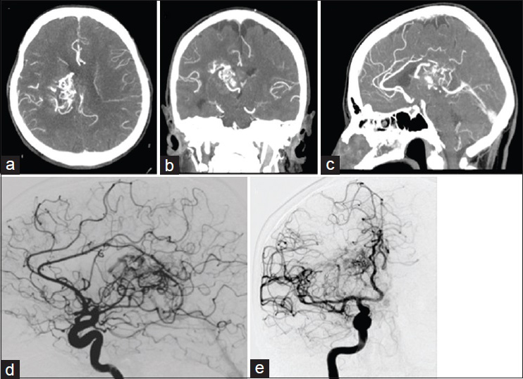
To further assess the vasculature, a computed tomography angiography (CTA) was obtained. Here, the mass was characterized by numerous dense calcifications, as well as by intrinsic hemorrhage posteriorly [
Figure 2
Multimodal angiography. (a) Cranial computed tomography angiography with axial sections displaying a highly vascularized lesion with posteriorly located hemorrhage and focal calcification. (b, c) Correlating coronal and sagittal images demonstrate the specific aspect of this lesion with dilated marginal vessels almost entirely surrounding and draining it. (d, e) Cerebral digital subtraction angiography confirms arteriovenous malformation-like morphology with sagittal and coronal images visualizing a vascular lesion with enlarged draining veins and multiple vessels feeding into a nidus at the posterior margin of the lesion (arrows)
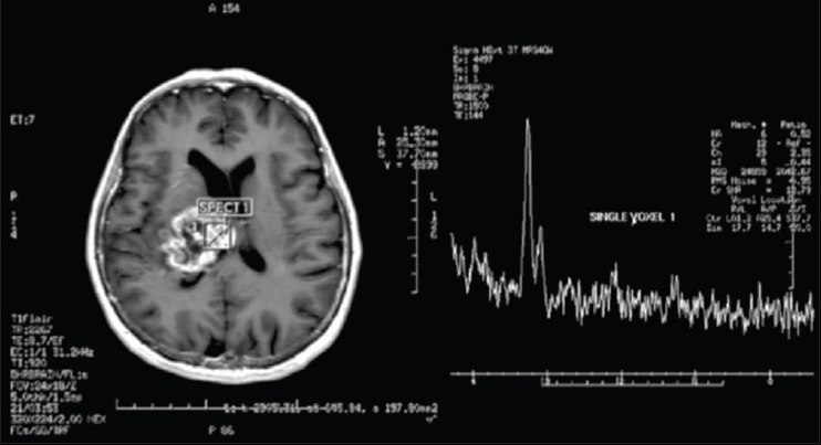
MR-perfusion was performed and showed high perfusion and markedly increased blood volume within the right thalamic lesion maintaining the concern for an AVM, whereas MR multivoxel spectroscopy indicated an increase in creatinine/choline peak ratio [
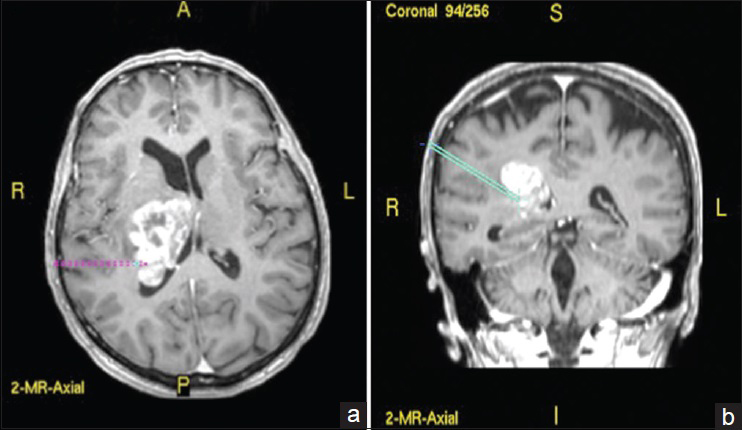
Because the prognosis and treatment algorithms for an AVM and a malignant glioma differ substantially, a definite tissue-based diagnosis was required, however, it was only hesitantly considered given the high risk of procedural bleeding. A stereotactic biopsy was finally performed under general anesthesia using a CRW frame (Radionics Inc., Burlington, MA). Preoperative T1 gadolinium MR as well as CTA-images were fused to the intraoperative frame-based CT using the StereoCalc® and NeuroSight® Arc software (Radionics Inc., Burlington MA) allowing three-dimensional planning of coordinates. Meticulous attention was paid to the delineation of a suitable target area for the biopsy at the inferior margin of the hypervascular zone but avoiding any prominent vessels [Figure
Figure 4
Three-dimensional planning of a frame-based stereotactic biopsy. (a, b) The target point was set to the lateral posterior margin of the lesion with respect to the major vascular aggregations for limiting the bleeding risk. The procedure was planned in a CRW frame using the Stereocalc software (Radionics, Burlington, MA)
Multiple serial stereotactic biopsy specimen were ascertained for histopathological analysis using a set of unique microforceps with a cubic bite size of 1 mm3 (Medical High Tech Inc., Emmendingen, Germany), as described by Warnke et al.[
Intraoperative fresh frozen analysis showed malignant glioma. Final histopathological analysis revealed a densely cellular, astrocytic glial fibrillary acidic protein positive tumor with necrosis, thrombosed vessels, and scattered mitosis among irregular hyperchromatic nuclei. The proliferative index (MIB-1) was above 10%. Thus, final histopathologic diagnosis confirmed GBM with profound hypervascularity.
After an uneventful biopsy and discharge home, subsequent treatment consisted of involved field radiation and standard chemotherapy with Temozolomide, as per Stupp et al.[
DISCUSSION
Simultaneous presentation of vascular lesions and glioma are rare encounters, notably the coincidence of AVM and GBM. Formerly the term “angioglioma,” originally used by Russell and Rubinstein to describe a composite intrinsic tumor of angiogenic and glial origin, included gliomas in combination with venous malformations, cavernous angioma or arteriovenous malformations.[
In a preeminent study addressing the association of AVM and glioma, 2277 patients with primary brain tumors and 222 patients with AVMs were reviewed.[
Contrasting with the low association of AVM and glioma found above, Ziyal et al. argued that such low incidence was likely an underestimate by the study because CT was the only imaging modality used. They, hence, concluded that the true incidence would be higher with modern imaging technologies such as MRI, emphasizing the importance of adequate diagnostic work up.[
In the previous cases, imaging included CT or MRI in combination with a conventional angiogram.[
In this case, the patient's management would have remained the same whether the diagnosis was hypervascularized GBM or AVM-associated GBM, however, treatment would have changed if it was an AVM only. Hence, obtaining histopathology was warranted to confirm the presence of the tumor and not to be misled by imaging suggestive of an AVM. However, distinguishing hypervascular GBM from AVM-associated GBM would have clinical implications only when considering complete surgical resection, which was impossible here. Instead we employed diligent three-dimensional stereotaxy, which allowed us to obtain the correct pathology without any procedural bleeding.
The reasoning for the pursuit of obtaining a tissue diagnosis is further illustrated by Gmeiner et al.[
Given the low number of reported cases, one might question any specific pathoetiology of cases with concurrent AVM and glioma, which remains controversial. One theory brought up the notion of an AVM derived process in which neoplastic changes occur in the perivascular glial tissue.[
Similarly, it has been shown that VEGF levels are increased in surgical specimens from brain AVMs.[
Although various reports have tried to address this specific brain pathology, individual constellations occur in the absence of any superior diagnostic or treatment scheme. Of the 13 reported cases, only 3 included treatment of the AVM as well as that of the glioma.[
CONCLUSION
Coincidental presentation of a vascular lesion and malignant glioma is rare and represents a major challenge in diagnostic imaging and therapy since surgery in this setting carries a significant risk. However, a tissue-based diagnosis remains crucial for any targeted therapeutic approach. Treatment decisions addressing either one of these lesions are complicated because suitable options (e.g., radiosurgery and/or open resection) carry specific risks. This requires standardization of diagnostic and therapeutic approaches to allow for the most appropriate therapy. Whether there is any role for primary treatment with antiangiogenesis agents (e.g., bevacizumab) in this unique setting may be suggested by much anticipated results from ongoing clinical trials of such drugs in brain AVMs.
Consent
Written informed consent was obtained from the patient's family for publication of this case report including any illustrating images. A copy of the written consent is available for review by the Editor of this journal. In accordance with the Ethical Principles for Medical Research Involving Human Subjects, WMA Declaration of Helsinki a continuing review IRB CR approval for “Evaluation of Outcomes Following Neurosurgery Procedures” was obtained by the BIDMC Committee on Clinical Investigations. The protocol number is 2013-P-000253/4.
Authors’ contributions
LNL edited and summarized the clinical data in addition to performing a Medline search and review of the existing literature. CS collected the clinical data and reviewed the patient's files. RR performed and interpreted imaging analysis. MA carried out histopathological analysis of the patient's specimen and thus completed preliminary imaging diagnosis. YL revised the manuscript and completed literature research. EMK initiated this case review, coordinated clinical data retrieval, and supervised manuscript edition.
Data sources and search strategy
We searched MEDLINE (through PubMed interface) to identify potentially relevant articles or abstracts. Our search included following search terms: Angioglioma, arterio-venous malformation associated glioma, hypervascular glioblastoma multiforme, astroglial tumor, and vascular lesions. Search terms were used both discretely and in combination with each other using the Boolean operator AND. There were no language restrictions. We reviewed the bibliographies of all selected articles to identify additional relevant studies.
Financial support and sponsorship
Nil.
Conflicts of interest
There are no conflicts of interest.
References
1. Aucourt J, Jissendi P, Kerdraon O, Baroncini M. Neuroimaging features and pathology of mixed glioblastoma--AVM complex: A case report. J Neuroradiol. 2012. 39: 258-62
2. Bonnin JM, Pena CE, Rubinstein LJ. Mixed capillary hemangioblastoma and glioma. A redefinition of the “angioglioma”. J Neuropathol Exp Neurol. 1983. 42: 504-16
3. Borges LR, Malheiros SM, Pelaez MP, Stavale JN, Santos AJ, Carrete H. Arteriovenous malformation-glioma association: Study of four cases. Arq Neuropsiquiatr. 2003. 61: 426-9
4. Cemil B, Tun K, Polat O, Ozen O, Kaptanoglu E. Glioblastoma multiforme mimicking arteriovenous malformation. Turk Neurosurg. 2009. 19: 433-6
5. Schlegel U, Westphal M. Stereotaktische Biopsie. Neuroonkologie, Thieme, Stuttgart. 1998. p. 75-84
6. Chen W, Choi EJ, McDougall CM, Su H. Brain arteriovenous malformation modeling, pathogenesis, and novel therapeutic targets. Transl Stroke Res. 2014. 5: 316-29
7. Dupuis-Girod S, Ginon I, Saurin JC, Marion D, Guillot E, Decullier E. Bevacizumab in patients with hereditary hemorrhagic telangiectasia and severe hepatic vascular malformations and high cardiac output. JAMA. 2012. 307: 948-55
8. Fischer EG, Sotrel A, Welch K. Cerebral hemangioma with glial neoplasia (angioglioma?). Report of two cases. J Neurosurg. 1982. 56: 430-4
9. Gazzeri R, De Bonis C, Carotenuto V, Catapano D, d’Angelo V, Galarza M. Association between cavernous angioma and cerebral glioma. Report of two cases and literature review of so-called angiogliomas. Neurocirugia. 2011. 22: 562-6
10. Gmeiner M, Sonnberger M, Wurm G, Weis S. Glioblastoma with the appearance of arteriovenous malformation: Pitfalls in diagnosis. Clin Neurol Neurosurg. 2013. 115: 501-6
11. Goodkin R, Zaias B, Michelsen WJ. Arteriovenous malformation and glioma: Coexistent or sequential? Case report. J Neurosurg. 1990. 72: 798-805
12. Lee TT, Landy HJ, Bruce JH. Arteriovenous malformation associated with pleomorphic xanthoastrocytoma. Acta Neurochir. 1996. 138: 590-1
13. Licata C, Pasqualin A, Freschini A, Barone G, Da Pian R. Management of associated primary cerebral neoplasms and vascular malformations: 2. Intracranial arterio-venous malformations. Acta Neurochir. 1986. 83: 38-46
14. Lombardi D, Scheithauer BW, Piepgras D, Meyer FB, Forbes GS. “Angioglioma” and the arteriovenous malformation-glioma association. J Neurosurg. 1991. 75: 589-566
15. Narayana A, Gruber D, Kunnakkat S, Golfinos JG, Parker E, Raza S. A clinical trial of bevacizumab, temozolomide, and radiation for newly diagnosed glioblastoma. J Neurosurg. 2012. 116: 341-5
16. Pallud J, Belaid H, Guillevin R, Vallee JN, Capelle L. Management of associated glioma and arteriovenous malformation--The priority is the glioma. Br J Neurosurg. 2009. 23: 197-8
17. Russell DR, LJ.editorsPathology of tumors of the nervous system. London: Arnold; 1971. p.
18. Stupp R, Mason WP, van den Bent MJ, Weller M, Fisher B, Taphoorn MJ. Radiotherapy plus concomitant and adjuvant temozolomide for glioblastoma. N Eng J Med. 2005. 352: 987-96
19. Walker EJ, Su H, Shen F, Degos V, Amend G, Jun K. Bevacizumab attenuates VEGF-induced angiogenesis and vascular malformations in the adult mouse brain. Stroke. 2012. 43: 1925-30
20. White RJ, Kernohan JW, Wood MW. A study of fifty intracranial vascular tumors found incidentally at necropsy. J Neuropathol Exp Neurol. 1958. 17: 392-8
21. Xhumari A, Rroji A, Enesi E, Bushati T, Sallabanda Diaz K, Petrela M. Glioblastoma after AVM radiosurgery. Case report and review of the literature. Acta Neurochir. 2015. 157: 889-95
22. Ziyal IM, Ece K, Bilginer B, Tezel GG, Ozcan OE. A glioma with an arteriovenous malformation: An association or a different entity?. Acta Neurochir. 2004. 146: 83-6
23. Zuccarello M, Giordano R, Scanarini M, Mingrino S. Malignant astrocytoma associated with arteriovenous malformation. Case report. Acta Neurochir. 1979. 50: 305-9


