

- Department of Orthopaedics, Naha City Hospital, Naha City, Japan.
Correspondence Address:
Hisashi Serikyaku, Department of Orthopaedics, Naha City Hospital, Naha City, Japan.
DOI:10.25259/SNI_561_2021
Copyright: © 2021 Surgical Neurology International This is an open-access article distributed under the terms of the Creative Commons Attribution-Non Commercial-Share Alike 4.0 License, which allows others to remix, tweak, and build upon the work non-commercially, as long as the author is credited and the new creations are licensed under the identical terms.How to cite this article: Hisashi Serikyaku, Shoichiro Higa, Tetsuya Yara. Intradural disc herniation at the L1-2 level. 12-Jul-2021;12:351
How to cite this URL: Hisashi Serikyaku, Shoichiro Higa, Tetsuya Yara. Intradural disc herniation at the L1-2 level. 12-Jul-2021;12:351. Available from: https://surgicalneurologyint.com/surgicalint-articles/10957/
Date of Submission
07-Jun-2021
Date of Acceptance
19-Jun-2021
Date of Web Publication
12-Jul-2021
Abstract
Background: Intradural disc herniations (IDHs) are rare, are difficult to diagnose on preoperative MR/CT imaging, and typically, are most readily confirmed at the time of surgery. However, one of the greatest challenges posed by these lesions, is the repair of the ventral dural rent.
Case Description: A 55-year-old male with a 20-year history of lumbago presented with low back pain and right lower extremity sciatica of 3 months’ duration. The MR and CT studies showed a compressive lesion at the L1-2 level. There was no original suspicion that this was an IDH. At surgery, performed under the operating microscope, a subtotal L1-L2 laminectomy was performed (i.e. while lysing severe adhesions between the posterior longitudinal ligament and the ventral dura, a traumatic durotomy occurred. White, spongious, friable, soft tissue, and free-floating disc fragments extruded through the durotomy site. Notably, it was initially considered to be a tumor rather than a disc. Once all fragments had been delivered, unsuccessful attempts were made to repair the ventral dura. Further efforts were curtailed due to concern that they would result in damage to multiple ventral nerve rootlets. Despite the lack of primary dural repair, the secondary measures resulted in no postoperative recurrent cerebrospinal fluid leakage (CSF) and a smooth postoperative surgical course.
Conclusion: IDH at the L1-2 level is rare, and preoperative MR/CT studies may not always document their intradural location. Ideally, ventral dural tears attributed to these lesions should be directly repaired and/or managed with additional adjunctive CSF leak repair techniques (i.e. muscle patch grafts, microfibrillar collagen, and fibrin sealants).
Keywords: Intradural disc herniation, Lumbar spine, Laminectomy, Durotomy, Suturing of ventral dura, Cerebrospinal fluid leakage, L1-2 level
INTRODUCTION
Intradural disc herniations (IDHs) are very rare (0.26% and 0.30%).[
CASE DESCRIPTION
A 55-year-old male presented with low back pain and right leg sciatica of 3 months’ duration. His preoperative neurological examination was normal.
X-rays, MR, and CT studies
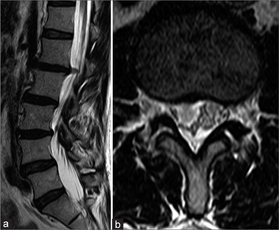
The preoperative X-rays, MR, and CT did not show pathognomonic findings for an IDH. Plain X-rays showed no widening of interpedicular distance or significant vertebral scalloping. The MR revealed a compressive lesion ventral to the dural sac at the L1-2 level (i.e. T2-weighted sagittal and axial images), while the gadolinium-enhanced MR showed peripheral enhancement of the lesion [


Surgery
A subtotal laminectomy of L1 and L2 was performed under a surgical microscope. Several attempts were made to retract the dural sac on the right side to expose the disc fragments. However, this was very difficult due to marked adhesions between the posterior longitudinal ligament (PLL) and the ventral dura. An inadvertent median durotomy occurred, resulting in the spontaneous extrusion of multiple white, spongious, friable, soft free-floating disc fragments. At first, this was thought to be a spinal tumor, but was later confirmed to constitute disc [

Attempt at ventral dural repair
Several attempts were made to suture the ventral dura, but were unsuccessful. Secondary measures including microfibrillar collagen and fibrin sealants were utilized for dural repair resulting in no apparent residual leak utilizing Valsalva maneuvers.
Pathology
The pathological was consistent with degenerated cartilaginous tissue consistent with disc (i.e. myxoid degeneration/edematous tissue).
Postoperative course
The postoperative course was uneventful, and there was no further CSF leakage. The MRI 3 months later showed confirmed no residual disc herniation, and the patient had fully recovered.
DISCUSSION
IDHs are rare, occurring from 0.26% to 0.30% of the time in the lumbar spine.[
Etiology of lumbar IDHs
Establishing the diagnosis of an IDH is often difficult. Discontinuity of the PLL and a “hawk-beak sign” on MRI may help diagnose IDH.[
Surgical documentation of IDH
The diagnosis of intradural herniation is often made at the time of surgery. Of the 122 cases, we identified in the literature, only eight were accurately diagnosed preoperatively.[
Surgery
Surgery is the only way to remove an IDH, noting that some may regress spontaneously like other discs. Here, however, the ventral durotomy, whether deliberate or traumatic, should be closed where feasible to avoid postoperative CSF fistulas, pseudomeningoceles, meningitis, and postural headaches.[
CONCLUSION
Here, we reported an IDH at the L1-2 level that was removed through an L1-L2 laminotomy without being able to perform a primary ventral dural repair; only secondary measures were utilized (i.e. microfibrillar collagen and fibrin sealants). Although this did not result in postoperative recurrent CSF leakage, other may have this complication, thus confirming the need to perform a primary repair of the attendant durotomy whenever feasible.
Declaration of patient consent
The authors certify that they have obtained all appropriate patient consent.
Financial support and sponsorship
Nil.
Conflicts of interest
There are no conflicts of interest.
References
1. Aprígio RM, Caramanti RL, Santos FO, Maia IP, Filipe FM, de Moraes DF. Intradural disc herniation at the L1-L2 level: A case report and literature review. Surg Neurol Int. 2019. 10: 196
2. Arnold PM, Wakwaya YT. Intradural disk herniation at L1-L2: Report of two cases. J Spinal Cord Med. 2011. 34: 312-4
3. Aydin MV, Ozel S, Sen O, Erdogan B, Yildirim T. Intradural disc mimicking: A spinal tumor lesion. Spinal Cord. 2004. 42: 52-54
4. Blikra G. Intradural herniated lumbar disc. J Neurosurg. 1969. 31: 676-9
5. Chaudary KS, Bapat M. Conus medullaris syndrome due to an intradural disc herniation: A case report. Indian J Orthop. 2008. 42: 94-6
6. Choi JY, Lee WS, Sung KH. Intradural lumbar disc herniationis it predictable preoperatively? A report of two cases. Spine J. 2007. 7: 111-7
7. Dandy WE. Serious complications of ruptured intervertebral disks. JAMA. 1942. 119: 474-7
8. D’Andrea G, Trillό G, Roperto R, Celli P, Orlando ER, Ferrante L. Intradural lumbar disc herniations: The role of MRI in preoperative diagnosis and review of the literature. Neurosurg Rev. 2004. 27: 75-80
9. Ducati LG, Silva MV, Brandão MM, Romero FR, Zanini MA. Intradural lumbar disc herniation: Report of five cases with literature review. Eur Spine J. 2013. 22: S404-8
10. Epstein NE. A review article on the diagnosis and treatment of cerebrospinal fluid fistulas and dural tears occurring during spinal surgery. Surg Neurol Int. 2013. 4: S301-17
11. Floeth F, Herdmann J. Chronic dura erosion and intradural lumbar disc herniation: CT and MR imaging and intraoperative photographs of a transdural sequestrectomy. Eur Spine J. 2012. 21: S453-7
12. Galhom AE, Elhadi M. Transdural lumbar disc herniation: Experiences often ten cases with review of literature. J Spine Surg. 2018. 4: 1-14
13. Han IH, Kim KS, Jin BH. Intradural lumbar disc herniations associated with epidural adhesion: Report of two cases. J Korean Neurosurg. 2009. 46: 168-71
14. Hidalgo-Ovejero ÁM, García-Mata S, Gozzi-Vallejo S, IzcoCabezón T, Martínez-Morentín J, Martínez-Grande M. Intradural disc herniation and epidural gas: Something more than a casual association?. Spine (Phila Pa 1976). 2004. 29: E463-7
15. Inoue T. Pure conus medullaris syndrome without lower extremity involvement caused by intradural disc herniation at L1/2: A case report. Spine Surg Relat Res. 2019. 3: 392-5
16. Iwamura Y, Onari K, Kondo S, Inasaka R, Horii H. Cervical intradural disc herniation. Spine (Phila Pa 1976). 2001. 26: 698-702
17. Koç RK, Akdemir H, Öktem IS, Menkü A. Intradural lumbar disc herniation: Report of two cases. Neurosurg Rev. 2001. 24: 44-7
18. Krajewski KL, Regelsberger J. Intradural lumbar disc herniation associated with degenerative spine disease and rheumatoid arthritis. Spine (Phila Pa 1976). 2013. 38: E763-5
19. Lee JS, Suh KT. Intradural disc herniation at L5-S1 mimicking an intradural extramedullary spinal tumor: A case report. J Korean Med Sci. 2006. 21: 778-80
20. Luo D, Ji C, Xu H, Feng H, Zhang H, Li K. Intradural disc herniation at L4/5 level causing cauda equina syndrome: A case report. Medicine (Baltimore). 2020. 99: e19025
21. Mailleux P, Marneffe V, Michel I, Dehullu JP The. “Crumble disc sign”: A specific MRI sign of intradural lumbar disc herniation, allowing a preoperative diagnosis. J Belg Soc Radiol. 2015. 99: 25-9
22. Matsumoto T, Toyoda H, Terai H, Dohzono S, Hori Y, Nakamura H. Utility of discography as a preoperative diagnostic tool for intradural lumbar disc herniation. Asian Spine J. 2016. 10: 771-5
23. Öztürk A, Avcl E, Yazgan P, Torun F, Yücetaş S, Karabağ H. Intradural herniation of intervertebral disc at the level of lumbar 1-lumbar 2. Turk Neurosurg. 2007. 17: 134-7
24. Ponzo G, Furnari M, Umana GE, Giuffrida M, Nicoletti GF, Scalia G. Intradural lumbar disc herniations at the L1-L2 level: A case study and literature review. Surg Neurol Int. 2020. 11: 67
25. Schisano G, Franco A, Nina P. Intraradicular and intradural lumbar disc herniation: Experiences with nine cases. Surg Neurol. 1995. 44: 536-43
26. Smith RV. Intradural disc rupture, Report of two cases. J Neurosrg. 1981. 55: 117-20
27. Tamaki Y, Sakai T, Miyagi R, Nakagawa T, Shimakawa T, Sairyo K. Intradural lumbar disc herniation after percutaneous endoscopic lumbar discectomy: Case report. J Neurosurg Spine. 2015. 23: 336-9
28. Tateiwa D, Yamasaki R, Tei R, Shin Y, Ariga K, Hayashida K. Intradural disk herniation mimicking a spinal tumor: Radiologic imaging, pathogenesis, and operative management. Case Rep Orthop. 2018. 2018: 9810762
29. Yildizhan A, Paşaoğlu Okten T, Ekinci N, Aycan K, Aral O. Intradural disc herniations pathogenesis, clinical picture, diagnosis and treatment. Acta Neurochir (Wien). 1991. 110: 160-5

