- Department of Neurosurgery, Iran University of Medical Sciences, Tehran, Iran
Correspondence Address:
Arash Fattahi
Department of Neurosurgery, Iran University of Medical Sciences, Tehran, Iran
DOI:10.4103/sni.sni_284_18
Copyright: © 2018 Surgical Neurology International This is an open access journal, and articles are distributed under the terms of the Creative Commons Attribution-NonCommercial-ShareAlike 4.0 License, which allows others to remix, tweak, and build upon the work non-commercially, as long as appropriate credit is given and the new creations are licensed under the identical terms.How to cite this article: Hossein Ghalaenovi, Arash Fattahi. Migrating spinal intradural schwannoma with adjacent disc herniation: A case report and brief literature review. 05-Nov-2018;9:228
How to cite this URL: Hossein Ghalaenovi, Arash Fattahi. Migrating spinal intradural schwannoma with adjacent disc herniation: A case report and brief literature review. 05-Nov-2018;9:228. Available from: http://surgicalneurologyint.com/surgicalint-articles/9058/
Abstract
Background:Schwannoma is the most common migrating tumor in the intradural space.
Case Description:We present a patient with migrating schwannoma of the cauda which admitted with buttock and thigh pain since 2 years ago. On two preoperative lumbosacral magnetic resonance imaging (MRI), he had a L2/3 disc herniation concomitant with the intradural extra-axial mass at level of the L1/2 disc on the first MRI and then on the second one, his mass was just posterior to the L3 vertebral body, 22 months later. During the surgical resection of the mass, we found it just posterior to the L2 body. In the literature, we found no accompaniment of migrating intradural spinal mass with adjacent intervertebral disc herniation caused by a double-level cerebro-spinal fluid (CSF) blockade, as it was with our case. On serial imaging, we have seen the mass above and then below the level of CSF blockade, resulting from an extra-dural disc herniation.
Conclusion:We think this rare case could promote a better understanding of the dynamic nature of the central nervous system and the peripheral nervous system within intradural space.
Keywords: CSF blockade, CSF dynamic, disc herniation, migrating schwannoma
BACKGROUND
Migrating schwannomas of the cauda equina are reported in the literature.[
CASE DESCRIPTION
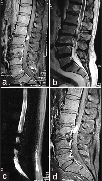
A 60-year-old male presented with 2 years of left buttock and posterior thigh pain that was exacerbated at night. On examination, he had muscle spasm, but no focal neurological deficits. The first lumbosacral magnetic resonance imaging (MRI) scan revealed an intradural extramedullary mass with homogeneous enhancement at the L1/2 level accompanied by an L2/3 disc herniation [Figure
Figure 1
Twenty-two months ago, the first lumbosacral magnetic resonance imaging (MRI) with and without gadolinium revealed a well-defined intradural extra-axial mass that homogenously enhanced posterior to the L1/2 disc space (a). There was also an L2/3 disc herniation (b and c). Twenty-two months later, the second MRI showed the mass migrated posterior to the L3 vertebral body, while the L2/3 disc herniation remained the same (d)
The patient underwent a laminectomy involving the L1, L2, and L3 levels. At surgery, the mass was behind the left L3 body, and its capsule was connected to one nerve root of the cauda. As the rootlet was intrinsically involved within the lesion, it was sacrificed in an en bloc fashion. Additionally, the L2/3 disc was removed. Three months later, the patient was asymptomatic. The histopathological exam was consistent with a schwannoma.
DISCUSSION
The literature showed 28 cases of migrating spinal intradural masses that were predominantly schwannomas, followed by ependymomas and neurenteric cysts.[
The etiology of the migration of schwannomas within the spinal neuraxis is difficult to explain, although likely attributable to the: “dynamic nature of the central nervous system and peripheral nervous system in the CSF space”.[
CONCLUSION
Here, we report a lumbar intradural schwannoma that migrated from the L1/2 to the L3 level over a 22-month period, accompanied by an L2/3 disc herniation.
Declaration of patient consent
The authors certify that they have obtained all appropriate patient consent forms. In the form the patient(s) has/have given his/her/their consent for his/her/their images and other clinical information to be reported in the journal. The patients understand that their names and initials will not be published and due efforts will be made to conceal their identity, but anonymity cannot be guaranteed.
Financial support and sponsorship
Nil.
Conflicts of interest
There are no conflicts of interest.
References
1. Friedman JA, Atkinson JL, Lane JI. Migration of an intraspinal schwannoma documented by intraoperative ultrasound: Case report. Surg Neurol. 2000. 54: 455-7
2. Khan RA, Rahman A, Bhandari PB, Khan SK. Double migration of a schwannoma of thoracic spine. BMJ Case Rep 2013. 2013. p.
3. Lee DY, Chung HJ. Mobile schwannoma of the cauda equina diagnosed by magnetic resonance imaging and magnetic resonance myelography: A case report and review of the literatures. Nerve. 2017. 3: 71-4
4. Marin-Sanabria EA, Sih IM, Tan KK, Tan JS. Mobile cauda equina schwannomas. Singapore Med J. 2007. 48: e53-6
5. Murai H, Arai M, Satoh K, Kataoka Y, Senma S. (Mobile spinal neurenteric cyst) A case report. Seikei Geka. 1991. 42: 1746-9
6. Naik S, Agarwal V, Kumar S. Migrating extramedullary intradural schwannoma of cervico-thoracic junction. J Spine. 2016. 5: 323-


