Tools
Nancy E. Epstein
  1. Department of Neurousrgery, Winthrop Neuroscience, Winthrop University Hospital, Mineola, New York, USA

Correspondence Address:
Nancy E. Epstein
Department of Neurousrgery, Winthrop Neuroscience, Winthrop University Hospital, Mineola, New York, USA

DOI:10.4103/2152-7806.174895

Copyright: © 2016 Surgical Neurology International This is an open access article distributed under the terms of the Creative Commons Attribution-NonCommercial-ShareAlike 3.0 License, which allows others to remix, tweak, and build upon the work non-commercially, as long as the author is credited and the new creations are licensed under the identical terms.

How to cite this article: Epstein NE. More nerve root injuries occur with minimally invasive lumbar surgery, especially extreme lateral interbody fusion: A review. Surg Neurol Int 25-Jan-2016;7:

How to cite this URL: Epstein NE. More nerve root injuries occur with minimally invasive lumbar surgery, especially extreme lateral interbody fusion: A review. Surg Neurol Int 25-Jan-2016;7:. Available from: http://surgicalneurologyint.com/surgicalint_articles/more-nerve-root-injuries-occur-with-minimally-invasive-lumbar-surgery-especially-extreme-lateral-interbody-fusion-a-review/

Date of Submission
31-Oct-2015

Date of Acceptance
02-Nov-2015

Date of Web Publication
25-Jan-2016

Abstract

Background:In the lumbar spine, do more nerve root injuries occur utilizing minimally invasive surgery (MIS) techniques versus open lumbar procedures? To answer this question, we compared the frequency of nerve root injuries for multiple open versus MIS operations including diskectomy, laminectomy with/without fusion addressing degenerative disc disease, stenosis, and/or degenerative spondylolisthesis.

Methods:Several of Desai et al. large Spine Patient Outcomes Research Trial studies showed the frequency for nerve root injury following an open diskectomy ranged from 0.13% to 0.25%, for open laminectomy/stenosis with/without fusion it was 0%, and for open laminectomy/stenosis/degenerative spondylolisthesis with/without fusion it was 2%.

Results:Alternatively, one study compared the incidence of root injuries utilizing MIS transforaminal lumbar interbody fusion (TLIF) versus posterior lumbar interbody fusion (PLIF) techniques; 7.8% of PLIF versus 2% of TLIF patients sustained root injuries. Furthermore, even higher frequencies of radiculitis and nerve root injuries occurred during anterior lumbar interbody fusions (ALIFs) versus extreme lateral interbody fusions (XLIFs). These high frequencies were far from acceptable; 15.8% following ALIF experienced postoperative radiculitis, while 23.8% undergoing XLIF sustained root/plexus deficits.

Conclusions:This review indicates that MIS (TLIF/PLIF/ALIF/XLIF) lumbar surgery resulted in a higher incidence of root injuries, radiculitis, or plexopathy versus open lumbar surgical techniques. Furthermore, even a cursory look at the XLIF data demonstrated the greater danger posed to neural tissue by this newest addition to the MIS lumbar surgical armamentariu. The latter should prompt us as spine surgeons to question why the XLIF procedure is still being offered to our patients?

Keywords: Extreme lateral interbody fusions (XLIF), minimally invasive surgery (MIS), nerve root injuries: lumbar surgery, percutaneous procedures, posterior lumbar interbody fusion (PLIF), posterolateral fusions (PLF), transforaminal lumbar interbody fusion (TLIF)

INTRODUCTION

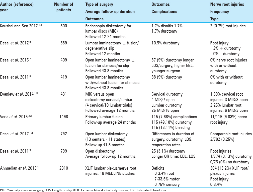
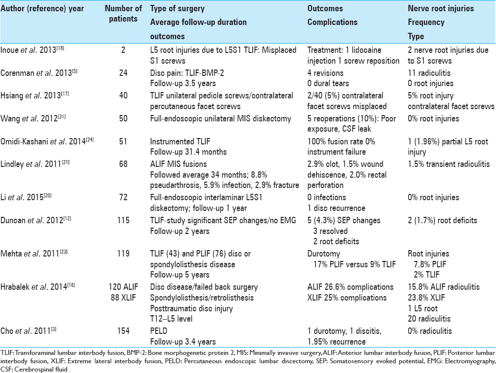
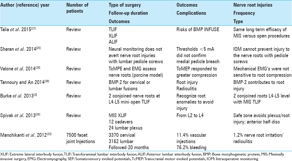
In the lumbar spine, do more nerve root injuries occur utilizing minimally invasive surgery (MIS) techniques versus open lumbar procedures? To answer this question, we compared the frequency of nerve root injuries for multiple open versus MIS operations including diskectomy, laminectomy with/without fusion addressing degenerative disc disease, stenosis, and/or degenerative spondylolisthesis [Tables 1 3 ].


Table 1

Nerve root injuries with lumbar surgery; series with 300 patients or more


Table 2

Nerve root injuries with lumbar surgery; series with 2-154 patients


Table 3

Review articles and other studies: Root injuries with MIS versus open procedures


Desai et al. large Spine Patient Outcomes Research Trial (SPORT) studies showed the frequency for nerve root injury following an open diskectomy ranged from 0.13% to 0.25%, for open laminectomy/stenosis with/without fusion it was 0%, and for open laminectomy/stenosis/degenerative spondylolisthesis with/without fusion it was 2% [ Table 1 ].[ 6 7 8 9 10 ] Another study compared the incidence of root injuries utilizing MIS transforaminal lumbar interbody fusion (TLIF) versus posterior lumbar interbody fusion (PLIF) techniques addressing disc disease or spondylolisthesis; 7.8% of PLIF versus 2% of TLIF patients sustained new root injuries [ Table 2 ].[ 23 ] A further TLIF study showed a 45.8% (11/24 patients) incidence of transient postoperative radiculitis attributed to the additional use of bone morphogenetic protein-2 (BMP-2), yet they claimed none sustained frank root injuries [ Table 1 ].[ 5 ] Data for other MIS anterior lumbar interbody fusion (ALIF) and extreme lateral interbody fusion (XLIF) series further demonstrated even higher and more unacceptable frequencies of radiculitis and/or nerve root injuries. One study compared the frequencies of radiculitis and nerve root injuries; 15.8% after ALIF experienced postoperative radiculitis, while 23.8% undergoing XLIF sustained new deficits (one L5 root injury and 20 cases of radiculitis/plexopathy) [ Table 1 ].[ 16 ] Another XLIF series showed a 13.2% incidence of plexus injuries versus a 0–3.4% incidence of root injuries [ Table 2 ].[ 1 ]

This review of open versus MIS lumbar surgical studies variously addressing degenerative lumbar disc disease, stenosis, stenosis/spondylolisthesis, indicates a higher overall incidence of root injuries, radiculitis, or plexopathy utilizing MIS approaches. Furthermore, the addition of BMP-2 to lumbar fusions (off-label) correlated with nearly a 50% frequency of new postoperative root-related complaints. After performing this analysis we then question, where is the value added for many of these MIS techniques? Certainly, even a cursory look at the XLIF demonstrates its significant threat to neural tissue. Should not we, therefore, reconsider whether it should remain part of the MIS lumbar surgical armamentarium? After all, we as spine surgeons must use our cumulative knowledge to question and change the procedures being offered to our patients.

NERVE ROOT COMPLICATIONS OF OPEN LUMBAR SURGERY

Nerve root complications of open lumbar stenosis surgery

Spine Patient Outcomes Research Trial: Does incidental durotomy affect long-term outcomes of spinal stenosis?

Desai in 2011 looked at the SPORT data regarding the incidence of durotomy and its impact on outcomes for 419 patients with spinal stenosis with/without spondylolisthesis undergoing initial open laminectomies with/without fusion [ Table 1 ].[ 6 ] Patients were followed an average of 43.8 months. Durotomy occurred in 38 (9%) patients and resulted in increased operative time/duration, blood loss, and length of stay (LOS). However, there were no differences in the frequency of nerve root injuries with (0/38) or without durotomy, mortality, reoperations, and outcomes. Notably, for those with/without durotomy, there were also no significant differences regarding age; sex; race; body mass index; the prevalence of smoking, diabetes mellitus, hypertension; decompression level; number of levels decompressed; or whether an additional fusion was performed.

Comments: In Desai et al. study in 2011, the SPORT data analysis revealed that for 419 patients undergoing open initial decompressive laminectomies with/without fusions, 9% developed dural tears, but there were no root injuries in either the durotomy or nondurotomy groups.[ 6 ] It is important to note that open procedures resulted in no root injuries (0%), whereas MIS lumbar procedures (other studies) carried varied higher risks of nerve root damage.

COMPLICATIONS OF OPEN LUMBAR SURGERY: DISKECTOMY

Spine Patient Outcomes Research Trial: Do outcomes vary across centers for surgery for lumbar disc herniation?

Desai et al. in 2012 noted that lumbar diskectomy is the most common spine operation; over 250,000 elective procedures are performed per year.[ 10 ] The SPORT study evaluated 792 patients undergoing initial disc excisions at 6 weeks, at 3, 6, and 12 months, and yearly thereafter (13 academic spine centers; 11 US states) [ Table 1 ].[ 10 ] Seven hundred and ninety-two patients underwent 1st time lumbar diskectomy. Between centers, there were significant differences in short-term outcomes, duration of surgery (47.1–97.7 min), estimated blood loss (EBL) (40.9–191.8 cc), durotomy (0–16%), LOS (0.77–1.6 days), and reoperation rates (5–20%). However, there were no significant differences in the frequency of nerve root injuries (0–1% or 2/792 total), postoperative mortality, or long-term functional outcomes (e.g., short-form 36 [SF-36] physical function scores and Oswestry Disability Index [ODI]) at an average of 4 postoperative years).

Comments: This is an excellent study in which 792 patients from 13 academic centers in 11 states entered data regarding the frequency of risks and complications following initial open diskectomy.[ 10 ] Although there were significant differences in short-term outcomes, duration of surgery, LOS, and reoperation rates between centers, there were no significant differences in the frequency of nerve root injuries (0–1% or 2/792 total), mortality, or in long-term functional outcomes over an average of 4 postoperative years.

COMPLICATIONS OF OPEN LUMBAR SURGERY: FUSION

Impact of complications on patient outcomes following spinal fusion surgery

Verla et al. in 2015 noted that 2–16% of patients undergoing spinal surgery developed complications; they questioned their impact on long-term results [ Table 1 ].[ 30 ] For the 1498 patients undergoing primary lumbar fusion for low back pain and/or radiculopathy, outcomes were comparable for both operative groups (back pain vs. radiculopathy) utilizing ODI, SF-36, and Visual Analog Scale (VAS) scores at 1 and 2 years postoperatively. Complications occurred in 115 (7.68%) of patients, and these included; cerebrospinal fluid (CSF) leak (49.18%), bleeding requiring transfusion (13.11%), and nerve root injury (9.83%; 11 patients).

Comments: This series of 1498 patients underwent primary lumbar fusions for back pain or radiculopathy[ 30 ] Lets stop right there; are these true indications for spinal surgery much less for performing instrumented fusions? We are not only informed that these patients had major spinal fusions but are also advised that 115 or 7.68% sustained major complications; nearly half of the 115 patients (49.18% or 43 patients) had developed new CSF fistulas, and 9.83% (11 patients) incurred new root injuries. In short, unnecessary surgery resulted in major new postoperative morbidity.

LUMBAR NERVE ROOT INJURIES WITH MINIMALLY INVASIVE SURGERY

Minimally invasive surgery for disc degeneration and nerve root injuries

Clinical and radiological outcomes of anterior-posterior fusion (APF) versus transforaminal lumbar interbody fusion (TLIF) for disc degeneration

Faundez et al. in 2009 compared the operative results, including frequency of nerve root injuries, for 133 patients with lumbar disc degeneration undergoing one or two level anterior/posterior spine fusions (anterior-posterior fusion [APF]: 68 patients versus TLIF: 65 patients).[ 14 ] Patients’ outcomes were assessed with SF-36 and ODI questionnaires, and they were followed at least for 2 years. They found TLIF resulted in reduced mean operating room time, LOS, and EBL (409 vs. 480 cc.). Although intraoperative complications were higher for the APF patients (e.g., mostly including vein lacerations resulting from the anterior retroperitoneal approach), more postoperative complications were encountered for TLIF patients. The latter were attributed to “graft material extruding against the nerve root or wound drainage.”

Comments: This study addressed degenerative lumbar disease variously treated with APF versus TLIF. Notably, patients did not clearly exhibit focal preoperative neurological deficits or neurodiagnostic evidence of neural compromise to warrant these operations in the first place. Nevertheless, of the total 133 procedures performed, APF resulted in major life-threatening venous lacerations, while TLIF were responsible for many new root deficits.[ 14 ] Again, one must question whether any of these operations was warranted in the first place.

Preventing postoperative dysesthesia in minimally invasive transforaminal percutaneous endoscopic lumbar discectomy for intracanalicular lumbar discs

Cho et al. in 2011 recorded perioperative complications attributed to MIS transforaminal percutaneous endoscopic lumbar discectomy (PELD) [ Table 1 ].[ 3 ] Any open versus MIS diskectomy may result in dural injury, infection, nerve root irritation, or disc recurrence. However, PELD was uniquely noted for contributing to postoperative dysesthesias (PODs) attributed to dorsal root ganglion (DRG) injuries incurred during the typical dissection/approach. In this study, 154 patients (160 discs) underwent PELD utilizing a “floating retraction technique” that offered, “gentle retraction of the root with perineural fat instead of direct compression of DRG.” The average operative time was 36 min, no operation had to be converted to an open procedure, LOS averaged 1.8 days, and patients were followed for a mean of 3.4 years. Postoperatively, all patients sustained symptomatic relief, 0% developed POD, 1 had a dural injury, 1 had discitis, and the disc recurrence rate was just 1.95%. The authors concluded this technique was safe and effective and helped avoid postoperative POD.

Comments: This study by Cho et al. in 2011 notes that of 154 patients undergoing MIS PELD utilizing a modified “floating method,” the frequency of POD (e.g., nerve root injuries) resulting from retraction of the DRG was reduced to 0%, although there was still 1 dural injury, 1 instance of discitis, and 3 cases (1.9%) of disc recurrence.[ 3 ] Whenever one encounters 100% or 0% in any article, one must question how thorough the evaluation was and who was performing the postoperative assessment. I would also add that a 36 min procedure might be of limited value.

Full-endoscopic interlaminar approach for lumbar discs, and when conversion to an open procedure becomes warranted

Wang et al. in 2012 utilized a full-endoscopic (FE) technique, including a unilateral portal interlaminar approach, for treating 50 patients with lumbar disc herniations (LDH) [ Table 2 ].[ 31 ] They classified the location of the nerve root after excision of ossification of the yellow ligament as: Type I (the nerve root starting point was higher than the incision) in 47 cases (94%) and Type II (the nerve root was lower than the incision) in 3 cases (6%). Five (10%) patients with discs at the L5-S1 level (4 cases), and one with an L4–L5 disc required conversion to open procedures. These conversions were warranted due to: poor placement of the MIS retractor, problematic root exposure/hemostasis/dural fistula, and inadequately exposed nerve roots warranting greater lateral recess exposure. The authors concluded, “proper patient selection and specific radiographic examinations are needed to obtain optimal outcomes using an FE technique for microdiscectomies.”

Comments: Wang et al. in 2012 observed a 10% conversion rate (5 of 50 patients) for patients initially undergoing FE unilateral portal interlaminar lumbar diskectomies.[ 31 ] Shortcomings of the limited exposure provided by this technique in these 5 cases included; poor placement of the MIS retractor, inadequate exposure/poor hemostasis resulting in a CSF fistula, and overall inadequate exposure warranting further dissection for the lateral recess to decompress the nerve root. In short, this article acknowledges how the severity of stenosis impacts the extent of decompression warranted to safely remove many lumbar discs.

Posterior minimally invasive endoscopic discectomy: Results in 300 patients

Kaushal and Sen in 2012 evaluated the results of 300 posterior lumbar MIS endoscopic discectomies performed for managing disc disease [ Table 1 ].[ 19 ] The results were evaluated utilizing MacNab's criteria after a minimum follow-up of 12 months and maximum follow-up of 24 months. Outcomes were excellent/good in 90% of patients, fair in 8%, and poor in 2%. Complications included discitis (5 cases: 1.7%), dural tears (5 cases: 1.7%), and nerve root injuries (2 cases: 0.7%). The authors concluded; endoscopic diskectomy provided a safe and minimal access corridor for lumbar diskectomy.

Comments: Kaushal and Sen in 2012 performed 300 posterior lumbar MIS endoscopic diskectomies and concluded the procedure was both “safe and effective.”[ 19 ] Nevertheless, of the 300 cases performed, 5 had dural tears, 5 had discitis, and 2 had new nerve root injuries. A total of 4% sustained significant morbidity. This complication rate is greater than that encountered with conventional open procedures[ 9 10 ] and, therefore, their conclusions and the safety/efficacy of this approach must be called into question.

Exiting root injury in transforaminal endoscopic discectomy

Choi et al. in 2013 evaluated the clinical/radiological features signaling the risks of root injuries for proposed transforaminal endoscopic discectomy.[ 4 ] In this retrospective analysis of 233 patients treated with PELD for disc disease, 20 (Group A) exhibited new postoperative exiting root injuries (e.g., dysesthesias or motor weakness), while the remainder did not. Patients sustaining root injuries in Group A showed a shorter distance between the exiting roots to the lower facet on magnetic resonance imaging studies (MR) (6.4 ± 1.5 mm vs. 4.4 ± 0.8 mm). They recommended that measurement of this distance on preoperative MR studies would allow surgeons to choose more optimal approaches (e.g., microdiscectomy or conventional open discectomy).

Comments: This MR-based study nicely documents that MIS procedures should be individually tailored to each patient's anatomy. Here, directly measuring on the preoperative MR a shorter distance between the exiting root to the lower facet (6.4 ± 1.5 mm vs. 4.4 ± 0.8 mm) would enable the surgeon to choose other safer approaches (e.g., microdiscectomy or conventional open discectomy) to avoid root injuries that occurred in 20 (4.3%) of 233 MIS transforaminal endoscopic diskectomies.[ 4 ] This is also another way of stating; be on the look-out for stenosis.

Meta-analysis of minimally invasive surgery versus open surgery for cervical and lumbar discectomy

In 2014, Evaniew et al. compared the safety/efficacy of MIS diskectomy versus open procedures addressing cervical or lumbar disc herniations (LDH) [ Table 1 ].[ 13 ] They utilized the MEDLINE, EMBASE and the Cochrane Library databases, and some randomized controlled trials. There were four trials in the cervical discectomy group (n = 431 patients): microendoscopic diskectomy and fusion (37 patients); FE cervical posterior foraminotomy (100 patients); full endoscopy anterior cervical decompression (60 patients); and tubular posterior diskectomy/foraminotomy (22 patients), among others. There were 10 studies in the lumbar discectomy group (n = 1159 patients); tubular diskectomy (167 + 66 patients); microscope assisted percutaneous nucleotomy (52 patients); microendoscopic diskectomy (55 + 10 + 21 + 15 + 70 patients); endoscopic interlaminar and transforaminal diskectomy (100 patients); and minimal access trocar microdiskectomy (30 patients), plus others. Of interest, only one of the four cervical series reported nerve root injuries; the frequency was 3 with MIS versus 3 and 3 with open procedures, for a total of 1.39%. There was also a higher durotomy rate for MIS (14 cases) versus open procedures (7 cases). Of the 10 lumbar series, (e.g., 4 did not report root injuries, and 5 did not report on durotomy) there were six root injuries with MIS versus 3 with open lumbar procedures, for a total of 2.25%. The frequency of lumbar durotomy was 25 with MIS versus 16 with open lumbar procedures. The authors concluded, “Current evidence does not support the routine use of MIS for cervical or lumbar discectomy.”

Comments: This study described the higher root injury and durotomy rates for different types of MIS versus open cervical and lumbar diskectomy procedures. Root injuries occurred in 1.39% of cervical and 2.25% of lumbar cases. Consistently, higher rates of neural injury were reported utilizing MIS techniques in both the cervical and lumbar regions.[ 13 ] Of interest, durotomies were also higher across the board in MIS cervical (MIS 14 vs. 7 open cases) or lumbar procedures (25 vs. 16 cases). It should also be noted that the authors themselves could not support the routine use of MIS for cervical or lumbar diskectomies due to these greater major morbidities; why should we?

Full-endoscopic interlaminar L5/S1 discectomy

Li et al. in 2015 evaluated the efficacy of fully endoscopic (no conversions to open procedures) interlaminar L5/S1 diskectomy in 72 patients with axillary, ventral, or shoulder types/locations of disc herniations [ Table 2 ].[ 20 ] Postoperative MR studies were utilized to assess the extent of resection, and outcomes were evaluated utilizing VAS, ODI, and MacNab scores 12-months after surgery. Surgery required an average of 45 min (20–80 min). Complications included; one disc recurrence (revised microendoscopically), no nerve root injuries, and no infections. Postoperative ODI/VAS scores were significantly decreased, while MacNab scores at 1 year showed; 44 excellent, 26 good, 1 fair, and 1 poor outcome(s).

Comments: In this evaluation of 72 patients undergoing endoscopic diskectomy at the L5–S1 level, the authors noted no root injuries, no infections, and only one disc recurrence.[ 20 ] Notably, the operative time ranged from 20 to 80 min, averaging 45 min. I simply ask, what did they accomplish in 20 min? Was that the patient with the recurrent disc or the original disc that was never adequately resected?

MINIMALLY INVASIVE FUSION AND NERVE ROOT INJURIES; TRANSFORAMINAL LUMBAR INTERBODY FUSION (TLIF) ALONE (VS. OTHER MINIMALLY INVASIVE SURGICAL FUSIONS)

Transforaminal lumbar interbody fusion (TLIF) versus posterior lumbar interbody fusion (PLIF) morbidity including frequency of root injuries

Over an average 5-year period, Mehta et al. in 2011 retrospectively compared the intraoperative and postoperative morbidity of 119 TLIF (43 patients) versus PLIF (76 patients) in patients undergoing spinal fusion for spondylolisthesis and degenerative disc disease [ Table 2 ].[ 23 ] Their hypothesis was that the unilateral exposure of the TLIF would decrease the incidence of durotomy and root injury. Patients had surgery addressing: mechanical back pain (109 [92%]), radicular pain (95 [80%]), and radicular motor weakness (10 [8%]). Those having PLIF had more root injuries versus TLIF (all transient) (6 [7.8%] vs. 1 [2%] for TLIF), and more durotomies (13 [17%] vs. 4 [9%] for TLIF) (neither statistically significant). Other factors including blood loss, and LOS were similar. Comparable rates of the following variables were also noted for TLIF versus PLIF: pseudarthrosis 2 (2.6% TLIF) versus 2 (4.6% PLIF), postoperative radicular pain (88% TLIF vs. 79% PLIF), and residual low back pain (74% TLIF vs. 80% PLIF). The authors concluded there was a trend but no documented significant reduction in nerve root injuries with TLIF versus PLIF. They further observed interbody fusions increased the rate of both neurological complications and durotomy. They, therefore, suggested “MIS TLIF and PLIF should only be considered when the goals of surgery cannot be addressed with decompression and traditional posterolateral fusion.”

Comments: The authors ultimately concluded that any interbody device, whether placed utilizing the TLIF or PLIF approaches, resulted in a higher incidence of not only nerve root injuries, but also durotomy.[ 23 ] They were completely “on target” when they suggested that MIS interbody fusions should, therefore, only be performed where posterolateral traditional decompressions/fusions will not suffice.

Minimally invasive one-level lumbar transforaminal lumbar interbody fusion (TLIF) utilizing both pedicle screw fixation and transpedicular facet screws

Hsiang et al. in 2013 presented a new minimally invasive (MI) technique for one-level TLIF utilizing ipsilateral pedicle screws alone with percutaneously placed contralateral tranpedicular fact screws [ Table 2 ].[ 17 ] For the series of 40 patients, the average operative time (124 min), EBL (140 cc), and LOS (3 days) were studied along with the 5% (2 patients) of patients who developed new radicular pain attributed to poor placement of the contralateral facet screws. The authors concluded this MI construct was of value as it minimized tissue injury while providing “biomechanical advantages of bilateral pedicle screw fixation,” additionally noting that the contralateral facet screws should be carefully placed.

Comments: This study presents a modification of the typical MI TLIF; they utilized ipsilateral pedicle screws, but contralateral percutaneously placed transpedicular facet screws to minimize soft tissue manipulation, and yet “preserve” biomechanical stabilization.[ 17 ] Nevertheless, the placement of the contralateral screws resulted in a 5% (2 patients) incidence or root injuries warranting screw removal. With such a high incidence of root injury, how can one conclude that the contralateral percutaneous transpedicular facet screws placement was safe and effective?

Nerve root anomalies: Implications for transforaminal lumbar interbody fusion (TLIF)

Burke et al. in 2013 noted 2 cases in which conjoined/confluent nerve roots were either anticipated based on preoperative MR studies or simply encountered during mini-open TLIF surgery at the L4–L5 level [ Table 3 ].[ 2 ] The 2 cases included; a 68-year-old with L4–L5 stenosis/spondylolisthesis, and a 60-year-old with severe left L4-L5 foraminal stenosis/focal scoliosis.

Comments: Recognition either preoperatively or intraoperatively of conjoined/confluent nerve root anomalies is critical particularly with more restricted operative exposures provided in MI/MIS, or mini-open TLIF. Indeed, these minimal exposures potentially subject these nerve roots to inadvertent injury.[ 2 ]

L5 spinal nerve injury caused by misplacement of outwardly inserted S1 pedicle screws

Inoue et al. in 2013 evaluated 2 cases in which L5 nerve root injuries (e.g., severe pain resulting) were caused during L5S1 pedicle screw instrumented TLIF procedures [ Table 2 ].[ 18 ] These injuries were attributed to “outwardly placed S1 pedicle screws” that perforated the anterior sacral cortex where it contacted the L5 root. Utilizing computed tomography following rootography, the L5 roots were found to be either (1) “compressed laterally by screw threads,” or (2) “crushed between the screw threads and the sacral body.” The first case was successfully treated with “three spinal nerve infiltrations,” while the latter required redirection of the screw.

Comments: This is an interesting analysis of what is often seen; anterior sacral perforation by S1 pedicle screws.[ 18 ] The observation that such perforations can contribute to L5 root syndromes is of interest. In this study, one L5 deficit responded to nerve blocks, while the other case warranted screw redirection/secondary surgery.

Instrumented transforaminal lumbar interbody fusion (TLIF) for recurrent discs

Omidi-Kashani et al. in 2014 noted that after surgery for LDH the frequency of recurrent discs ranged from 5% to 15% [ Table 2 ].[ 24 ] Here they asked what that rate would be following TLIF addressing the same pathology. They retrospectively evaluated 51 patients, and followed them for an average of 31.4 postoperative months (25–50). Preoperative versus postoperative assessment included respective ODI (7.4–3.4) and VAS (72.1–27.5) scores. Only 1 patient sustained a surgery-related partial L5 nerve root injury. They claimed a 100% fusion rate and observed no instrumentation failures.

Comments: There are several major questions raised here. First, why for LDH, is a TLIF required? Why not a focal decompression with diskectomy. Second, it is very difficult to believe the 100% TLIF fusion rate, particularly when many other series cite higher frequencies of pseudarthrosis. For example, Gologorsky et al. in 2014 cited a 17.5% pseudarthrosis rate for TLIF using unilateral pedicle instrumentation, and 2.5% for bilateral TLIF instrumented procedures.[ 15 ] Similarly, Faundez et al. in 2009, cited a much higher 23.1% pseudarthrosis rate for 65 patients undergoing TLIF. Additionally, if we question the pseudarthrosis rate presented for this TLIF series, do we infer that the 1 of 51 patients with a partial L5 root injury rate was also underestimated?

Increased pseudarthrosis rate after unilateral instrumented transforaminal lumbar interbody fusion (TLIF) for lumbar spondylosis

Gologorsky et al. in 2014 evaluated the utility of TLIF in 80 prospective patients utilizing bilateral (40 patients) or unilateral (40 patients) pedicle screw instrumentation over an average of 52 postoperative months.[ 15 ] Data (demographic/surgical) were largely similar for both groups. However, the pseudarthrosis rate was much greater for those undergoing unilateral (7 patients [17.5%]) versus bilateral instrumentation (1 patient [2.5%]). Notably, additional surgery was offered to 8 patients following unilateral versus only 1 patient after bilateral surgery. The authors concluded that TLIF with bilateral instrumentation effectively manages lumbar spondylosis, but unilateral constructs are 7 times more likely to fail requiring reoperations.

Comments: This article highlights the results of unilateral versus bilateral pedicle screw placement in TLIF constructs in a prospective patient population.[ 15 ] My question here would be, why should not the appropriate biomechanical data have first been obtained from animal studies to more clearly indicate that unilateral pedicle/screw instrumentation with TLIF would result in a such a higher (e.g., 7 fold greater) pseudarthrosis rate? In short, why not first perform this study in an animal model rather than a clinical patient population?

MINIMALLY INVASIVE FUSION AND NERVE ROOT INJURIES; EXTREME LATERAL INTERBODY FUSION (XLIF)

Review: Safe zone for retractor placement to the lumbar spine via the transpsoas approach or extreme lateral interbody fusion (XLIF)

Spivak et al. in 2013 discussed the variable anatomy of the lumbar plexus with respect to the psoas muscle at the L23, L34, and L45 disc levels so that retractors for XLIF/transpsoas procedures could be safely placed [ Table 3 ].[ 26 ] The study involved 12 cadavers and 24 lumbar plexuses/psoas muscles (ages 7–35). In these specimens, the L2–L4 roots were identified. They found: “The anterior-posterior diameter of the psoas increased from L2 to L4, with mean vertebral body coverage of 80%, 86%, and 85% at L2/3, L3/4, and L4/5, respectively.” They concluded, the “safe” area to avoid lumbar nerve root/plexus injury was the anterior half of the disc.

Comments: As XLIF are typically performed for patients with pain alone without focal neurological or neuroradiological deficits, they should be considered unnecessary. Furthermore, this cadaveric study documents that the lumbar plexus and nerve roots from the L23 through the L45 levels are at great risk during these procedures.[ 26 ] The proclivity for plexus/nerve root injuries with XLIF should prompt spinal surgeons to strongly questions whether these procedures are safe and even warranted?

Review: Analysis of lumbar plexopathies and nerve injury after extreme lateral interbody fusion (XLIF) (lateral retroperitoneal transpsoas approach)

Ahmadian et al. in 2013 noted that the most typical complication of XLIF (lateral retroperitoneal transpsoas approach) included lumbar plexus/nerve root injuries [ Table 1 ].[ 1 ] Using MEDLINE they found 18 studies involving 2310 patients; 304 patients exhibited potential plexus-related injuries. Documented nerve and/or root injuries occurred in from 0% to 3.4% of patients; motor (0.7–33.6%) and sensory deficits (0–75%) resulted. They concluded, “There is underreporting of postoperative lumbar plexus nerve injury and a lack of standardization of clinical findings of neural complications related to the MI lateral retroperitoneal transpsoas approach.”

Comments: In Ahmadian et al. study in 2013, they evaluated 18 studies involving 2310 patients, 304 (13.2%) of whom sustained root/plexus injuries.[ 1 ] Documented root injuries occurred in up to 3.4% of cases, with up to 33.6% showing motor, and up to 75% demonstrating sensory deficits. Again for patients undergoing XLIF for pain alone without preoperative documented focal neurological deficits or radiographic impingement on neural structures, this would appear to be a high price to pay. With such a high morbidity, why aren’t XLIF procedures being withdrawn from the lexicon of offered lumbar therapies?

A comparison of complication rates for anterior versus lateral approaches to the lumbar spine; minimally invasive anterior lumbar interbody fusion (ALIF) versus minimally invasive extreme lateral interbody fusion (XLIF)

Hrabalek et al. in 2015 compared the complication rates of traditional MIS ALIF (120 patients) versus newer MIS XLIF (88 patients) procedures to approach disc herniations at the T12–L5 levels [ Table 2 ].[ 16 ] Preoperatively patients exhibited; “degenerative disc disease, failed back surgery syndrome, spondylolisthesis, retrolisthesis, and posttraumatic disc injury.” For patients undergoing ALIF, the major complication was lumbar postsympathectomy syndrome in 19 patients (15.8%) plus 35 demonstrated minor intra- and post-operative complications (32 patients [26.6%]). Notably, howevr, for XLIF patients, there were 26 complications (22 patients [25%]). One major complication was partial/transient injury to an L5 nerve root (1.1%). Twenty of the other 25 minor complications involved root or plexus injuries; 11 included transient pain of the left groin or anterior thigh (12.5%), and 9 included numbness in the same dermatomes (10.2%). Despite the high incidence of root/plexus injuries described above, the authors still concluded that for ALIF and XLIF; “MIS approaches to levels T12–L5 disc spaces are safe procedures” but fully recommended the use of intraoperative neural monitoring (IOM).

Comments: Again and again, we see studies in which the data clearly point out a vast number of major and/or minor complications attributed to minimally invasive operations, yet the authors/surgeons conclude the procedures are safe and effective. Nevertheless, the high frequency of nerve root/plexus-related complications noted in this XLIF study alone would seriously call into question whether these procedures should be performed at all.[ 16 ] Why didn’t the authors come to the same conclusion?

Comparison of lumbar interbody fusion techniques: Transforaminal lumbar interbody fusion (TLIF), anterior lumbar interbody fusion (ALIF), and extreme lateral interbody fusion (XLIF): A review

Talia et al. in 2015 reviewed the different surgical techniques offered for interbody lumbar fusion; anterior, lateral, transforaminal, and posterior approaches [ Table 3 ].[ 27 ] They noted that although MIS approaches have been offered, “good fusion rates and low complication rates” are often “limited” by the necessity for thecal and nerve root retraction. TLIF they felt avoided some of the retraction issues and was considered particularly useful for revision procedures. ALIF, although avoiding the neural elements, had the risk of visceral and vascular exposure. Extreme lateral XLIF lumbar interbody fusions put the lumbar plexus at risk while dissecting the psoas muscle. Overall, despite the multitude of MIS procedures, and their reduced EBL, there were no adequate long-term data confirming the benefit/efficacy of these MIS approaches. Furthermore, they raised the issue of iliac crest as the gold standard for bone grafting/fusion and offered that alternatives such as BMPs (BMP/INFUSE) are being frequently used but carry significant risks/complications.

Comments: This review advisedly questions the safety/efficacy of TLIF, ALIF, and XLIF versus open procedures, while also raising the controversy regarding the use of BMP/INFUSE versus the gold standard; iliac crest autograft.[ 27 ] Here, the authors concluded that there were no adequate long-term data confirming the benefit/efficacy of MIS surgery; why should we disagree?

COMPLICATIONS OF MINIMALLY INVASIVE ANTERIOR LUMBAR INTERBODY FUSIONS (ALIF) INCLUDING ROOT INJURIES

In 2011, Lindley et al. reviewed the complications of 68 MIS ALIF performed at the L1-L5, and L5-S1 levels over an average postoperative period of 34 months [ Table 2 ].[ 21 ] Eighteen (26.5%) complications were observed in 16 (23.5%) patients; these included “pseudarthrosis (8.8%), superficial infection (5.9%), sacral fracture (2.9%), pelvic hematoma (2.9%), failure of wound closure (1.5%), transient nerve root irritation (1.5%), and rectal perforation (2.9%).” Of interest, the authors concluded that the 26.5% complication rate was “relatively low” and that many could be avoided with improved patient selection and surgical planning.

Comments: In this 2011 study, Lindley et al. found a 26.5% complication rate for 68 MIS ALIF performed at the L4–L5 and L5–S1 levels. Notably, there was a 1.5% incidence of transient nerve root irritation among a myriad of other significant complications.[ 21 ] If we keep in mind that ALIF are performed in patients who do not warrant surgery in the first place (e.g., no focal neurological deficits and no significant neurodiagnostic evidence of thecal sac or nerve root intrusion), then one would have to conclude that this overall complication rate was too high and should never have occurred.

DUROTOMY AND LUMBAR NERVE ROOT INJURIES

Outcomes after incidental durotomy during 1st time open posterior lumbar diskectomy

Desai et al. in 2011 utilized the SPORT data to evaluate the frequency of durotomy and its accompanying complications following 799 1st time open diskectomy utilizing a “standard” open posterior lumbar diskectomy (use of the microscope was at the surgeon's discretion and was not recorded) [ Table 1 ].[ 9 ] Patients were followed an average of 12 postoperative months (13 spine clinics in 11 US states). The incidental durotomy rate was 25 (3.1%); notably, there were no significant differences between the durotomy and nondurotomy groups regarding the incidence of root injuries (1/774 [0.13%] no durotomy vs. 0/25 durotomy [0%]), mortality, reoperation rates, outcomes, and other variables (age, sex, race, body mass index, herniation level/type, smoking, diabetes, or hypertension). Nevertheless, they required longer operative time, blood loss, and LOS.

Comments: The morbidity of open lumbar diskectomy as confirmed in this SPORT trial was extremely low. Out of 799 patients undergoing initial open surgery, the incidental durotomy rate was only 3.1%, and the frequency of neural injury was 1/774 (0.13%) without durotomy and 0/25 (0%) with durotomy.[ 9 ] The frequencies of nerve root and dural injuries were clearly very low with open operative procedures, and were much lower than with MIS procedures. Still, of interest, is the comment that the use of the operating microscope was at the surgeon's discretion; perhaps less discretion and more utilization of the microscope as part of the standard of care would further minimize the incidence of both of these injuries.

Surgery for lumbar degenerative spondylolisthesis in Spine Patient Outcomes Research Trial: Does incidental durotomy affect the outcome?

Desai et al. in 2012 retrospectively reviewed a multi-institutional (e.g., SPORT) database looking at the impact of durotomy on outcome in 389 patients undergoing initial decompressive lumbar laminectomy for degenerative spondylolisthesis with/without fusion [ Table 1 ].[ 8 ] Patients were followed up to 12 postoperative months; the study included 13 spine clinics in 11 US states. A 10.5% frequency of dural injury occurred; those with/without durotomy showed similar clinical/comorbid factors and other variables (e.g., no significant differences in age, race, smoking, diabetes, hypertension, decompression level, number of levels, and presence/absence of fusion). Notably, there were no differences in the incidence of nerve root injury (0% for no durotomy; 2% [1 patient] with durotomy), mortality, need for additional surgery, SF-36 physical function scores, or ODI.

Comments: In this SPORT trial by Desai et al. in 2012, for the 389 patients undergoing decompressive lumbar laminectomy for degenerative spondylolisthesis with/without fusion (not a MIS study), the overall incidence or durotomy was 10.5%.[ 8 ] Of interest, the frequency of nerve root injuries was nearly comparable with durotomy (2%) or without (0%) durotomy.

Incidental durotomy impact on long-term outcomes for spinal stenosis

Desai et al. in 2015, as part of another SPORT study evaluated patients with spinal stenosis without spondylolisthesis who underwent 1st time laminectomies with/without fusions [ Table 1 ].[ 7 ] Patients were followed for a total of 12 postoperative months, and yearly thereafter for an average of 43.8 postoperative months (13 spine clinics; 11 US state). Of 409 patients undergoing open laminectomy, 37 (9%) had traumatic durotomy. Notably, the methods clearly stated, “The use of a microscope was at the surgeon's discretion but was not recorded as a SPORT data element.” The frequency of durotomy was the same irrespective of the number of stenotic levels, location/severity of stenosis, and prior lumbar epidural steroid injections (ESI). Durotomy, increased the operative time by 29% (161.7 vs. 125.2 min), EBL by 85% (534.4 vs. 288.9 mL), and LOS by 39% (4.3 vs. 3 days). Durotomy did not, however, increase wound dehiscence, graft complications, neurological complications (no root injuries in either group), fusion failure rates, wound infections, mortality, additional surgeries, or primary outcomes (SF-36 bodily pain or physical function scores or ODI).

Comments: The frequency of durotomy in this series appeared to be extraordinarily high at 9%; indeed there were 13 centers in 11 states involved in the study. Likely, many of these centers were major teaching hospitals, and the higher incidence of dural tears with less experienced surgeons likely reflected the number of residents involved.[ 7 ] However, why wasn’t an operating microscope uniformly used to reduce these injuries? Aren’t we beyond “the use of a microscope was at the surgeon's discretion?” And, how convenient was it that that they did not record who did/did not use the microscope as a part of the “SPORT data element.”

ELECTROPHYSIOLOGICAL MONITORING OF LUMBAR SURGERY AND NERVE ROOT INJURIES

Intraoperative decrease in amplitude of somatosensory evoked potentials of the lower extremities with interbody fusion cage placement during lumbar fusions

Duncan et al. in 2012 retrospectively analyzed over a 2 year period how often decreases in somatosensory evoked potential (SEP) amplitudes occurred without electromyographic (EMG) or MEP changes during 115 TLIF procedures [ Table 2 ].[ 12 ] The underlying assumption in this study was that SEP and MEP typically monitor cord function, while EMGs are best at detecting root injuries. In this study, 5 patients developed intraoperative SEP but no EMG changes during the placement of interbody fusion cages. Although cage removal reversed all SEP changes, 2 of the 5 patients exhibited new postoperative deficits. The import of this study was that the development of SEP changes without EMG changes can occur when these interbody devices are applied, and as in this case, despite SEP resolution, 2 of 5 patients sustained permanent neurological deficits.

Comments: This study underscores the need to better monitor not only TLIF but also any lumbar decompression with/without fusion with SEPs and EMGs (often MEP are not used as there is no cord involvement).[ 12 ] During typical lumbar decompressive procedures without interbody fusion (e.g., diskectomy/laminectomy for stenosis), there may be very transient EMG and occasional sphincteric changes which typically immediately resolve with cessation of dissection. However, significant SEP changes almost never occur unless there is major manipulation of the cauda equina as with the application of interbody devices. This study confirmed the potential increased morbidity of TLIF/PLIF or the placement of any interbody devices and provides physiological SEP evidence as to why these procedures are not really “safe.”

Electrophysiological monitoring of lumbar fusion

Sharan et al. in 2014 observed that IOM is often utilized during lumbar fusion surgery to avert nerve root injuries attributed to pedicle screw placement [ Table 3 ].[ 25 ] Nevertheless, the authors found “no evidence to date that IOM can prevent injury to the nerve roots;” in fact they determined that once an injury occurs, it is permanent, and repositioning of the screws does not reverse these deficits. They further determined that low thresholds (e.g., below 5 mA) when stimulating screws did not clearly confirm a medial pedicle breach.

Comments: This article states that IOM of pedicle screw placement during lumbar spinal instrumentation does not avoid nerve root injuries, and that low thresholds encountered following screw placement (e.g., M5 mA) did not clearly reflect medial pedicle breaches.[ 25 ] Nevertheless, I would counter that continuous IOM, but only with the interpreting physiologist is in the room, does provide critical early warnings regarding real-time “changes” (e.g., EMG monitoring, SEP monitoring, and sphincter funciton). These may occur during the most critical phases of these operations, and with immediate feedback in the operating room, directly from the interpreting monitoring specialist the majority of these nerve root injuries may be avoided.

Efficacy of transcranial motor evoked potentials and electromyography to assess nerve root function during compression in a porcine model

Valone et al. in 2014 observed that lumbar nerve root injury/weakness, variously attributed to operative manipulation/decompression, occurs in up to 30% of spinal deformity cases [ Table 3 ].[ 29 ] To evaluate this, they utilized transcranial motor evoked potentials (TcMEPs), mechanically elicited EMG responses, and evoked EMG responses’ to evaluate nerve root compression changes in a porcine model. They observed that compression at “1 and 2 N produced consistent changes in TcMEPs and EMG responses.” While TcMEP monitoring readily responded to greater compression, “mechanically elicited EMG responses were not sensitive to nerve root compression.”

Comments: Valone et al. in 2014 observed that lumbar nerve root injury/weakness, variously attributed to operative manipulation/decompression, occurs in up to 30% of spinal deformity cases [ Table 3 ].[ 29 ] They found that TcMEP monitoring readily responded to greater compression, while “mechanically elicited EMG responses were not sensitive to nerve root compression.”

NERVE ROOT INJURIES DUE TO BONE MORPHOGENETIC PROTEIN IN LUMBAR FUSIONS

Bone morphogenetic protein-2 and spinal arthrodesis: Protein interaction with the nervous system

Dmitriev et al. in 2011 noted the high frequency of off-label use of BMP-2 (rhBMP-2) in spinal fusions. In theory, rhBMP-2, a potent growth factor, when placed near neural structures, can cause direct cord and root injury (e.g., DRG).[ 11 ] The authors noted that early animal studies on the safety of rhBMP-2 did not clearly record injurious effects. However, in this study they found: “rhBMP-2 does elicit a profound signaling response within the spinal cord and the peripheral ganglia. Recent preclinical studies indicate that rhBMP-2, if provided direct access to the spinal cord parenchyma or the DRG, can trigger significant inflammation and morphologic changes within these tissues that could be deleterious to neurologic recovery.”

Comments: There is a significant negative impact for applying rhBMP-2 near neural structures as documented by Dmitriev et al. in this 2011 article.[ 11 ] Specifically, the “article provides rather clear anatomical and pathophysiological evidence regarding the negative and deleterious impact of rhBMP-2 when utilized clinically to perform spinal fusions.”

Bone morphogenetic protein-2 (INFUSE) transforaminal lumbar interbody fusion for discogenic pain

Corenman et al. in their 2013 retrospective study, evaluated the outcomes, complications, and reoperation rates for TLIF performed in patients with discogenic pain syndrome (DPS) over a 2-year period utilizing BMP-2 [ Table 2 ]. Complications of BMP-2 included; osteolysis, heterotopic bone, and unexplained postoperative radiculitis (BMPP).[ 5 ] Thirty-six (80%) of the original 45 patients completed both the preoperative and postoperative questionnaires and were followed an average of 41.9 ± 11.9 months. They demonstrated significant improvement on the ODI, SF-12 (physical component), and numeric rating scale for back pain. There were, however, “3 perioperative complications, 4 revision surgical procedures, and 11 (30.6%) cases of benign BMPP, despite the absence of intraoperative dural tears or frank nerve root injury. Despite the 11 (30.6%) of 36 patients with BMPP, the authors concluded: “BMP with TLIF (bTLIF) is a reasonable treatment option for patients who experience DPS.”

Commentary: Corenman et al. 2013 retrospectively evaluated the results of TLIF with BMP-2 performed for patients with DPSs.[ 5 ] A major problem for this study is that DPS basically describes pain alone without significant neuorlogical deficits or radiographic findings. Nevertheless, these patients were subjected to TLIF plus the off-label use of BMP-2. Next, postoperatively, we were told that patients, who never required surgery in the first place, sustained 3 postoperative complications, with 4 requiring revision surgery, and 11 exhibiting BMPP. It is inexplicable how the authors then concluded that this procedure bTLIF/BMP-2 was a reasonable treatment option. Furthermore, how can they end with the favorite statement that a larger study is warranted when they already have shown an enormous 30.6% frequency of postoperative radiculities (BMPP).

Complications with the use of bone morphogenetic protein 2 in spine surgery (review)

Tannoury and An in 2014 reviewed literature regarding the morbidity/adverse events whey utilizing rhBMP-2 to perform cervical and lumbar spine fusions [ Table 3 ]. Adverse events included not only postoperative nerve root injury and radiculitis, but also contributed to “retrograde ejaculation, antibodies formation, ectopic bone formation, vertebral osteolysis/edema, dysphagia and neck swelling, hematoma formation, interbody graft lucency, and wound healing complications”.[ 28 ] The authors concluded, “Despite the excellent spinal fusion rates promoted by this powerful molecule, the increasingly reported adverse outcomes associated with BMP usage have created real concerns.”

Comments: This review article by Tannoury and An in 2014 underscores the now commonly held belief that the use of rhBMP-2 resulted in adverse events including nerve root injury/radiculitis when utilized to perform cervical or lumbar fusions.[ 28 ] Note the complications included; “retrograde ejaculation, antibodies formation, ectopic bone formation, vertebral osteolysis/edema, dysphagia and neck swelling, hematoma formation, interbody graft lucency, and wound healing.” It is all well and good to cite “real concerns”, but where is the momentum to remove this product from the spinal surgeons’ armamentarium?

ROOT INJURIES ATTRIBUTED TO FACET JOINT NERVE BLOCKS

Complications of fluoroscopically directed facet joint nerve blocks: A prospective evaluation of 7500 episodes with 43,000 nerve blocks

Manchikanti et al. in 2012 studied the adverse effects and complications of ambulatory fluoroscopically guided facet joint injection (FJI) performed over 20 months in the cervical spine (3370 patients), in the lumbar spine (3162 patients), and thoracic spine (950 patients) [ Table 3 ].[ 22 ] Although there were no major complications the following “minor” complications occurred: intravascular penetration 11.4% (20% cervical, 4% lumbar, 6% in thoracic reqion), local bleeding (76.3%), oozing (19.6%), and local hematoma/nerve root irritation (1.2% including profuse bleeding, bruising, soreness, and nerve root irritation).

Comments: Considering there is no long-term documented efficacy for epidural steroid injections (ESIs) or facet joint injections (FJs), the frequency of minor complications appeared quite significant.[ 22 ] First and foremost, the intravascular penetration rate of 11.4% with the majority being performed in the cervical region, can result in brain stem stroke/paralysis/death-not a minor complication by any means. Furthermore, the 1.2% conglomerate frequency of bleeding and nerve root irritation complications is also disturbing; I wonder how many of these resulted in significant postoperative neurological deficits that were simply not observed/recorded as those performing the injections were not typically the on-going treating physicians (e.g. spine specialists).

Financial support and sponsorship

Nil.

Conflicts of interest

There are no conflicts of interest.

References

1. Ahmadian A, Deukmedjian AR, Abel N, Dakwar E, Uribe JS. Analysis of lumbar plexopathies and nerve injury after lateral retroperitoneal transpsoas approach: Diagnostic standardization. J Neurosurg Spine. 2013. 18: 289-97

2. Burke SM, Safain MG, Kryzanski J, Riesenburger RI. Nerve root anomalies: Implications for transforaminal lumbar interbody fusion surgery and a review of the Neidre and Macnab classification system. Neurosurg Focus. 2013. 35: E9-

3. Cho JY, Lee SH, Lee HY. Prevention of development of postoperative dysesthesia in transforaminal percutaneous endoscopic lumbar discectomy for intracanalicular lumbar disc herniation: Floating retraction technique. Minim Invasive Neurosurg. 2011. 54: 214-8

4. Choi I, Ahn JO, So WS, Lee SJ, Choi IJ, Kim H. Exiting root injury in transforaminal endoscopic discectomy: Preoperative image considerations for safety. Eur Spine J. 2013. 22: 2481-7

5. Corenman DS, Gillard DM, Dornan GJ, Strauch EL. Recombinant human bone morphogenetic protein-2-augmented transforaminal lumbar interbody fusion for the treatment of chronic low back pain secondary to the homogeneous diagnosis of discogenic pain syndrome: Two-year outcomes. Spine (Phila Pa 1976). 2013. 38: E1269-77

6. Desai A, Ball PA, Bekelis K, Lurie J, Mirza SK, Tosteson TD. SPORT: Does incidental durotomy affect long-term outcomes in cases of spinal stenosis?. Neurosurgery. 2011. 69: 38-44

7. Desai A, Ball PA, Bekelis K, Lurie J, Mirza SK, Tosteson TD. SPORT: Does incidental durotomy affect longterm outcomes in cases of spinal stenosis?. Neurosurgery. 2015. 76: S57-63

8. Desai A, Ball PA, Bekelis K, Lurie J, Mirza SK, Tosteson TD. Surgery for lumbar degenerative spondylolisthesis in spine patient outcomes research trial: Does incidental durotomy affect outcome?. Spine (Phila Pa 1976). 2012. 37: 406-13

9. Desai A, Ball PA, Bekelis K, Lurie JD, Mirza SK, Tosteson TD. Outcomes after incidental durotomy during first-time lumbar discectomy. J Neurosurg Spine. 2011. 14: 647-53

10. Desai A, Bekelis K, Ball PA, Lurie J, Mirza SK, Tosteson TD. Spine patient outcomes research trial: Do outcomes vary across centers for surgery for lumbar disc herniation?. Neurosurgery. 2012. 71: 833-42

11. Dmitriev AE, Lehman JT, Symes AJ. Bone morphogenetic protein-2 and spinal arthrodesis: The basic science perspective on protein interaction with the nervous system. Spine J. 2011. 11: 500-5

12. Duncan JW, Bailey RA, Baena R. Intraoperative decrease in amplitude of somatosensory-evoked potentials of the lower extremities with interbody fusion cage placement during lumbar fusion surgery. Spine (Phila Pa 1976). 2012. 37: E1290-5

13. Evaniew N, Khan M, Drew B, Kwok D, Bhandari M, Ghert M. Minimally invasive versus open surgery for cervical and lumbar discectomy: A systematic review and meta-analysis. CMAJ Open. 2014. 2: E295-305

14. Faundez AA, Schwender JD, Safriel Y, Gilbert TJ, Mehbod AA, Denis F. Clinical and radiological outcome of anterior-posterior fusion versus transforaminal lumbar interbody fusion for symptomatic disc degeneration: A retrospective comparative study of 133 patients. Eur Spine J. 2009. 18: 203-11

15. Gologorsky Y, Skovrlj B, Steinberger J, Moore M, Arginteanu M, Moore F. Increased incidence of pseudarthrosis after unilateral instrumented transforaminal lumbar interbody fusion in patients with lumbar spondylosis: Clinical article. J Neurosurg Spine. 2014. 21: 601-7

16. Hrabalek L, Adamus M, Gryga A, Wanek T, Tucek P. A comparison of complication rate between anterior and lateral approaches to the lumbar spine. Biomed Pap Med Fac Univ Palacky Olomouc Czech Repub. 2014. 158: 127-32

17. Hsiang J, Yu K, He Y. Minimally invasive one-level lumbar decompression and fusion surgery with posterior instrumentation using a combination of pedicle screw fixation and transpedicular facet screw construct. Surg Neurol Int. 2013. 4: 125-

18. Inoue M, Inoue G, Ozawa T, Miyagi M, Kamoda H, Ishikawa T. L5 spinal nerve injury caused by misplacement of outwardly-inserted S1 pedicle screws. Eur Spine J. 2013. 22: S461-5

19. Kaushal M, Sen R. Posterior endoscopic discectomy: Results in 300 patients. Indian J Orthop. 2012. 46: 81-5

20. Li ZZ, Hou SX, Shang WL, Song KR, Zhao HL. The strategy and early clinical outcome of full-endoscopic L5/S1 discectomy through interlaminar approach. Clin Neurol Neurosurg. 2015. 133: 40-5

21. Lindley EM, McCullough MA, Burger EL, Brown CW, Patel VV. Complications of axial lumbar interbody fusion. J Neurosurg Spine. 2011. 15: 273-9

22. Manchikanti L, Malla Y, Wargo BW, Cash KA, Pampati V, Fellows B. Complications of fluoroscopically directed facet joint nerve blocks: A prospective evaluation of 7,500 episodes with 43,000 nerve blocks. Pain Physician. 2012. 15: E143-50

23. Mehta VA, McGirt MJ, Garcés Ambrossi GL, Parker SL, Sciubba DM, Bydon A. Trans-foraminal versus posterior lumbar interbody fusion: Comparison of surgical morbidity. Neurol Res. 2011. 33: 38-42

24. Omidi-Kashani F, Ghayem Hasankhani E, Noroozi HR. Instrumented transforaminal lumbar interbody fusion in surgical treatment of recurrent disc herniation. Med J Islam Repub Iran. 2014. 28: 124-

25. Sharan A, Groff MW, Dailey AT, Ghogawala Z, Resnick DK, Watters WC. Guideline update for the performance of fusion procedures for degenerative disease of the lumbar spine. Part 15: Electrophysiological monitoring and lumbar fusion. J Neurosurg Spine. 2014. 21: 102-5

26. Spivak JM, Paulino CB, Patel A, Shanti N, Pathare N. Safe zone for retractor placement to the lumbar spine via the transpsoas approach. J Orthop Surg (Hong Kong). 2013. 21: 77-81

27. Talia AJ, Wong ML, Lau HC, Kaye AH. Comparison of the different surgical approaches for lumbar interbody fusion. J Clin Neurosci. 2015. 22: 243-51

28. Tannoury CA, An HS. Complications with the use of bone morphogenetic protein 2 (BMP-2) in spine surgery. Spine J. 2014. 14: 552-9

29. Valone F, Lyon R, Lieberman J, Burch S. Efficacy of transcranial motor evoked potentials, mechanically elicited electromyography, and evoked electromyography to assess nerve root function during sustained compression in a porcine model. Spine (Phila Pa 1976). 2014. 39: E989-93

30. Verla T, Adogwa O, Fatemi P, Martin JR, Gottfried ON, Cheng J. Clinical implication of complications on patient perceived health status following spinal fusion surgery. J Clin Neurosci. 2015. 22: 342-5

31. Wang B, Lü G, Liu W, Cheng I, Patel AA. Full-endoscopic interlaminar approach for the surgical treatment of lumbar disc herniation: The causes and prophylaxis of conversion to open. Arch Orthop Trauma Surg. 2012. 132: 1531-8

Leave a Reply

Your email address will not be published. Required fields are marked *