

- Department of Neurosurgery, Neurozone 3D Research Center, Lima, Peru.
Correspondence Address:
Cristian Eugenio Salazar Campos, Department of Neurosurgery, Neurozone 3D Research Center, Lima, Peru.
DOI:10.25259/SNI_936_2023
Copyright: © 2024 Surgical Neurology International This is an open-access article distributed under the terms of the Creative Commons Attribution-Non Commercial-Share Alike 4.0 License, which allows others to remix, transform, and build upon the work non-commercially, as long as the author is credited and the new creations are licensed under the identical terms.How to cite this article: Christian Alexander Yataco-Wilcas, Alberto Salazar-Ascurra, Bruno Eduardo Diaz-Llanes, Yosimar Salomón Coasaca-Tito, Luis Alberto Lengua-Vega, Cristian Eugenio Salazar-Campos. Morphometric analysis of the foramen magnum in the Peruvian population. 12-Jan-2024;15:9
How to cite this URL: Christian Alexander Yataco-Wilcas, Alberto Salazar-Ascurra, Bruno Eduardo Diaz-Llanes, Yosimar Salomón Coasaca-Tito, Luis Alberto Lengua-Vega, Cristian Eugenio Salazar-Campos. Morphometric analysis of the foramen magnum in the Peruvian population. 12-Jan-2024;15:9. Available from: https://surgicalneurologyint.com/surgicalint-articles/12704/
Date of Submission
22-Nov-2023
Date of Acceptance
08-Dec-2023
Date of Web Publication
12-Jan-2024
Abstract
Background: The foramen magnum, as an anatomical structure, holds clinical and functional significance due to its strategic location in the craniovertebral transition. A detailed understanding of its dimensions and shapes is crucial for better comprehension of related pathologies and for enhancing neurosurgical techniques within a specific population. The objective is to measure precise morphometric reference points of the foramen magnum in individuals of Peruvian ancestry, aiming to establish specific anatomical patterns and potential variations within this population.
Methods: The study was conducted on 17 unidentified skulls donated to the NeuroZone3D Research Center, utilizing an inelastic and soft measuring tape as the tool. Our report considered direct anthropometric measurement techniques with data collection performed by a single researcher.
Results: Distinct morphometric characteristics were observed in the foramen magnum of the Peruvian population compared to other studies. The average measurements of the skull base revealed a foramen magnum with a mean length of 33.80 mm and a width of 28.70 mm, along with right condyles measuring 25 mm in length and 14.10 mm in width, and left condyles measuring 23.80 mm in length and 13.90 mm in width.
Conclusion: The morphometric analysis of the foramen magnum in the Peruvian population provides valuable insights into specific anatomical features within this ethnic group. These findings could have significant implications across various medical and surgical disciplines, from interpreting diagnostic images to designing more precise therapeutic interventions tailored to this population.
Keywords: Anatomical variations, Anthropometric measurements, Foramen magnum, Morphometric analysis, Neurosurgical techniques
INTRODUCTION
The foramen magnum, an anatomical structure located at the base of the skull, has sparked notable multidisciplinary interest across various academic fields, ranging from forensic anthropology to neurosurgery and evolutionary biology.[
The foramen magnum is a fundamental anatomical region that serves as a critical point of connection between the spinal column and the central nervous system.[
A detailed understanding of the dimensions and morphological variations of the foramen magnum is crucial in medical practice, especially in neurology and neurosurgery.[
Variability in the dimensions and characteristics of the foramen magnum in different ethnic populations has been documented,[
MATEIALS AND METHODS
Study sample
The study sample comprises 17 cadaveric specimens from institutional osteological collections in Peru. These specimens, mostly donated to the NeuroZone3D Research Center, represent various geographical origins.
Classification methods
For study purposes, the 17 specimens were classified by the NeuroZone3D Research Center and its collaborators without considering age or sex. This decision was made due to the complexity of tracking and the lack of evidence supporting these characteristics.
Data collection
Measurements were conducted using conventional measurement techniques with a soft, inelastic measuring tape (Perfect Measuring Tape Company, USA). A single researcher assessed each skull, determining six cranial measurements according to widely recognized nomenclature[
Statistical analysis
The collected data were tabulated and analyzed using Microsoft Excel. The results of cranial measurements are presented as mean, median, standard deviation, minimum, and maximum values.
Cranial reference points: Neurozone3D skull base protocol
The NeuroZone3D protocol represents a cranial reference system used to measure human skull dimensions. Based on recognized literature in neurosurgery, its primary objective is to unify consistent definitions that contribute to consensus in broad discussions about neuroanatomy, thus providing a supportive tool for the scientific community.
Within this protocol, six cranial measurements were prioritized at the skull base, including the length and width of the foramen magnum, as well as the condyle of the occipital bone. Detailed information on cranial measurements, their abbreviations, and specimen identification are provided in
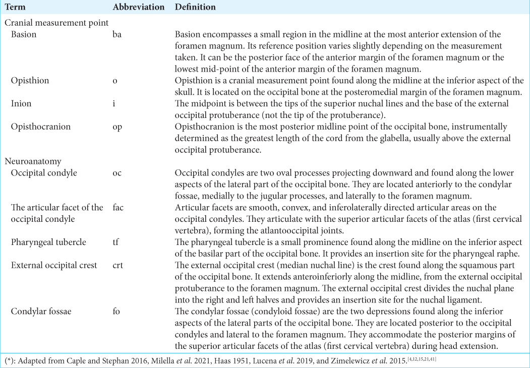
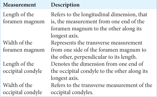
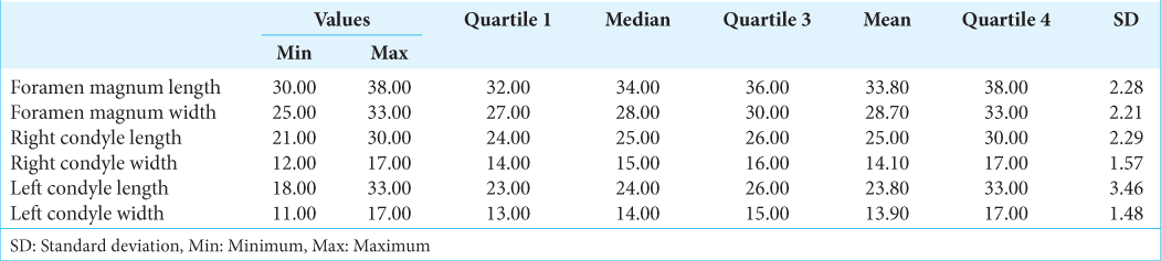
Figure 1:
Inferior view of the skull with anatomical landmarks and measurements between the average distances of cranial points. (a) Skull base in an inferior view: cranial reference points are highlighted in green, red dashed lines indicate length measurements (ba, basion; o, opisthion; i, inion; y op, opisthocranion), and a yellow line measures the width of the occipital condyles (oc) and the foramen magnum. The shaded orange color delineates the articular facet of the left occipital condyle (fac), while a light blue tone outlines the pharyngeal tubercle (tf) on the occipital bone. Additionally, a red shading marks the right condylar fosase (fo), and a dark blue hue highlights the external occipital crest (crt)of the occipital bone. (b) The left occipital condyle is shown with approximate measurements of length (red dashed line) and width (yellow line). (c) The right occipital condyle is depicted with approximate measurements of length (red dashed line) and width (yellow line) of the structure.
RESULTS
Cranial measurements from 17 skulls revealed significant variations in the dimensions of the foramen magnum and the condyles. For the foramen magnum, lengths ranged from 30.00 mm to 38.00 mm, with widths varying between 25.00 mm and 33.00 mm. Regarding the condyles, the average length of the right condyle was 25.00 mm, ranging from 21.00 mm to 30.00 mm, while the left condyle showed an average length of 23.80 mm, varying between 18.00 mm and 33.00 mm. Concerning width, the right condyle had a mean of 14.10 mm, fluctuating between 12.00 mm and 17.00 mm, whereas the left condyle averaged a width of 13.90 mm, ranging from 11.00 mm to 17.00 mm [
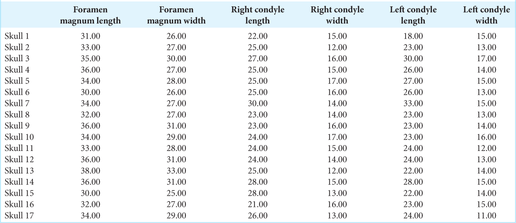
When evaluating the skull base, the foramen magnum exhibited an average length of 33.80 ± 2.28 mm and a width of 28.70 ± 2.21 mm. Regarding the condyles, the right condyle showed a length of 25.00 ± 2.29 mm and a width of 14.10 ± 1.57 mm, while the left condyle recorded an average length of 23.80 ± 3.46 mm and a width of 13.90 ± 1.48 mm. The overall average length measurements for both condyles yielded a value of 19.68 ± 2.90 mm, while the average width was 11.18 ± 1.54 mm [
DISCUSSION
For the first time, the collection at the NeuroZone3D Research Center in Peru provides detailed measurements of the foramen magnum and condyles in cadaveric specimens [
Anatomical studies of the foramen magnum in Nigeria revealed significant measurements in African specimens. Osunwoke and Oladipo[
Anatomical studies conducted across various regions of Asia have unveiled intriguing patterns in the measurements of the foramen magnum and condyles, particularly when examining differences between men and women in these measurements. In the study conducted in Oman by Kumar et al.[
Conversely, multiple investigations in India[
A recent study conducted in Thailand demonstrated similar results, showing a clear disparity between men and women in measurements.[
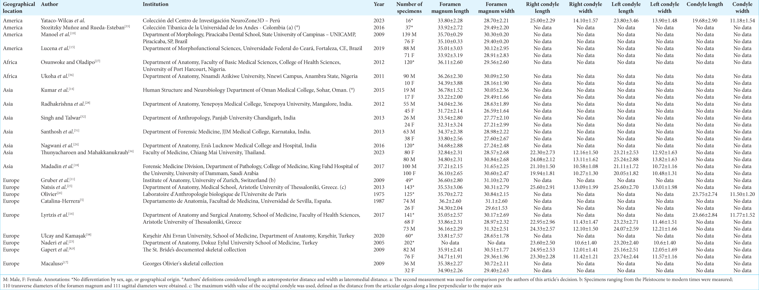
The anatomical analysis of the foramen magnum and condyles has been the subject of numerous studies in Europe, revealing significant variability in the measurements obtained. In Olivier’s work,[
The addition of this present study conducted in Peru provides specific data on the foramen magnum and condyles in this region of America. The obtained values rank on the lower average compared to other previously mentioned studies, both in America and other regions. This observation emphasizes the need to consider anatomical variability not only among different geographical locations but also within the same region, highlighting the importance of future research to understand morphological diversity in craniovertebral morphology.
The significance of craniotomy varies considerably depending on the geographical region and specific anatomical characteristics of each population. Detailed anatomical studies in different countries have revealed significant variations in the dimensions of the foramen magnum and condyles, emphasizing the need to consider local anatomical peculiarities in neurosurgical interventions.[
The creation of indices tailored to each type of skull based on the region or country emerges as an innovative and crucial strategy in the field of neurosurgery. These indices could establish specific parameters for each region, considering the anatomical differences found in detailed anatomical studies.[
CONCLUSION
The morphometric analysis of the foramen magnum and condyles in a sample of the Peruvian population revealed distinctive anatomical characteristics compared to studies conducted in other regions of the world. Significant variations were observed in the dimensions of the foramen magnum and condyles, showing slightly smaller average measurements in this population. These findings highlight the importance of considering anatomical diversity within a specific region when planning neurosurgical interventions and clinical diagnoses. The development of indices tailored to the craniovertebral morphology of each population could be crucial to enhancing the precision and effectiveness of surgical techniques, reducing risks, and optimizing postoperative outcomes in neurosurgical procedures.
Ethical approval
The Institutional Review Board approval is not required.
Declaration of patient consent
Patients’ consent not required as patients’ identities were not disclosed or compromised.
Financial support and sponsorship
Nil.
Conflicts of interest
There are no conflicts of interest.
Use of artificial intelligence (AI)-assisted technology for manuscript preparation
The authors confirm that there was no use of artificial intelligence (AI)-assisted technology for assisting in the writing or editing of the manuscript and no images were manipulated using AI.
Disclaimer
The views and opinions expressed in this article are those of the authors and do not necessarily reflect the official policy or position of the Journal or its management. The information contained in this article should not be considered to be medical advice; patients should consult their own physicians for advice as to their specific medical needs.
References
1. Bahşi İ, Adanır SS, Orhan M, Kervancıoğlu P, Büyükbeşe ZS, Beger O. Anatomical evaluation of the foramen magnum on cone-beam computed tomography images and review of literature. Cureus. 2021. 13: e19385
2. Brennan CM, Taylor GA. Sonographic imaging of the posterior fossa utilizing the foramen magnum. Pediatr Radiol. 2010. 40: 1411-6
3. Burkle FM, Hadley KS, Ridge LL, Herman JK, Kobeissy FH. Delayed-onset neuropathological complications from a foramen magnum and occipital crest focused traumatic brain injury of the Vietnam war and other conflicts: Part I, case report. Mil Med. 2022. 187: e921-5
4. Caple J, Stephan CN. A standardized nomenclature for craniofacial and facial anthropometry. Int J Legal Med. 2016. 130: 863-79
5. Catalina-Herrera CJ. Study of the anatomic metric values of the foramen magnum and its relation to sex. Acta Anat (Basel). 1987. 130: 344-7
6. Chethan P, Prakash KG, Murlimanju BV, Prashanth KU, Prabhu LV, Saralaya VV. Morphological analysis and morphometry of the foramen magnum: An anatomical investigation. Turk Neurosurg. 2012. 22: 416-9
7. Demir BT, Eşme S, Patat D, Bilecenoğlu B. Clinical and anatomical importance of foramen magnum and craniocervical junction structures in the perspective of surgical approaches. Anat Cell Biol. 2023. 56: 342-9
8. Gapert R, Black S, Last J. Sex determination from the foramen magnum: Discriminant function analysis in an eighteenth and nineteenth century British sample. Int J Legal Med. 2009. 123: 25-33
9. Gapert R, Black S, Last J. Sex determination from the occipital condyle: Discriminant function analysis in an eighteenth and nineteenth century British sample. Am J Phys Anthropol. 2009. 138: 384-94
10. Goel A. Can foramen magnum decompression surgery become historical?. J Craniovertebr Junction Spine. 2015. 6: 49-50
11. Gruber P, Henneberg M, Böni T, Rühli FJ. Variability of human foramen magnum size. Anat Rec (Hoboken). 2009. 292: 1713-9
12. Haas LL. The posterior condylar fossa, foramen and canal, and the jugular Foramen. Radiology. 1957. 69: 546-52
13. Kamath VG, Asif M, Shetty R, Avadhani R. Binary logistic regression analysis of foramen magnum dimensions for sex determination. Anat Res Int. 2015. 2015: e459428
14. Kumar A, Dave M, Anwar S. Morphometric evaluation of foramen magnum in dry human skulls. Int J Anat Res. 2015. 3: 1015-23
15. Lucena JD, Sanders JV, Brito HM, Cerqueira GS, Silva IB, Oliveira AS. Morphometric analysis of the foramen magnum in dry human skulls in northeastern Brazil. J Morphol Sci. 2019. 36: 97-104
16. Lyrtzis C, Piagkou M, Gkioka A, Anastasopoulos N, Apostolidis S, Natsis K. Foramen magnum, occipital condyles and hypoglossal canals morphometry: Anatomical study with clinical implications. Folia Morphol. 2017. 76: 446-57
17. Macaluso PJ. Metric sex determination from the basal region of the occipital bone in a documented french sample. Bull Mém Société Anthropol Paris. 2011. 23: 19-26
18. Madadin M, Menezes RG, Al Saif HS, Abu Alola H, Al Muhanna A, Gullenpet AH. Morphometric evaluation of the foramen magnum for sex determination: A study from Saudi Arabia. J Forensic Leg Med. 2017. 46: 66-71
19. Manoel C, Prado F, Caria P, Groppo F. Morphometric analysis of the foramen magnum in human skulls of Brazilian individuals: Its relation to gender. Braz J Morphol Sci. 2009. 26: 104-8
20. Mehta M, Saini V, Patel MN, Menon SK. Applicability and reliability of foramen magnum for sex determination in contemporary Western Indian population: A computed tomographic study. J Forensic Radiol Imaging. 2019. 17: 31-5
21. Milella M, Franklin D, Belcastro MG, Cardini A. Sexual differences in human cranial morphology: Is one sex more variable or one region more dimorphic?. Anat Rec. 2021. 304: 2789-810
22. Muthukumar N, Swaminathan R, Venkatesh G, Bhanumathy SP. A morphometric analysis of the foramen magnum region as it relates to the transcondylar approach. Acta Neurochir (Wien). 2005. 147: 889-95
23. Naderi S, Korman E, Çıtak G, Güvençer M, Arman C, Şenoğlu M. Morphometric analysis of human occipital condyle. Clin Neurol Neurosurg. 2005. 107: 191-9
24. Nagwani M, Rani A, Rani A. A morphometric and comparative study of foramen magnum in North Indian population. J Anat Soc India. 2016. 65: S11-5
25. Natsis K, Piagkou M, Skotsimara G, Piagkos G, Skandalakis P. A morphometric anatomical and comparative study of the foramen magnum region in a Greek population. Surg Radiol Anat. 2013. 35: 925-34
26. Olivier G. Biometry of the human occipital bone. J Anat. 1975. 120: 507-18
27. Osunwoke E, Oladipo G. Morphometric analysis of the foramen magnum and jugular foramen in adult skulls in southern Nigerian population. Am J Sci Ind Res. 2012. 3: 446-8
28. Radhakrishna SK, Shivarama CH, Ramakrishna A, Bhagya B. Morphometric analysis of foramen magnum for sex determination in south Indian population. J Health Allied Sci NU. 2012. 2: 20-2
29. Rhoton AL. The foramen magnum. Neurosurgery. 2000. 47: S155
30. Richards GD, Jabbour RS. Foramen magnum ontogeny in Homo sapiens: A functional matrix perspective. Anat Rec (Hoboken). 2011. 294: 199-216
31. Santhosh C, Vishwanathan K, Ashok G, Siddesh R, Tejas J. Morphometry of the foramen magnum: An important tool in sex determination. Res Rev J Med Health Sci. 2013. 2: 88-91
32. Singh G, Talwar I. Morphometric analysis of foramen magnum in human skull for sex determination. Hum Biol Rev. 2013. 2: 29-41
33. Stozitzky Muñoz N, Rueda-Esteban RJ. Morphometric study of five constant skull base foramina in the muisca population of the tibanica anthropological collection of the universidad de los andes. Int J Morphol. 2016. 34: 1313-7
34. Thunyacharoen S, Mahakkanukrauh P. Craniometric study and anatomical variations of base of skull in a Thai population associated with clinical implications. Appl Sci. 2023. 13: 2046
35. Turamanlar O, Horata E, Kaya F, Boyaci MG, Kiyak O, Oren FN. Does foramen magnum morphometry influence the development of chiari malformation?. Turk Neurosurg. 2021. 31: 704-9
36. Ukoha U, Egwu OA, Okafor IJ, Anyabolu AE, Ndukwe GU, Okpala I. Sexual dimorphism in the foramen magnum of Nigerian adult. Int J Biol Med Res. 2011. 2: 878-81
37. Ulcay T, Kamaşak B, Görgülü Ö, Uzun A, Aycan K. A golden ratio for foramen magnum: An anatomical pilot study. Folia Morphol. 2022. 81: 220-6
38. Ulcay T, Kamaşak B. Evaluation of craniometric measurements in human skulls. J Health Sci Med. 2021. 4: 38-44
39. Vinutha SP, Suresh V, Shubha R. Discriminant function analysis of foramen magnum variables in south Indian population: A study of computerised tomographic images. Anat Res Int. 2018. 2018: 2056291
40. Zdilla MJ, Russell ML, Bliss KN, Mangus KR, Koons AW. The size and shape of the foramen magnum in man. J Craniovertebr Junction Spine. 2017. 8: 205-21
41. Zimelewicz Oberman D, Pérez Zabala J, López T. Morphometry of the posterior cranial fossa: Importance in retrocondylar approaches. Rev Argent Anat Online. 2015. 6: 87-92

