

- Department of Neurosurgery, Rutgers Robert Wood Johnson Medical School, 1 Robert Wood Johnson Pl, New Brunswick, New Jersey, USA
Correspondence Address:
Alexa Semonche
Department of Neurosurgery, Rutgers Robert Wood Johnson Medical School, 1 Robert Wood Johnson Pl, New Brunswick, New Jersey, USA
DOI:10.4103/sni.sni_353_17
Copyright: © 2019 Surgical Neurology International This is an open access journal, and articles are distributed under the terms of the Creative Commons Attribution-NonCommercial-ShareAlike 4.0 License, which allows others to remix, tweak, and build upon the work non-commercially, as long as appropriate credit is given and the new creations are licensed under the identical terms.How to cite this article: Danika Paulo, Alexa Semonche, Rachana Tyagi. Novel method for stepwise reduction of traumatic thoracic spondyloptosis. 27-Feb-2019;10:23
How to cite this URL: Danika Paulo, Alexa Semonche, Rachana Tyagi. Novel method for stepwise reduction of traumatic thoracic spondyloptosis. 27-Feb-2019;10:23. Available from: http://surgicalneurologyint.com/surgicalint-articles/9207/
Date of Submission
19-Sep-2017
Date of Acceptance
27-Oct-2017
Date of Web Publication
27-Feb-2019
Abstract
Background:Spondyloptosis involving complete subluxation of spinal vertebrae resulting in permanent spinal cord damage is rarely caused by high-force trauma. Rapid re-stabilization of the spine is crucial for maximizing chances of neural recovery and can significantly improve the patient's quality of life. In this case study, we describe the challenges associated with the surgical management of traumatic thoracic spondyloptosis, and propose a novel, safe, step-wise, spinal reduction method employing an inflatable beanbag.
Case Description:A 17-year-old male fell 25 feet from a tree, resulting in anterior spondyloptosis at the T11/12 level. He presented with para plegia and a T11 sensory level to pin below the umbilicus. Surgical management involved a posterior-anterior-posterior approach with initial posterior decompression, then T12 corpectomy and reconstruction and finally pedicle screw fixation. We utilized an inflatable beanbag to realign the spinal column in a stepwise fashion, thereby minimizing the risk of damage to the surrounding structures, including the thecal sac and great vessels. Postoperatively, the patient regained some sensory function below his injury level of T11 but remained plegic. X-ray imaging confirmed successful spinal fusion.
Conclusion:Early spinal realignment and stabilization following spondyloptosis at the T11/T12 level resulted in some improvement in sensory function without resolution of motor plegia. Here, we described how to utilize a novel beanbag reduction method to safely achieve stepwise spinal realignment.
Keywords: Fracture dislocation, pediatric, spine, spondyloptosis, subluxation, trauma
INTRODUCTION
Sagittal spondyloptosis, defined as total subluxation (≥100%) of one vertebra on another, is rare, especially in the thoracic region. The underlying mechanism of injury is typically high-energy/impact trauma (e.g., motor vehicle collisions or critical falls) causing complete cord transection, resulting in paralysis in approximately 80% of the cases.[
CASE DESCRIPTION
History and examination
A 17-year-old male sustained a 25-foot fall from a tree, resulting in multiple posterior spinal fractures from T9-12 with sagittal, anterior spondyloptosis at the T11/12 level. He presented with a full motor/sensory paraplegia at the T11 level (ASIA A spinal cord injury). Computed tomography (CT) of the thoracic spine showed acute fracture dislocation at the T11/T12 level, suggesting complete cord transection, and an epidural hematoma from T4-T12 [Figure

Operation
The patient underwent T10-L1 laminectomies with Ponte ostomies at T11/12 to facilitate reduction of the fracture from the lateral approach. This was performed to prevent fracture fragments from injuring the spinal cord during reduction. In the lateral decubitus position on a beanbag, a thoracotomy was performed to expose T10-T12 and complete a T11 corpectomy with adjacent discectomies. To reduce the dislocation, we used a Cobb to elevate T10 superiorly while manually pushing the distal portion of the spine from the patient's back. This was repeated multiple times for stepwise reduction of the bony elements, with deflation and re-inflation of the sandbag to maintain reduction, until adequate alignment was achieved [Figure

Postoperative course
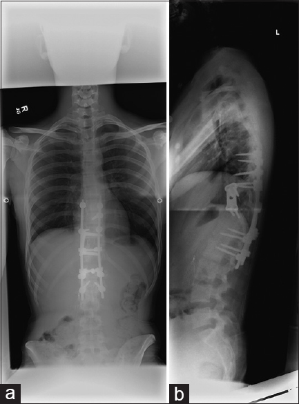
His length of stay was 11 days. On postoperative day one, he exhibited neurogenic shock. His complete spinal cord injury with a T11 sensory level remained stable throughout to postoperative day 10. He began to regain some patchy sensation to the bilateral lower extremities levels starting 2 weeks after surgery, with return of sensation at the T11-T12 levels by 2 months post-operation. Although sensory function continues to improve, he remains paraplegic below T12. Three months post-operation, he started to develop a flexible scoliosis of his lumbar spine, which progressed [Figure

DISCUSSION
Traumatic thoracic spondyloptosis is an uncommon injury. There have been four case series and seven case reports published involving a total of 38 patients with traumatic spondyloptosis [

Here, we utilized a new technique employing sequential sandbag deflation and reinflation to attain a stepwise correction of alignment. With multiple cycles of inflating and deflating the sandbag in lateral decubitus position, with light Cobb distraction, the dislocation could be incrementally reduced. Advantages of this technique included minimizing risk of the spinal cord injury and accidental durotomy. The 360-degree decompression, reduction, and circumferential reconstruction also increased construct stability.
CONCLUSION
Thoracic spondyloptosis is an uncommon injury. Stepwise decompression, step-wise reduction utilizing a sandbag method, and fusion allows for 360-degree correction of the deformity while reducing the risk of potential complications.
Financial support and sponsorship
Nil.
Conflicts of interest
There are no conflicts of interest.
References
1. Cappuccio M, Corghi A, De Iure F, Amendola L. Traumatic expulsion of T4 vertebral body into the spinal canal treated by vertebrectomy and spine shortening. Spine (Phila Pa 1976). 2014. 39: E748-51
2. Chandrashekhara SH, Kumar A, Gamanagatti S, Kapoor K, Mukund A, Aggarwal D. Unusual traumatic spondyloptosis causing complete transaction of spinal cord. Int Orthop. 2011. 35: 1671-5
3. El Masri WS, Silver JR. Traumatic spondyloptosis of the dorsal spine with incomplete neurological deficit. Injury. 1983. 15: 35-7
4. Gitelman A, Most MJ, Stephen M. Traumatic thoracic spondyloptosis without neurologic deficit, and treatment with in situ fusion. Am J Orthop (Belle Mead NJ). 2009. 38: E162-5
5. Hasturk AE, Ilik K, Coven I, Ozdemir O. Unusual traumatic midthoracic spondyloptosis and its surgical management: Case report. Neurol Med Chir (Tokyo). 2013. 53: 887-9
6. Lee CW, Hwang SC, Im SB, Kim BT, Shin WH. Traumatic thoracic spondyloptosis: A case report. J Korean Neurosurg Soc. 2004. p. 622-4
7. Mishra A, Agrawal D, Gupta D, Sinha S, Satyarthee GD, Singh PK. Traumatic spondyloptosis: A series of 20 patients. J Neurosurg Spine. 2015. 22: 647-52
8. Rahimizadeh A, Rahimizadeh A. Management of traumatic double-level spondyloptosis of the thoracic spine with posterior spondylectomy: Case report. J Neurosurg Spine. 2015. 23: 715-20
9. Sandquist L, Paris A, Fahim DK. Definitive single-stage posterior surgical correction of complete traumatic spondyloptosis at the thoracolumbar junction. J Neurosurg Spine. 2015. 22: 653-7
10. Sekhon LH, Sears W, Lynch JJ. Surgical management of traumatic thoracic spondyloptosis: Review of 2 cases. J Clin Neurosci. 2007. 14: 770-5
11. Shapiro S, Abel T, Rodgers RB. Traumatic thoracic spinal fracture dislocation with minimal or no cord injury, Report of four cases and review of the literature. J Neurosurg. 2002. 96: 333-7
12. Yadla S, Lebude B, Tender GC, Sharan AD, Harrop JS, Hillbrand AS. Traumatic spondyloptosis of the thoracolumbar spine. J Neurosurg Spine. 2007. 9: 145-51

