Tools

Srihari Sridharan, D. Balasubramanian
  1. Department of Neurosurgery, Institute of Neurosurgery, Madras Medical College, Chennai, Tamil Nadu, India

Correspondence Address:
Srihari Sridharan
Department of Neurosurgery, Institute of Neurosurgery, Madras Medical College, Chennai, Tamil Nadu, India

DOI:10.4103/sni.sni_495_16

Copyright: © 2017 Surgical Neurology International This is an open access article distributed under the terms of the Creative Commons Attribution-NonCommercial-ShareAlike 3.0 License, which allows others to remix, tweak, and build upon the work non-commercially, as long as the author is credited and the new creations are licensed under the identical terms.

How to cite this article: Srihari Sridharan, D. Balasubramanian. Primary calvarial tuberculosis. 21-Jun-2017;8:126

How to cite this URL: Srihari Sridharan, D. Balasubramanian. Primary calvarial tuberculosis. 21-Jun-2017;8:126. Available from: http://surgicalneurologyint.com/surgicalint-articles/primary-calvarial-tuberculosis/

Date of Submission
30-Dec-2016

Date of Acceptance
19-Mar-2017

Date of Web Publication
21-Jun-2017

Abstract

Background:Tuberculosis is a very common disease in India. Its prevalence, capacity to affect every other organ, and atypical presentations make it an important part of the differential diagnosis for cases presenting in the rural setup. Primary tuberculous caries of the calvarium is a rare manifestation of this common entity.

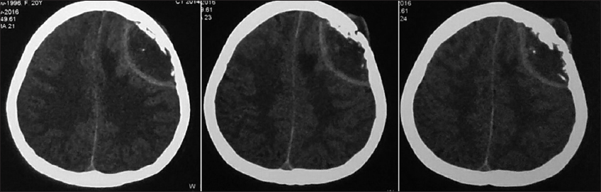
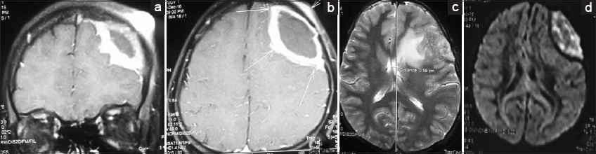
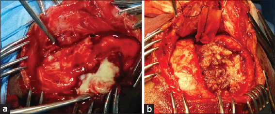
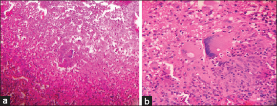
Case Description:A young lady presented with headache and discharging sinus of the scalp. Imaging revealed epidural abscess with midline shift. She underwent surgery, and histopathological diagnosis of tuberculous etiology was made. The pre and intraoperative images, radiological images, and histopathological images are reported and discussed.

Conclusion:Tuberculosis should be included in the differential diagnosis of the osteolytic lesions of skull with discharging sinus. A high degree of suspicion, especially in patients from low socioeconomic status, is warranted in managing such cases.

Keywords: Calvarium, skull tuberculosis, TB, tuberculosis

INTRODUCTION

Tuberculosis is a major public health problem with a significant effect on the economy of developing countries. The social stigma associated with the disease cannot be understated. Malnutrition, overcrowding, poor sanitation, lack of hygiene, lack of awareness, and delay in seeking medical care contribute significantly to the spread of the disease. Extrapulmonary tuberculosis is the term used to describe isolated occurrence of tuberculosis at body sites other than the lung.[ 10 ] Primary skull involvement is a rare form of skeletal tuberculosis with few retrospective series offering insights.[ 3 8 9 ] We report a rare case of primary tuberculous osteomyelitis with epidural abscess causing midline shift and discharging sinus of scalp. The disease progression was arrested with anti-tuberculous therapy, and the patient improved symptomatically.

CASE HISTORY

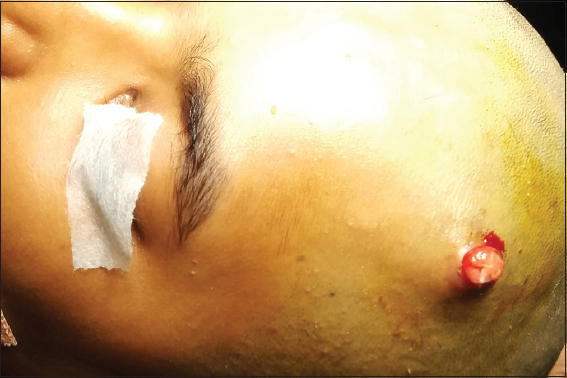
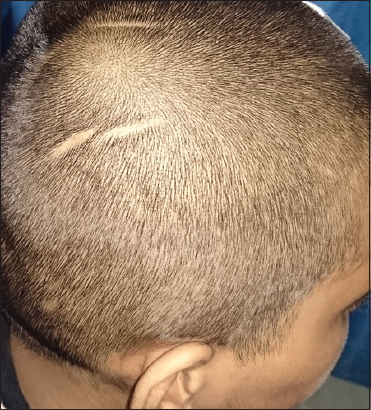
A 20-year-old lady from the rural parts of Tamil Nadu, south India presented with complaints of headache for the past 4 months and persistent discharging sinus from the left frontal scalp region for 1 month [ Figure 1 ]. There was no history suggestive of trauma. Serology for human immunodeficiency virus (HIV) was negative. She was neurologically intact. Computed tomography (CT) scan showed a biconvex hypodense lesion in the left frontal region with hyperdense rim causing midline shift. Osteolytic changes in the left frontal bone with soft tissue swelling of the scalp over osteolytic focus were also noted [ Figure 2 ]. Contrast-enhanced magnetic resonance imaging (MRI) [Figure 3a and b ] revealed osteolytic focus in the left frontal bone with irregular erosions of the inner table. A 2.5-cm thick extra-axial collection measuring 5 cm in the anteroposterior direction and 6 cm in the craniocaudal direction with thick irregular enhancing walls was visualized adjacent to the osteolytic focus with restricted diffusion [ Figure 3d ]. Enhancement of dura adjacent to extra-axial collection was noted. The left frontal cortex showed a small nodular enhancement with edema of white matter [ Figure 3c ]. Mass effect in the form of regional sulcal effacement, compression of underlying left frontal lobe, and subfalcine herniation to the right (6 mm) were seen. Surgical intervention was planned. Skin incision included the sinus tract. Bone was soft and necrotic with cheesy material underneath [ Figure 4a ]. Debridement of the diseased bone was done till healthy bone was encountered [ Figure 4b ]. Extradural collection was evacuated. Sinus tract was excised. Dura was found to be intact, debrided off the granulation tissue, and washed with hydrogen peroxide and povidone–iodine solution. Histopathological evaluation showed spicules of bone and fibrous tissue with clusters of epithelioid cells, Langerhans giant cells, and lymphocytes and areas of caseation necrosis [ Figure 5 ]. Patient was started on anti-tuberculous therapy and was advised to continue that for 24 months. Follow-up of 1 month showed a well-healed wound and the patient was relieved of headache.


Figure 1

Discharging sinus of the scalp in the left frontal region


Figure 2

Plain axial computed tomography showing lentiform shaped collection in the extradural space of left frontal region. The destruction of bone along with scalp involvement is also noted


Figure 3

Contrast enhanced MRI. (a) Coronal cuts showing enhancement of the extradural collection and soft tissue in the scalp. (b) Axial images showing the dural enhancement. (c) T2 weighted images showing oedema of left frontal cortex. (d) Diffusion restriction is seen


Figure 4

Intra operative images showing (a) Necrotic bone and cheesy material underneath it. (b) Margins of healthy bone


Figure 5

Histopathology images: (a) 10X images showing granuloma and langhan's giant cells. (b) 40X images showing Langhan's giant cells


DISCUSSION

Reid reported the first case of calvarial tuberculosis.[ 6 ] There is no sex predilection but 80% of the cases are younger than 20 years.[ 9 ] Parietal and frontal bones are usually involved due to high cancellous portion. This also explains the absence of disease in infancy. Hematogenous spread or direct inoculation to the diploe has been proposed to be a cause.[ 2 ] Our case did not have any history of recent trauma nor any primary focus. On further questioning, it was revealed that she had a scalp injury when she was 5 years of age. However, this was on the opposite side of the scalp [ Figure 6 ]. This could have been a route of inoculation, however, involvement of the opposite side after a gap of 15 years reduces the probability of such an occurrence. In the series published by Raut et al.[ 9 ] none of the patients had a primary focus and three of the patients had prior surgery of the scalp, though the site is not mentioned. Once inoculated and after the granulation starts, spread occurs through the inner and outer table of the skull, although rare case of spread through diploe has also been reported.[ 1 ] Sutures are no barriers for the spread of granulation tissue[ 9 ] but dura can offer resistance. Our case had extensive destruction of both the inner and outer table of the frontal bone crossing the coronal suture and involving parts of the parietal and temporal bone. Skin involvement was evident in the form of discharging sinus. Examination of the discharge could have been used for diagnosis. However, the presence of extradural collection causing midline shift warranted immediate surgery. The involvement of the frontal cortex with white matter edema may be due to the penetration of bacilli through the dura. Because the lesion was small and dura was intact, surgical intervention was limited to dural debridement alone. However, anticonvulsants were started due to cortical involvement. CT imaging with contrast can show bone destruction as well as extradural and parenchymal spread of disease with meningeal enhancement. These findings are not specific for calvarial tuberculosis. Epidural collection has low attenuation with the characteristic lentiform shape.[ 9 ] CT imaging cannot usually differentiate between pyogenic osteomyelitis, metastasis, myeloma, hemangioma, and Langerhans cell histiocytosis.[ 4 ] In absence of pulmonary tuberculosis, calvarial tuberculosis is usually a last suspect. MRI can show high signal intensity soft tissue mass and peripheral capsular contrast enhancement.[ 9 ] CT imaging can be a cost-effective modality of scanning, however, MRI has an edge in delineating subtle parenchymal involvement. A definitive diagnosis is only through invasive methods and is a definitive need for starting chemotherapy. Histological examination reveals infiltration with lymphocytes, plasma cells, and focal polymorph nuclear cells with proliferating blood vessels. Epithelioid granulomas with Langerhans type giant cells are also seen with necrotic material.[ 6 ]


Figure 6

Old Scar in the right parietal prominence


Anti-tuberculous therapy forms the mainstay of the treatment[ 7 ] with or without surgical intervention.[ 3 8 9 ] The need for surgery in our case was inevitable with an epidural collection causing midline shift and discharging sinus. Most cases in the rural setup present like the reported case. Seeking medical care is often delayed, and thus, a preventable disease becomes a disease of neurosurgical emergency. Surgery is a need in large lesions and can play a role in establishing the diagnosis. It is also useful in small lesions with poor response to chemotherapy.[ 5 ]

Financial support and sponsorship

Nil.

Conflicts of interest

There are no conflicts of interest.

References

1. Barton . Tuberculosis of the vault of the skull. Br J Radiol. 1961. 34: 286-90

2. Biniwale SN, Rajshekhar V. Tuberculous osteomyelitis of bone flap following craniotomy for a glioma. Neurol India. 2000. 48: 91-2

3. Diyora B, Kumar R, Modgi R, Sharma A. Calvarial tuberculosis: A report of eleven patients. Neurol India. 2009. 57: 607-12

4. Gupta PK, Sastry-Kolluri VR, Chandramouli BA, Venkatramana NK, Das BS. Calvarial tuberculosis: A report of 2 cases. Neurosurgery. 1989. 25: 830-33

5. Ip M, Tsui E, Wong KL, Jones B, Pung CF, Ngan H. Disseminated skeletal tuberculosis with skull involvement. Tuberc Lung Dis. 1993. 74: 211-4

6. Jadhav RN, Palande DA. Calvarial tuberculosis. Neurosurgery. 1999. 45: 1345-50

7. LeRoux PD, Griffin GE, Marsh HT, Winn HR. Tuberculosis of the skull; A rare condition-Case report and review of the literature. Neurosurgery. 1990. 26: 851-5

8. Ramdurg SR, Gupta DK, Suri A, Sharma BS, Mahapatra AK. Calvarial tuberculosis: Uncommon manifestation of common disease – A series of 21 cases. Br J Neurosurg. 2010. 24: 572-7

9. Raut AA, Nagar AM, Muzumdar D, Chawla AJ, Narlawar RS, Fattepurkar S. Imaging features of calvarial tuberculosis: A study of 42 cases. Am J Neuroradiol. 2004. 25: 409-14

10. Sharma SK, Mohan A. Extrapulmonary tuberculosis. Indian J Med Res. 2004. 120: 316-53

Leave a Reply

Your email address will not be published. Required fields are marked *