- Department of Neurosurgery, Grant Medical Center, OhioHealth, Columbus, USA
- Department of Neurosciences, Genesis HealthCare System, Zanesville, Ohio, USA
- Department of Pathology, Genesis HealthCare System, Zanesville, Ohio, USA
Correspondence Address:
Christopher E. Stewart
Department of Pathology, Genesis HealthCare System, Zanesville, Ohio, USA
DOI:10.4103/sni.sni_63_17
Copyright: © 2017 Surgical Neurology International This is an open access article distributed under the terms of the Creative Commons Attribution-NonCommercial-ShareAlike 3.0 License, which allows others to remix, tweak, and build upon the work non-commercially, as long as the author is credited and the new creations are licensed under the identical terms.How to cite this article: Victor Awuor, Christopher E. Stewart, Albert Camma, Julie Renner, J. M. Tongson. Rare case of an extraosseous cervical chordoma with both intradural and extensive extraspinal involvement. 13-Oct-2017;8:250
How to cite this URL: Victor Awuor, Christopher E. Stewart, Albert Camma, Julie Renner, J. M. Tongson. Rare case of an extraosseous cervical chordoma with both intradural and extensive extraspinal involvement. 13-Oct-2017;8:250. Available from: http://surgicalneurologyint.com/surgicalint-articles/rare-case-of-an-extraosseous-cervical-chordoma-with-both-intradural-and-extensive-extraspinal-involvement/
Abstract
Background:Chordomas must be considered among the differential diagnoses for extradural spinal tumors, especially involving the clival or sacrococcygeal regions. They are often locally invasive and destructive to the osseous structures from which they arise, but rarely extend intradurally. Here, we report a unique chordoma that was intradural and spanned nearly four subaxial cervical vertebral levels.
Case Description:We report the case of an atypical intradural chordoma that spanned four subaxial levels of the cervical spine in an 81-year-old female. It also extended through multiple neural foramina but did not invade or destroy the bony elements of the cervical vertebrae. Notably, it demonstrated sizable extension into the deep carotid triangle abutting the internal jugular vein.
Conclusion:This case involved an extraosseous, intradural, four-level subaxial cervical chordoma that demonstrated significant extraspinal extension into the anterior soft tissues of the neck.
Keywords: Cervical chordoma, chordoma, intradural chordoma
INTRODUCTION
An 81-year-old female presented with a rare, extraosseous subaxial four-level cervical chordoma that demonstrated both intradural and extraspinal extension. Spinal chordomas are predominantly osseodestructive tumors originating from the embryonic notochord remnants within vertebral bodies.[
CASE REPORT
An 81-year-old female presented with complaints of shortness of breath and generalized weakness. She was treated for a pericardial effusion, urinary tract infection, lymphadenopathy, and a cervical mass that was discovered during her work up. Her neurological exam revealed an unsteady gait (without dysmetria), with motor strength of 4+/5 throughout the upper and lower extremities except for a left triceps deficit of 4-/5.
Magnetic resonance/computed tomography preoperative assessment
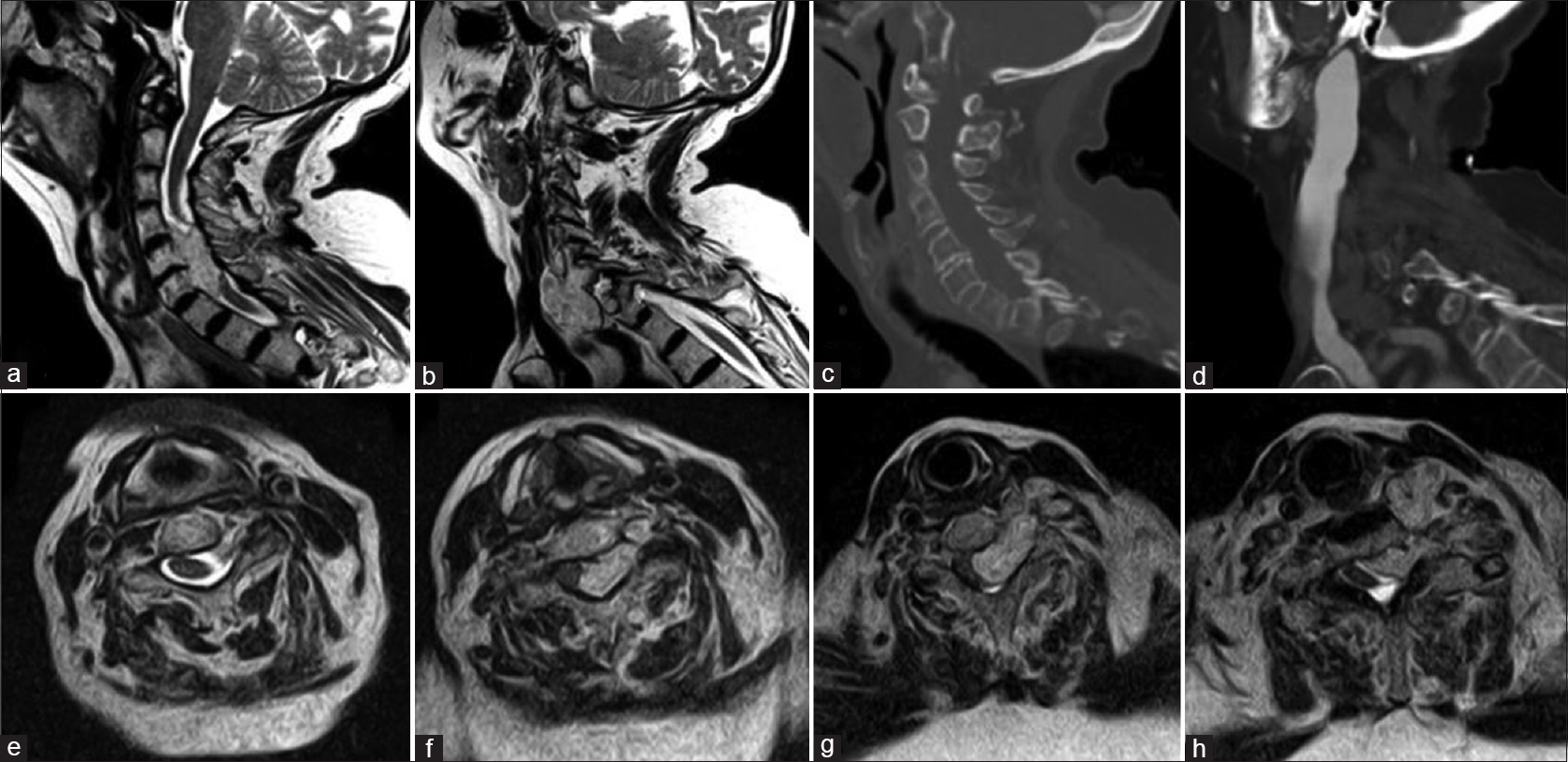
Magnetic resonance imaging (MRI) and computed tomography (CT) studies of the cervical spine with and without contrast demonstrated a left-sided extraosseous mass within the central canal extending from C4–7 with widening of the left C4–5, C5–6, and C6–7 foramina and invading the soft tissues of the carotid triangle. It also expanded anteriorly, abutting the posterior aspect of the carotid sheath causing focal narrowing of the internal jugular vein resulting in distal dilation [
Figure 1
(a) T2WI showing the intraspinal portion of the mass. (b) T2WI showing extraspinal portion of the mass. (c) CT showing intact vertebral bodies without bone involvement. (d) CTA showing dilation of the internal jugular vein. (e-h) T2WI axial cuts at C4, C5, C6, and C7, respectively. Abbreviations: CT, computed tomography; CTA, computed tomography angiography; T2WI, T2-weighted image
Surgery
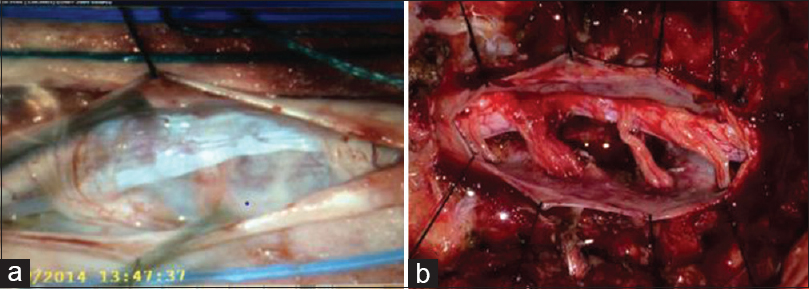
A C4–T1 laminectomy was performed and no mass was encountered in the epidural space. However, a midline durotomy exposed a well encapsulated grayish mass with severe compression of the cervical spinal cord. It was grossly resected in a piecemeal manner using both ronguers and an ultrasonic aspirator until the spinal cord was sufficiently decompressed. The durotomy was closed primarily and the posterior cervical wound was closed in the standard manner [
Postoperative course
Postoperatively, the patient regained her motor strength and was able to ambulate. Due to her advanced age and unwillingness to undergo more extensive surgery, the extraspinal portion of the tumor was not resected. She declined adjuvant therapy and elected to pursue only comfort measures.
Pathology
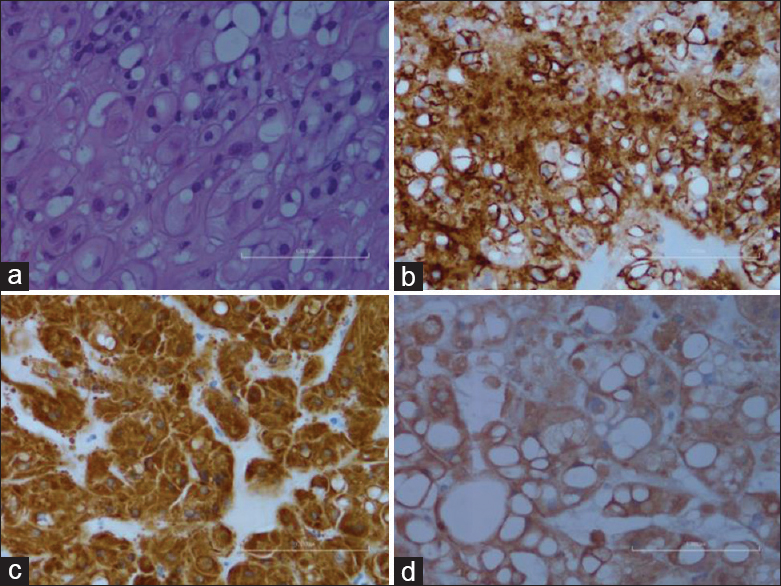
Histopathology revealed an epithelioid neoplasm with physaliphorous cells in a myxoid/mucoid matrix. Immunohistochemical staining showed positivity for AE1/AE3, EMA, and S100, supporting the diagnosis of chordoma [
DISCUSSION
Chordomas arise from the remnants of embryonic notochord. Notochord remnants are most commonly located intraosseously; hence, chordomas are considered primarily in the differential diagnosis of extradural spinal tumors. Extraosseous, intradural spinal chordomas are exceedingly rare. We identified only nine reported cases of similar chordomas; only two were intradural cervical lesions with extension outside the spinal canal.[
Financial support and sponsorship
Nil.
Conflicts of interest
There are no conflicts of interest.
References
1. Badwal S, Pal L, Basu A, Saxena S. Multiple synchronous spinal extra-osseous intradural chordomas: Is it a distinct entity?. Br J Neurosurg. 2006. 20: 99-103
2. Bergmann M, Abdalla Y, Neubauer U, Schildhaus H, Probst-Cousin S. Primary intradural chordoma: Report on three cases and review of the literature. Clin Neuropathol. 2010. 29: 169-76
3. Samadian M, Shafizad M. An intradural cervical chordoma mimicking schwannoma. J Injury Violence. 2012. 4: 1-
4. Steenberghs J, Kiekens C, Menten J, Monstry J. Intradural chordoma without bone involvement: Case report and review of the literature. J Neurosurg (Spine 1). 2002. 97: 94-7


Kiyoshi Ito
Posted November 2, 2017, 3:33 pm
Dear Dr. Christopher E. Stewart
Thank you for your very rare case report. Most chordoma is originated in the midline of the vertebral body. This mass is not. I would like to know the origin of this tumor. Would it be migrated into the intradural region?