

- Department of Neurosurgery, Kawasaki Saiwai Hospital, Kanagawa, Japan.
Correspondence Address:
Kohei Yamamoto, Department of Neurosurgery, Kawasaki Saiwai Hospital, Kanagawa, Japan.
DOI:10.25259/SNI_701_2023
Copyright: © 2023 Surgical Neurology International This is an open-access article distributed under the terms of the Creative Commons Attribution-Non Commercial-Share Alike 4.0 License, which allows others to remix, transform, and build upon the work non-commercially, as long as the author is credited and the new creations are licensed under the identical terms.How to cite this article: Kohei Yamamoto, Hidenori Matsuoka, So Ohashi, Kyohei Yamashiro, Kenta Kazami, Yusuke Hirokawa, Michihisa Narikiyo, Hirokazu Nagasaki, Yoshifumi Tsuboi. Retroperitoneal hematoma: A rare complication of percutaneous pedicle screw in an osteoporotic patient. 22-Sep-2023;14:345
How to cite this URL: Kohei Yamamoto, Hidenori Matsuoka, So Ohashi, Kyohei Yamashiro, Kenta Kazami, Yusuke Hirokawa, Michihisa Narikiyo, Hirokazu Nagasaki, Yoshifumi Tsuboi. Retroperitoneal hematoma: A rare complication of percutaneous pedicle screw in an osteoporotic patient. 22-Sep-2023;14:345. Available from: https://surgicalneurologyint.com/surgicalint-articles/12564/
Date of Submission
22-Aug-2023
Date of Acceptance
07-Sep-2023
Date of Web Publication
22-Sep-2023
Abstract
Background: Percutaneous pedicle screw (PPS) placement is an established technique for minimally invasive surgery. However, life-threatening hematomas may occur in osteoporotic patients undergoing percutaneous screw placement.
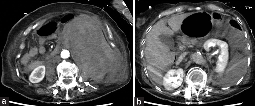
Case Description: An 80-year-old female with an osteoporotic T10 chance fracture developed a life-threatening hematoma following a T8–L3 posterior fusion performed with PPS. Prompt angiography diagnosed a life-threatening hematoma attributed to laceration of the left third lumbar artery occurring following pedicle screw (PS) placement into an osteoporotically fractured left L3 transverse process. This was immediately and successfully embolized.
Conclusion: An 80-year-old female with multiple lumbar osteoporotic fractures developed a life-threatening hematoma during a T8–L3 PS fusion. When the lumbar computed tomography angiography diagnosed a laceration of the left L3 lumbar artery, immediate transarterial embolization proved life-saving.
Keywords: Lumbar artery injury, Percutaneous pedicle screw, Transarterial embolization, Transverse process fracture, Retroperitoneal hematoma
INTRODUCTION
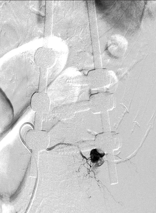
Lumbar artery injuries are rare following percutaneous pedicle screw (PPS) placement. Here, an 80-year-old female with multiple lumbar osteoporotic vertebral fractures developed a life-threatening retroperitoneal hematoma following pedicle screw (PS) laceration of the left L3 lumbar artery at the site of a left L3 osteoporotic transverse process (TP) fracture. Once the computed tomography (CT) angiogram diagnosed the site of the vascular injury, the patient successfully underwent lift-saving transarterial embolization (TAE).
CASE DESCRIPTION
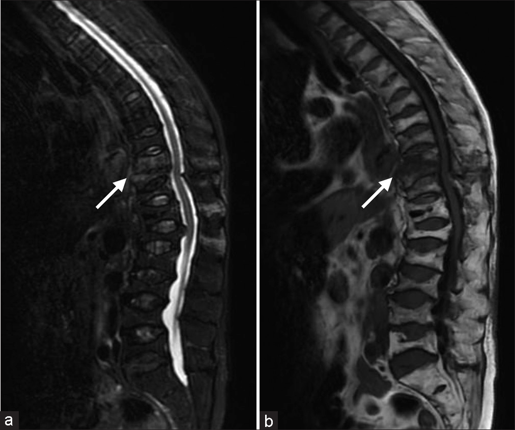
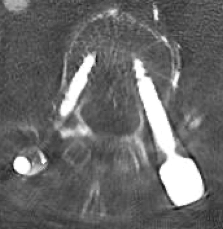
An 80-year-old osteoporotic female with a prior history of a T12 balloon kyphoplasty had fallen 10 days earlier. The thoracolumbar CT showed the left TP fractures from L1 to 3, a T10 chance fracture, and multiple osteoporotic fractures involving the T11, T12, L2, L3, and L4 vertebrae [
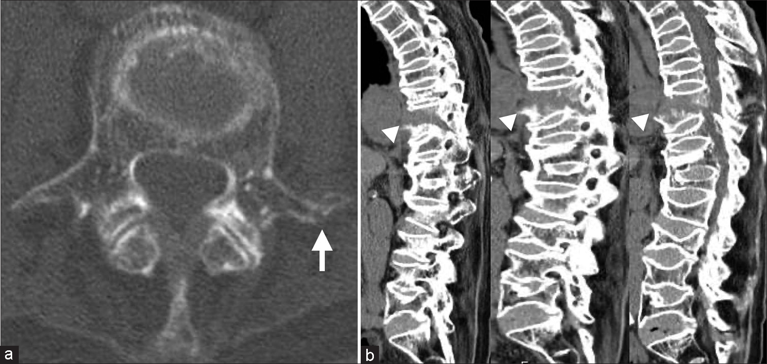
PPS T8–L3 fusion resulting in left L3 lumbar artery laceration (site of left L3 TP osteoporotic/traumatic fracture)
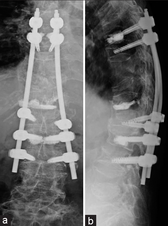
With an unstable vertebral fracture of T10, the patient underwent a minimally invasive thoracolumbar T8–L3 PPS fusion under C-arm fluoroscopy [
DISCUSSION
TP fractures can lead to lumbar artery injuries
TP fractures can lead to lumbar artery injuries, including pseudoaneurysm formation, following spinal trauma and/or spine surgery.[
History of iatrogenic lumbar artery injury with lumbar PS insertion
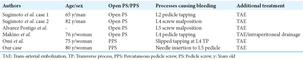
Although iatrogenic lumbar artery injuries due to PS insertion are rare, we found 6 such life-threatening complications in the literature [
CONCLUSION
An 80-year-old female developed a life-threatening retroperitoneal hematoma following a PPS/fusion (T8–L3) that included placing a PS into an osteoporotic left L3 TP; immediate resuscitation and TAE proved lifesaving.
Declaration of patient consent
Patient’s consent not required as patient’s identity is not disclosed or compromised.
Financial support and sponsorship
Nil.
Conflicts of interest
There are no conflicts of interest.
Use of artificial intelligence (AI)-assisted technology for manuscript preparation
The author(s) confirms that there was no use of artificial intelligence (AI)-assisted technology for assisting in the writing or editing of the manuscript and no images were manipulated using AI.
Disclaimer
The views and opinions expressed in this article are those of the authors and do not necessarily reflect the official policy or position of the Journal or its management. The information contained in this article should not be considered to be medical advice; patients should consult their own physicians for advice as to their specific medical needs.
References
1. Alvarez Postigo M, Pizones Arce J, Izquierdo Nunez E. Lumbar segmental artery pseudoaneurysm after L5 pedicle screw placement. A rare vascular complication. Rev Esp Cir Ortop Traumatol. 2017. 61: 436-40
2. Lee JS, Kim CW, Suh KT. Lumbar artery injury combined with a transverse process fracture of the lumbar spine presenting with hypovolemic shock after a fall: A case report. J Korean Orthop Assoc. 2008. 43: 400-3
3. Makino T, Kaito T, Sakai Y, Takenaka S, Yoshikawa H. Iatrogenic arteriovenous fistula and retroperitoneal hemorrhage after tapping of lumbar pedicle screws: A case report. JBJS Case Connect. 2019. 9: e0477
4. Oh YM, Choi HY, Eun JP. Delayed retroperitoneal hemorrhage due to lumbar artery pseudoaneurysm after lumbar posterolateral fusion. J Korean Neurosurg Soc. 2013. 54: 344-6
5. Omi H, Tomita T, Ichinohe M, Harada Y, Sato H, Ito J. Lumbar artery injury related to percutaneous pedicle screw insertion. Acta Med Okayama. 2022. 76: 85-8
6. Schlegel RN, Fitzgerald M, Reilly GO, Clements W, Goh GS, Groombridge C. The injury patterns, management and outcomes of retroperitoneal haemorrhage caused by lumbar arterial bleeding at a Level-1 Trauma Centre: A 10-year retrospective review. Injury. 2023. 54: 145-9
7. Sugimoto Y, Tanaka M, Gobara H, Misawa H, Kunisada T, Ozaki T. Management of lumbar artery injury related to pedicle screw insertion. Acta Med Okayama. 2013. 67: 113-6

