- Neurosurgeon, Sao Paulo Federal University, Sao Paulo, Brazil
- Neurologist, Hospital das Clinicas Sao Paulo, Sao Paulo, Brazil
- Neurosurgeon, Villa Lobos Hospital, Sao Paulo, Brazil
- Neuropathologist, Sao Paulo Federal University, Sao Paulo, Brazil
Correspondence Address:
Franz Jooji Onishi
Neuropathologist, Sao Paulo Federal University, Sao Paulo, Brazil
DOI:10.4103/sni.sni_467_16
Copyright: © 2017 Surgical Neurology International This is an open access article distributed under the terms of the Creative Commons Attribution-NonCommercial-ShareAlike 3.0 License, which allows others to remix, tweak, and build upon the work non-commercially, as long as the author is credited and the new creations are licensed under the identical terms.How to cite this article: Franz Jooji Onishi, Flavio Augusto Sekeff Salem, Diogo Luis de Melo Lins, Rafi Felicio Bauab Dauar, João Norberto Stavale. Spinal thoracic extradural angiolipoma manifesting as acute onset of paraparesis: Case report and review of literature. 18-Jul-2017;8:150
How to cite this URL: Franz Jooji Onishi, Flavio Augusto Sekeff Salem, Diogo Luis de Melo Lins, Rafi Felicio Bauab Dauar, João Norberto Stavale. Spinal thoracic extradural angiolipoma manifesting as acute onset of paraparesis: Case report and review of literature. 18-Jul-2017;8:150. Available from: http://surgicalneurologyint.com/surgicalint-articles/spinal-thoracic-extradural-angiolipoma-manifesting-as-acute-onset-of-paraparesis-case-report-and-review-of-literature/
Abstract
Background:Angiolipomas are benign tumors most commonly found in the thoracic spine. They are composed of mature adipocytes and abnormal vascular elements that usually present with a slowly progressive course of neurological deterioration.
Case Description:A 35-year-old female, with a prior history of back pain, acutely developed paraparesis. When the thoracic magnetic resonance imaging (MRI) revealed a dorsal epidural mass at the T3-T5 level, she underwent a laminectomy for gross total excision of the lesion that proved to be an angiolipoma. On the second postoperative day, the patient was again able to ambulate.
Conclusion:The angiolipomas of spine are rare causes of spinal cord compression, and those presenting with acute neurological deficits should be immediately treated.
Keywords: Angiolipoma, extradural spinal tumor, spinal cord compression, spinal tumor
INTRODUCTION
Angiolipomas are benign tumors consisting of mature fat cells and proliferating abnormal blood vessels. They generally occur in the subcutaneous tissue of the trunks and limbs. They can be further categorized into two subtypes – noninfiltrating (more common) and infiltrating. They account for 0.004–1.2% of all spinal axis tumors and 2–3% of extradural spinal lesions.[
CASE REPORT
A 35-year-old female, with 4 weeks of bilateral lower limb numbness, acutely presented with 24-hour evolution of an acute paraparesis, accompanied urinary retention. Physical examination revealed 4/5 motor strength in both the legs, diffuse lower extremity hyperreflexia, and superficial hypoesthesia below the T4 level.
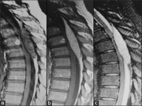
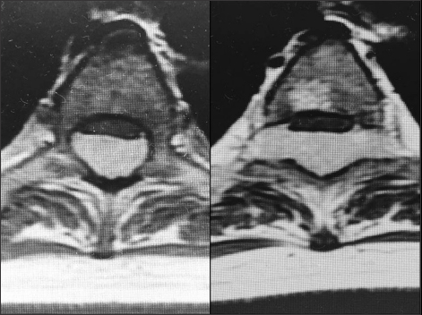
The magnetic resonance imaging (MRI) scan showed a large fusiform dorsal lesion measuring 8.0 cm × 2.8 cm × 1.1 cm compressing the thoracic spinal cord extending over the T3-T5 levels [
Figure 1
Sagittal MRI images showing heterogeneous epidural posterior mass compressing thoracic cord over three body segments between T3-T5. (a) T1-weighted MRI shows inhomogeneous iso to hypointensity mass; (b) Post-contrast T1-weightted MRI shows enhancing mass; (c) T2-weighted MRI shows a hyperintense tumor
Surgery
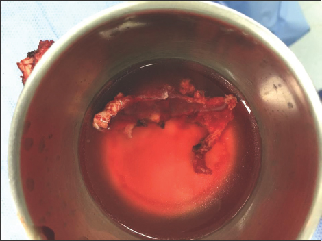
A laminotomy of T3-T5 was performed under general anesthesia with intraoperative neurophysiological monitoring. The posterior epidural space was filled with a fatty, highly vascular brown-pink mass that was extremely hemorrhagic. It was easily mobilized away from the compressed dura, and a gross total resection was accomplished (e.g., in one piece). There were no changes in the intraoperative motor evoked potentials [
Postoperative course
On postoperative day 2, the patient was able to ambulate, and she had fully recovered within 2 weeks. After 2 years, the patient remains asymptomatic and shows no signs of spinal deformity. There is also no evidence of radiographic recurrence.
Pathology
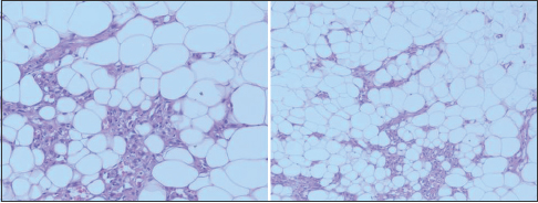
The analysis of the tumor revealed a lesion with adipocytes and capillary sized vessels [
DISCUSSION
Histology
Berenbruch in 1890 was the first to report extradural spinal angiolipoma; the first pathological report was made by Howard in 1960.[
These lesions consist of varying portions of mature fat cells and abnormal capillary, sinusoidal, venous, or arterial vascular elements. The ratio of fat to vessels is variable and ranges from 1:3 to 2:3. Tumors with an abundance of smooth muscle proliferation are further subclassified as angiomyolipomas. Angiolipoma generally arise from the dorsal aspect of spinal canal at thoracic levels compressing the spinal cord and causing symptoms.[
Origin
The origin and pathogenesis of angiolipomas are through to arise from pluripotential mesenchymal stem cells with secretory activity. Early inclusion of pluripotent stem cells during the developmental ossification of neural arch is believed to be a prerequisite for spinal angiolipoma formation. Degenerative changes may be presenting in some longstanding cases but malignant transformation and neural tissue infiltration have never been reported.[
Clinical presentation
Angiolipomas predominantly occur in females (female:male ratio = 22:17) in their fifties who typically suddenly develop paraparesis. The rapid onset of symptoms in usually due to vascular factors; e.g., anomalous vessels, intralesional thrombosis, and hemorrhage or steal phenomena.[
Demyelinating disease must be considered among the differential diagnoses as occasionally these lesions present with relapsing/remitting course.[
Imaging
MRI, T1-weighted images typically demonstrate a high signal accompanied by an inhomogeneous mass. On T2-weighted images, the signal intensity seems to be similar to adipose tissue. The lesion markedly enhances with gadolinium administration [
Treatment and prognosis
Spinal angiolipomas are treated exclusively by surgical removal, and most may be completely excised with good clinical results (e.g., normal postoperative examinations).[
Financial support and sponsorship
Nil.
Conflicts of interest
There are no conflicts of interest.
References
1. Boockvar JA, Black K, Malik S, Stanek A, Tracey KJ. Subacute paraparesis induced by venous thrombosis of a spinal angiolipoma: A case report. Spine. 1997. 22: 2304-8
2. Dogan S, Arslan E, Sahin S, Aksoy K, Aker S. Lumbar spinal extradural angiolipomas. Two case reports. Neurol Med Chir. 2006. 46: 157-60
3. Gelabert-González M, García-Allut A. Spinal extradural angiolipoma: Report of two cases and review of the literature. Eur Spine J. 2009. 18: 324-35
4. Guzey FK, Bas NS, Ozkan N, Karabulut C, Bas SC, Turgut H. Lumbar extradural infiltrating angiolipoma: A case report and review of 17 previously reported cases with infiltrating spinal angiolipomas. Spine J. 2007. 7: 739-44
5. Konya D, Ozgen S, Kurtkaya O, Pamir NM. Lumbar spinal angiolipoma: Case report and review of the literature. Eur Spine J. 2006. 15: 1025-8
6. Meng J, Du Y, Yang HF, Hu FB, Huang YY, Li B, Zee CS. Thoracic epidural angiolipoma: A case report and review of the literature. World J Radiol. 2013. 5: 187-92
7. Samdani AF, Garonzik IM, Jallo G, Eberhart CG, Zahos P. Spinal angiolipoma: Case report and review of the literature. Acta Neurochir. 2004. 146: 299-302


Nabeel ali
Posted August 1, 2017, 10:55 am
I have a patient with the same course (spastic paresis of both lower limbs) with mri reveals D5,6 extradural mass, with the same pic of your patient, totaly removed surgically and waiting for histopathology results