

- Department of Neurosurgery, International University of Health and Welfare, Narita, Japan.
Correspondence Address:
Tatsuya Tanaka, Department of Neurosurgery, International University of Health and Welfare, Narita, Japan.
DOI:10.25259/SNI_729_2022
Copyright: © 2022 Surgical Neurology International This is an open-access article distributed under the terms of the Creative Commons Attribution-Non Commercial-Share Alike 4.0 License, which allows others to remix, transform, and build upon the work non-commercially, as long as the author is credited and the new creations are licensed under the identical terms.How to cite this article: Tatsuya Tanaka, Yuhei Michiwaki, Fumitaka Yamane, Tomihiro Wakamiya, Ryohei Sashida, Ren Fujiwara, Yoshinori Takaya, Kazuaki Shioji, Eiichi Suehiro, Keisuke Onoda, Masatou Kawashima, Akira Matsuno. Stent retriever angioplasty for acute atherosclerotic occlusion of internal carotid artery: A case report. 21-Oct-2022;13:482
How to cite this URL: Tatsuya Tanaka, Yuhei Michiwaki, Fumitaka Yamane, Tomihiro Wakamiya, Ryohei Sashida, Ren Fujiwara, Yoshinori Takaya, Kazuaki Shioji, Eiichi Suehiro, Keisuke Onoda, Masatou Kawashima, Akira Matsuno. Stent retriever angioplasty for acute atherosclerotic occlusion of internal carotid artery: A case report. 21-Oct-2022;13:482. Available from: https://surgicalneurologyint.com/surgicalint-articles/11943/
Date of Submission
12-Aug-2022
Date of Acceptance
06-Oct-2022
Date of Web Publication
21-Oct-2022
Abstract
Background: Despite the proven benefit of stent retriever thrombectomy for acute ischemic stroke caused by large-vessel embolic occlusion, acute revascularization in the setting of underlying intracranial, atherosclerosis-related, and emergent large-vessel occlusion remains to be a challenge. In this case report, we present a novel revascularization technique that can be used to treat acute ischemic stroke caused by suspected intracranial, atherosclerosis-related, and emergent large-vessel occlusion of the internal carotid artery (ICA).
Case Description: This case report presents two patients with intracranial, atherosclerosis-related, and emergent large-vessel occlusion of the ICA: a 73-year-old man with a right-sided hemiparesis and aphasia and a 60-year-old man with altered level of consciousness. These patients were treated using the prolonged deployment and partial resheath method with a stent retriever, using the following devices: Solitaire Platinum, Trevo Trak 21, and AXS catalyst 6 for suction. On prolonged deployment of the Solitaire Platinum device, underlying focal atherosclerotic disease was noted. The device remained in place for more than 10 min, until the blood vessel was occluded. Next, the device was partially resheathed into the Trevo Trak 21 to reduce the radial force and minimize vessel injury during the pull. The partially constrained device was then retrieved under continuous aspiration at the lesion site and blood flow was successfully restored. Both patients recovered without any new deficits.
Conclusion: The prolonged deployment and partial resheath method using a stent retriever may be safe and effective in the treatment of intracranial, atherosclerosis-related, and emergent large-vessel occlusion of the ICA.
Keywords: Internal carotid artery occlusion, Intracranial atherosclerosis, Percutaneous transcatheter angioplasty, Plaque, Stent retriever
INTRODUCTION
The effectiveness of mechanical thrombectomy using a stent retriever or aspiration catheter for emergent large-vessel occlusion caused by cardiogenic embolic stroke has been established since the publication of several large, randomized, and controlled studies, and indications for this treatment are gradually expanding.[
We report the cases of two patients treated with mild angioplasty using the prolonged deployment and partial resheath technique with a stent retriever. This method enables avoidance of injury to the vascular endothelium and is excellent at dilating blood vessels for intracranial, atherosclerosis-related, and emergent large-vessel occlusion of the internal carotid artery (ICA).
CASE DESCRIPTION
Case one
A 73-year-old man was transferred to our hospital by ambulance for evaluation and treatment of complete right-sided hemiparesis and aphasia that occurred 3 days after he first perceived transient weakness in his right leg. His National Institutes of Health Stroke Scale (NIHSS) at admission was 14 points. On presentation, cranial magnetic resonance imaging (MRI)/magnetic resonance angiography (MRA) showed ICA occlusion and acute and subacute infarctions in the left ICA territory on the MRI diffusion-weighted image sequences [
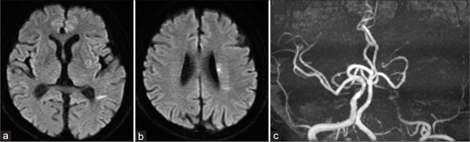
Figure 1:
Case 1: Initial magnetic resonance imaging, diffusion-weighted image (a and b), and magnetic resonance arteriography (c) results at stroke onset. Acute lesions are visualized in the left corona radiata and temporal lobe. Magnetic resonance arteriography shows a left internal carotid artery occlusion. MCA: Middle cerebral artery.
For endovascular recanalization, an 8-Fr long sheath was inserted into the right femoral artery, and an 8-Fr Optimo (Tokai Medical Products, Aichi, Japan) was delivered to the left ICA. Angiography revealed a tapered occlusion of the left ICA and moderate pial anastomosis in the left middle cerebral artery (MCA) territory from the left anterior and posterior cerebral arteries [

Initially, a Trevo Trak 21 microcatheter (Stryker, Kalamazoo, MI, USA) was delivered to the distal side of the occluded site, and Solitaire Platinum 6 × 40 mm (Medtronic, Minneapolis, MN, USA) was thereafter deployed. Immediate restoration of flow was confirmed, although the stent retriever could not completely open. This event similarly indicated atherothrombotic disease as the underlying pathology [

Figure 3:
Case 1: Angiography of the left ICA. The stent retriever fails to open completely at the occluded point (a). After prolonged deployment with stent retriever for 30 min, the left ICA is occluded (b). Following the removal of the stent retriever and microcatheter, the left ICA recanalizes with stenosis (c). ICA: Internal carotid artery.
The patient’s immediate postoperative course was uneventful. Pharmacologic treatment was continued with aspirin 100 mg/day and the cilostazol was increased to 200 mg/day. At 1-month follow-up, the MRA showed persistent mild stenosis in the right ICA. Having completed 3 months of rehabilitation, the patient was discharged with a modified Rankin Scale (mRS) score of 2 at 90 days.
Case two
A 60-year-old man was transferred to our hospital by ambulance for evaluation and treatment of altered level of consciousness that occurred 5 h after he first perceived gait disturbance. His NIHSS at admission was 30 points. On presentation, cranial MRI/MRA showed ICA occlusion and acute and subacute infarctions in the left watershed territories between the anterior cerebral artery, MCA, and posterior cerebral arteries on the MRI diffusion-weighted image sequences [

Figure 4:
Case 2: Initial magnetic resonance imaging, diffusion-weighted image (a), and magnetic resonance arteriography (b) images at stroke onset. Acute lesions are visualized in the left watershed territories between anterior cerebral artery, middle cerebral artery, and posterior cerebral artery. Magnetic resonance arteriography shows a proximal left internal carotid artery occlusion.
For endovascular recanalization, an 8-Fr long sheath was inserted into the right femoral artery, and an 8-Fr Optimo was delivered to the left ICA. Initially, a Trevo Trak 21 microcatheter was delivered to the distal side of the occluded site. The Solitaire Platinum 6 × 40 mm was deployed. Immediate restoration of flow was confirmed, although the stent retriever could not completely open. The Solitaire was gently retrieved; however, no significant clot was identified and the lesion recoiled and occluded a second time. These angiographic findings, the clinical course, and no existence of atrial fibrillation indicated the possibility of gradually progressive, intracranial, atherosclerosis-related, and emergent large-vessel occlusion of the ICA. Aspirin 200 mg and clopidogrel 300 mg were initiated through a nasogastric tube, sodium ozagrel 80 mg was administered intravenously for immediate effect, and the target activated clotting time was set at ≥250 s with a heparin dose afterward as atherothrombotic stroke was diagnosed after starting the treatment.
During the second attempt at endovascular recanalization, PTA with a balloon was considered; however, there was concern in this case that PTA could induce dissection and thrombosis and occlude it. Therefore, the Solitaire was deployed to the occluded site for mild forced angioplasty for approximately 10 min with contrast every 10 min. After occlusion for a 3rd time, the Solitaire was partially resheathed into the microcatheter to reduce the radial force and minimize vessel injury during the pull [

Figure 5:
Case 2: Angiography of the left ICA. The stent retriever fails to open completely at the occluded point (a). After prolonged deployment with stent retriever for 10 min, the left ICA is occluded (b). Following the removal of the stent retriever and microcatheter, the left ICA recanalizes with stenosis (c). ICA: Internal carotid artery.
The patient’s altered level of consciousness remarkably improved. Pharmacologic treatment was continued with aspirin 100 mg/day and cilostazol 200 mg/day. At the 2-week postoperative evaluation, MRA showed persistent mild stenosis in the right ICA, and the patient was discharged without rehabilitation because of no further symptoms or progression, but is being monitored. The mRS was 0 at 90 days.
DISCUSSION
Although endovascular treatment for acute occlusion of the main intracranial arteries associated with an embolic source has already been well established, aggressive therapy for intracranial, atherosclerosis-related, and emergent large-vessel occlusion remains challenging as well as controversial.[
In contrast, the effectiveness of the stenting without retrieval using the Solitaire was reported for intracranial, atherosclerosis-related, and emergent large-vessel occlusion.[
Based on these considerations, we attempted to perform mild angioplasty by the prolonged deployment and partial resheath method using the stent retriever, with the intention of avoiding injury to the vascular endothelium. Prolonged deployment of the stent retriever safely produces an angioplasty effect and maintains cerebral blood flow during the procedure. Stent retriever angioplasty has been identified to be safer than balloon PTA, even in tortuous vessels, such as the siphon of the ICA. Even if an in-stent thrombus develops, it can be retrieved, and the same procedure can be repeated if reocclusion occurs. To the best of our knowledge, to date, some reports have described revascularization in atheromatous disease with the deployment and resheath method.[
The technique used for these cases was relatively simple and the damage to the blood vessel was small. Consequently, the prolonged deployment and partial resheath methods may be particularly effective for lesions with unstable soft plaques near the important perforating arteries, although it is extremely palliative. The technique cost was relatively low, creating favorable conditions for obtaining a good prognosis, and it was a clinically beneficial technique. Because of the relatively small number of cases, a larger sample size is needed to confirm these findings, and the restenosis rates and long-term effects also require further investigation.
CONCLUSION
The prolonged deployment and partial resheath technique using a stent retriever may be safe and effective in the treatment of acute ischemic stroke caused by suspected, intracranial, atherosclerosis-related, and emergent large-vessel occlusion of ICA.
Declaration of patient consent
Patients’ consent not required as patients’ identities were not disclosed or compromised.
Financial support and sponsorship
Nil.
Conflicts of interest
There are no conflicts of interest.
Disclaimer
The views and opinions expressed in this article are those of the authors and do not necessarily reflect the official policy or position of the Journal or its management. The information contained in this article should not be considered to be medical advice; patients should consult their own physicians for advice as to their specific medical needs.
References
1. Ahmed SU, Mann J, Houde J, Barber E, Kelly ME, Peeling L. Permanent implantation of the solitaire device as a bailout technique for large vessel intracranial occlusions. J Neurointerv Surg. 2019. 11: 133-6
2. Baek JH, Kim BM, Kim DJ, Heo JH, Nam HS, Song D. Importance of truncal-type occlusion in stentriever-based thrombectomy for acute stroke. Neurology. 2016. 87: 1542-50
3. Campbell BC, Hill MD, Rubiera M, Menon BK, Demchuk A, Donnan GA. Safety and efficacy of solitaire stent thrombectomy: Individual patient data meta-analysis of randomized trials. Stroke. 2016. 47: 798-806
4. Gascou G, Lobotesis K, Machi P, Maldonado I, Vendrell JF, Riquelme C. Stent retrievers in acute ischemic stroke: Complications and failures during the perioperative period. AJNR Am J Neuroradiol. 2014. 35: 734-40
5. Kim JH, Jung YJ, Chang CH. Feasibility and safety of the strategy of first stenting without retrieval using solitaire FR as a treatment for emergent large-vessel occlusion due to underlying intracranial atherosclerosis. J Neurosurg. 2021. 135: 1091-9
6. Kwon HJ, Lim JW, Koh HS, Park B, Choi SW, Kim SH. Stent-retriever angioplasty for recurrent post-subarachnoid hemorrhagic vasospasm-a single center experience with long-term follow-up. Clin Neuroradiol. 2019. 29: 751-61
7. Moteki Y, Shimizu A, Nakamura A, Yamahata H, Kobayash T. Mild angioplasty with a stent retriever for acute atherothrombotic middle cerebral artery occlusion: A case report. Interdiscip Neurosurg. 2020. 22: 100856
8. Woo HG, Sunwoo L, Jung C, Kim BJ, Han MK, Bae HJ. Feasibility of permanent stenting with solitaire FR as a rescue treatment for the reperfusion of acute intracranial artery occlusion. AJNR Am J Neuroradiol. 2018. 39: 331-6

