- Department of Medicine, Pontifical Catholic University of Goiás, Goiânia, Goiás, Brazil.
- Department of Medicine, Catanduva Medical School (FAMECAUNIFIPA), Catanduva, Brazil.
- Department of Medicine, Faculty of Medical Sciences, Santa Casa de São Paulo, Brazil.
- Department of Medicine, Faculdade de Medicina do ABC, Brazil.
- Department of Neurosurgery, Santa Paula Hospital, São Paulo, Brazil.
- Department of Neurology and Neurosurgery, University of Caxias do Sul, Rio Grande do Sul, Brazil.
- Department of Neurology, Pontifical Catholic University of São Paulo, Sao Paulo, Brazil.
Correspondence Address:
Rafael Caiado-Vencio, Department of Medicine, Pontifical Catholic University of Goiás, Goiânia, Goiás, Brazil.
DOI:10.25259/SNI_853_2021
Copyright: © 2022 Surgical Neurology International This is an open-access article distributed under the terms of the Creative Commons Attribution-Non Commercial-Share Alike 4.0 License, which allows others to remix, transform, and build upon the work non-commercially, as long as the author is credited and the new creations are licensed under the identical terms.How to cite this article: Rafael Caiado-Vencio1, Paulo Eduardo Albuquerque Zito Raffa2, Bruna Marques Lopes3, Fernanda Lopes Rocha Cobucci4, Raphael Vinícius Gonzaga Vieira5, Paulo Roberto Franceschini6, Paulo Henrique Pires de Aguiar7. Success of lateral cervical spinal cord stimulation for the treatment of chronic neuropathic refractory pain. 11-Feb-2022;13:52
How to cite this URL: Rafael Caiado-Vencio1, Paulo Eduardo Albuquerque Zito Raffa2, Bruna Marques Lopes3, Fernanda Lopes Rocha Cobucci4, Raphael Vinícius Gonzaga Vieira5, Paulo Roberto Franceschini6, Paulo Henrique Pires de Aguiar7. Success of lateral cervical spinal cord stimulation for the treatment of chronic neuropathic refractory pain. 11-Feb-2022;13:52. Available from: https://surgicalneurologyint.com/surgicalint-articles/11379/
Abstract
Background: Spinal cord stimulation (SCS) is traditionally performed by implanting surgical leads along the midline of the spinal cord, over the dorsal columns. Here, we present a patient who successfully underwent lateral cervical SCS to treat chronic refractory neuropathic pain.
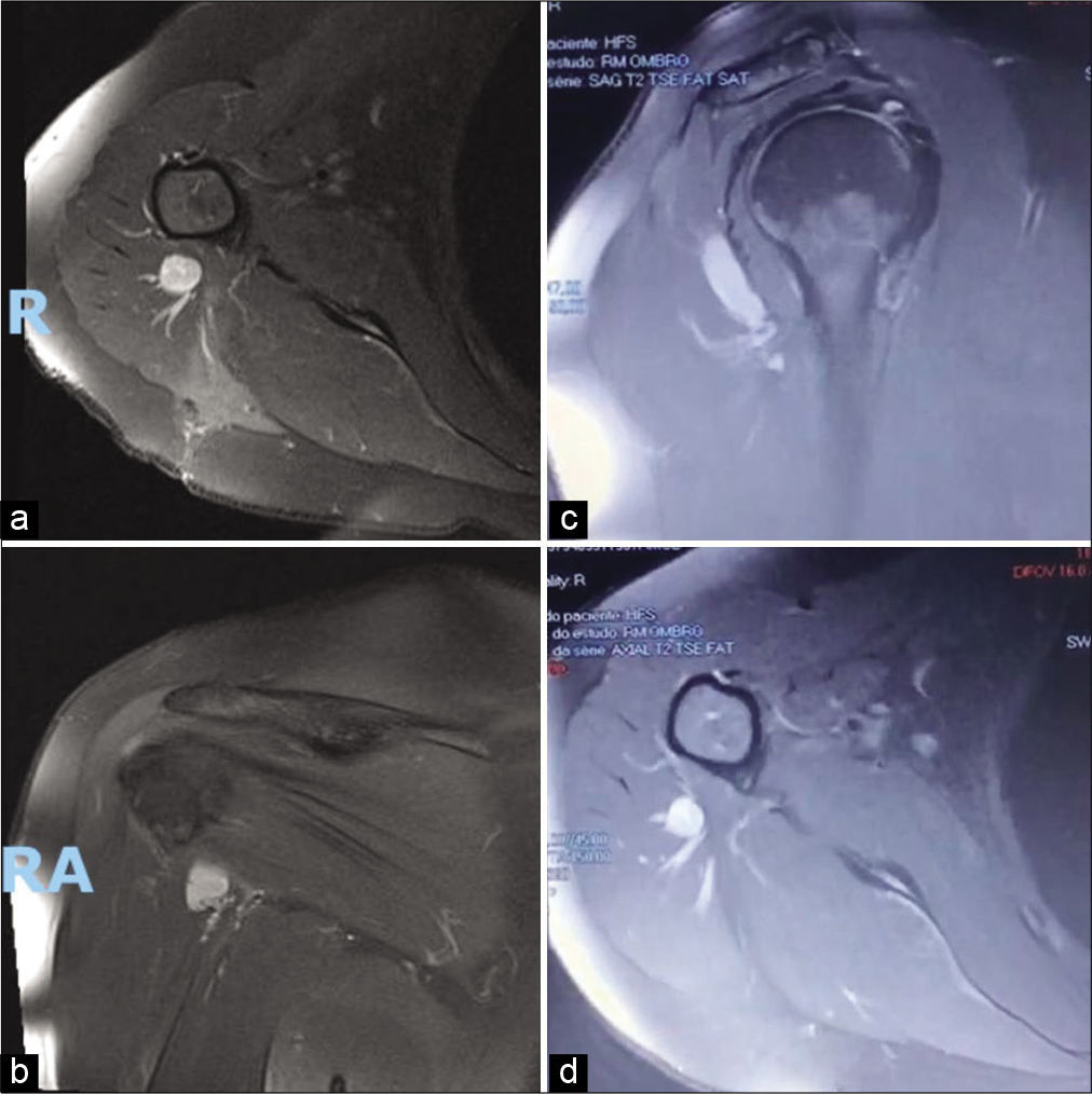
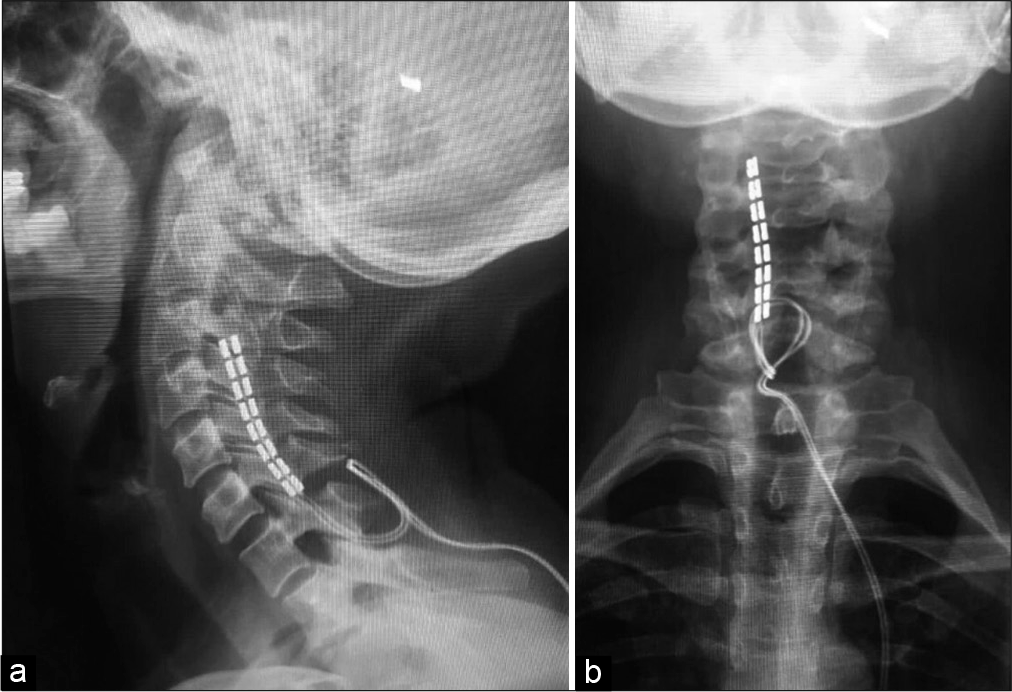
Methods: A 46-year-old female, with a schwannoma involving the right axillary nerve, presented with a chronic refractory right upper extremity pain syndrome. The tumor was located between the fibers of the teres minor and the posterior deltoid, and measured 2.2 cm in diameter. After 8 months of analgesics, opioids, physiotherapy, and acupuncture, the patient underwent surgery; however, the tumor was unresectable (i.e., due to significant adjacent vascular/neural structures). Three months later, she had a midline C6-C7 laminectomy for placement of a right-sided epidural SCS lead (i.e., containing 16 electrode contacts).
Results: Within 4 days following this SCS procedure, the patient’s pain completely resolved; at 10 postoperative months, she still remains pain free.
Conclusion: Lateral SCS at the C6-C7 level provided a safe and effective option for the relief of chronic neuropathic pain attributed to an unresectable schwannoma of the right axillary nerve in a 46-year-old female.
Keywords: Epidural lead, Lateral spinal cord, Neurostimulation, Schwannoma, Spinal cord stimulation
INTRODUCTION
Spinal cord stimulation (SCS) is widely used to treat chronic neuropathic pain.[
MATERIALS AND METHODS
A 46-year-old female with a schwannoma of the right axillary nerve (i.e., between the fibers of the teres minor and posterior deltoid muscles; it measured 2.2 cm in diameter) presented with chronic refractory pain in the upper right limb. The tumor was located in the right axillary nerve [
RESULTS
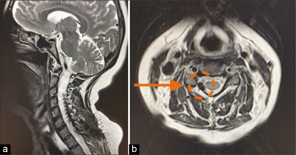
The postoperative MR confirmed the appropriate positioning of the surgical lead at the C6-C7 level in the right lateral epidural space [
DISCUSSION
SCS is traditionally performed by the insertion of a surgical lead along the posterior midline of the spinal cord, over the dorsal columns.[
Efficacy of lateral spinal cord stimulation
Lateral SCS is an effective treatment for neuropathic pain. Although dorsal root ganglion (DRG) stimulation is also an effective treatment for these pain syndrome,[
CONCLUSION
Lateral cervical epidural SCS proved to be an effective and safe treatment for managing chronic neuropathic pain in a 46-year-old female with an unresectable schwannoma of the right axillary nerve.
Declaration of patient consent
The authors certify that they have obtained all appropriate patient consent.
Financial support and sponsorship
Nil.
Conflicts of interest
Paulo Henrique Pires de Aguiar and Paulo Roberto Franceschini have received speaker honorarium from Medtronic. Other authors declare that they have no conflict of interest.
References
1. Chandrasekaran S, Nanivadekar AC, McKernan G, Helm ER, Boninger ML, Collinger JL. Sensory restoration by epidural stimulation of the lateral spinal cord in upper-limb amputees. eLife. 2020. 9: e54349
2. Colloca L, Ludman T, Bouhassira D, Baron R, Dickenson AH, Yarnitsky D. Neuropathic pain. Nat Rev Dis Primers. 2017. 3: 17002
3. Colombo EV, Mandelli C, Mortini P, Messina G, de Marco N, Donati R. Epidural spinal cord stimulation for neuropathic pain: A neurosurgical multicentric Italian data collection and analysis. Acta Neurochir (Wien). 2015. 157: 711-20
4. Deer TR, Levy RM, Kramer J, Poree L, Amirdelfan K, Grigsby E. Dorsal root ganglion stimulation yielded higher treatment success rate for complex regional pain syndrome and causalgia at 3 and 12 months: A randomized comparative trial. Pain. 2017. 158: 669-81
5. Esposito MF, Malayil R, Hanes M, Deer T. Unique characteristics of the dorsal root ganglion as a target for neuromodulation. Pain Med. 2019. 20: S23-30
6. Garg A, Danesh H. Neuromodulation of the cervical dorsal root ganglion for upper extremity complex regional pain syndrome-case report. Neuromodulation. 2015. 18: 765-8
7. Liem L, Russo M, Huygen FJ, van Buyten JP, Smet I, Verrills P. One-year outcomes of spinal cord stimulation of the dorsal root ganglion in the treatment of chronic neuropathic pain. Neuromodulation. 2015. 18: 41-8
8. Lynch PJ, McJunkin T, Eross E, Gooch S, Maloney J. Case report: Successful epiradicular peripheral nerve stimulation of the C2 dorsal root ganglion for postherpetic neuralgia. Neuromodulation. 2011. 14: 58-61


