- Department of Neurosurgery,
- Department of Neurology, Faculty of Medicine, Assiut University, Assiut, Egypt.
Correspondence Address:
Mohamed Khallaf
Department of Neurology, Faculty of Medicine, Assiut University, Assiut, Egypt.
DOI:10.25259/SNI-178-2019
Copyright: © 2019 Surgical Neurology International This is an open-access article distributed under the terms of the Creative Commons Attribution-Non Commercial-Share Alike 4.0 License, which allows others to remix, tweak, and build upon the work non-commercially, as long as the author is credited and the new creations are licensed under the identical terms.How to cite this article: Mohamed Khallaf, Mohamed Abdelrahman. Supratentorial cavernoma and epilepsy: Experience with 23 cases and literature review. 25-Jun-2019;10:117
How to cite this URL: Mohamed Khallaf, Mohamed Abdelrahman. Supratentorial cavernoma and epilepsy: Experience with 23 cases and literature review. 25-Jun-2019;10:117. Available from: https://surgicalneurologyint.com/surgicalint-articles/9405/
Abstract
Background: The current study aimed to assess the role of microsurgical treatment of patients with supratentorial cavernoma with epilepsy based on analysis of our patients.
Methods: This retrospective study included 23 patients with supratentorial cavernoma on computed tomography (CT) scan and magnetic resonance imaging (MRI) of the brain admitted to the Department of Neurosurgery, Assiut University Hospitals (single tertiary hospital) between January 2014 and January 2018 (minimum 12-month follow-up). Deep-seated hemispheric and multiple cavernomas were excluded. Radiographs and hospital data of the patients were gathered and analyzed. All patients underwent the surgical procedure by one experienced neurosurgeon and the diagnosis was confirmed by pathologic evaluation.
Results: A total of 23 patients underwent surgical intervention consist of 15 (65%) men and 8 (35%) women. Their age varies from 11 to 59 year with an average of 36.6 years. All patients presented with seizure. The supratentorial cavernomas were located commonly in temporal lobes; 9 patients (39.1%). 19 (83%) of cavernoma located in the left side. 18 (78%) of cavernoma had a size
Conclusion: Our retrospective population study demonstrates an insight into the supratentorial cavernoma and suggests that microsurgical removal of the symptomatic cavernoma is generally accepted as the most effective and safe method.
Keywords: Cavernoma, Microsurgery, Seizures, Supratentorial
INTRODUCTION
Intracranial cavernous vascular malformations are variously known as cavernous angiomas, cavernous hemangiomas, or, more simply, cavernomas. Cavernomas are congenital low flow vascular lesions. It composed of irregular sinusoidal vascular channels, lacking smooth muscle, and elastic fibers. They lack feeding arteries or draining veins and contain no neural tissue.
The first description of an intracranial cavernoma was given by Virchow, in 1863. For over a century, it was considered to be an extremely rare malformation, usually found at autopsy, and exceptionally diagnosed during life. The prevalence of cerebral cavernous vascular malformations is estimated to be 0.4–0.9%.[
They can be found in several locations in the brain, but 70–80% of them are supratentorial. Supratentorial cavernoma most frequently present with new-onset seizures and progresses to medically refractory epilepsy in 40% of patients.[
Cavernous malformations are dynamic lesions that may exhibit enlargement, regression, or even de novo formation.[
A true breakthrough in cavernoma diagnostics began with the widespread use of MRI. MRI allows cavernomas to be reliably diagnosed not only after acute neurological decline but also in asymptomatic incidental cases. Angiography is only able to detect the existence of abnormal venous drainage associated with cavernoma.
Surgical resection of symptomatic cavernoma lesions located in noneloquent areas is always recommended, as it has been shown to be safe as well as effective in treating epilepsy and preventing future hemorrhages.[
Complete removal of the lesion is required to prevent recurring hemorrhagic events but depends on the neurosurgeon’s experience.[
This article reviews the basic surgical management of supratentorial cavernous malformations, reflecting the treatment strategies used at our department and analyze the factors that influence the outcome.
PATIENTS AND METHODS
General information
A total of 23 patients were included in this study underwent surgical excision from January 2014 to January 2018 in the Neurosurgery Department, Assiut University (single tertiary hospital). Diagnosis was assisted by computed tomography (CT) brain and MRI brain and confirmed by pathological evaluation. Indications for cavernoma excision were made on inclusion criteria (supratentorial symptomatic cavernoma lesions located in easily accessible; safely removable and noneloquent areas with low morbidity associated with surgery). Deeply seated hemispheric and multiple cavernomas were excluded. Early surgery may be considered in situations with a high risk of bleeding, in patients unable to be compliant with antiepileptic drugs (AEDs) treatment, and in patients with a strong desire to eventually stop antiepileptic medication. Informed consent according to the criteria set by the local research ethics committee in our center had to be obtained in writing before surgery. The age of the patients ranged from 11 to 59 years with an average of 36.6 years [
Clinical symptoms
All patients enrolled in this study were presented by seizures either focal in 17 patients or focal with secondary generalization in six patients.
Imaging examination
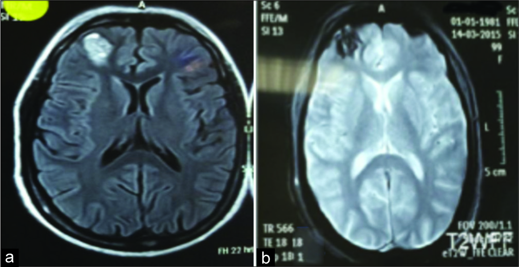
All cases were diagnosed based on CT scan and MRI on the brain. On CT, unless large, these lesions are difficult to see on CT. They do not enhance. If large, then a region of hyperdensity can be seen. If there has been a recent bleed, then it is more conspicuous and may be surrounded by a mantle of edema. In addition, all patients underwent MR scanning of brain and the entire spinal cord using the 1.5 T MRI system [
As could find on MRI, MRI is the modality of choice, demonstrating a characteristic “popcorn” or “berry” appearance with a rim of signal loss due to hemosiderin, which demonstrates prominent blooming on susceptibility weighted sequences. T1 and T2 signal are varied internally depending on the age of the blood products and small fluid- fluid levels may be evident. Size of the cavernoma can be estimated and divided into <2 cm (18 cases) and more than 2 cm (5 cases) with an average of 1.8 cm.
Surgical procedure
Removal of a cavernoma in patients with epilepsy should be assessed in the context of epilepsy surgery, implying indications for tailored surgery of the epileptogenic brain tissue. Failure to control epilepsy after an operation can be linked to incomplete resection and/or the persistence of a hemosiderin fringe.
The surgical procedure was chosen by the author neurosurgeon in a single center on the basis of comprehensive assessment of preoperative patients and intraoperative conditions. After careful analysis of MRI, we always carry out localizing procedures, to place the craniectomy exactly over the lesion. For that purpose, we sometimes use a frameless stereotactic device or neuronavigation system. We did not have motor, sensitive, or cranial nerves evoked potentials. Excision was done by craniotomy and was assisted by operative microscope [
Based on the information from the MRI, the surgical approach was directed on the shortest way to the lesion. We used a minimally invasive transsulcal approach to minimize cortical damage and to expose the lesion in a “keyhole” fashion. However, transcortical excision using intersulcal dissection may be used. The key method is careful dissection around the lesion, according to the principles of microsurgery. The microsurgical technique included sharp dissection and piecemeal resection or one-piece resection where possible in more superficial lesions. In the course of the dissection, afferent arterioles should be exposed, gently lifted up, and electrocoagulated one after the other. Neighboring veins should not be coagulated unless one is certain they drain the cavernoma exclusively. The lesion should be completely resected, including surrounding epileptogenic hemosiderin rich gliotic ring because subtotal removal of a cerebral cavernous malformation is associated with a high risk of recurrences.
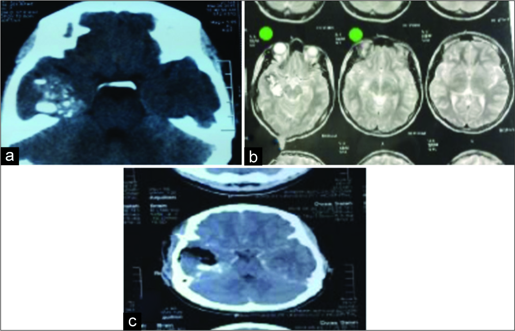
The patients were followed up for at least 1 year. Clinical symptoms and signs were evaluated. Postoperative investigations (CT-MRI brain images) were asked routinely for the patients [
Figure 3
Male patient 35 years presented by focal seizures for more than 1 year in the left side. Computed tomography (a) and magnetic resonance imaging (b) showed the right temporal lesion. Excision was done and postoperative computed tomography (c) showed complete excision and histopathology revealed cavernoma.
Postoperative evaluation indexes
To evaluate the therapeutic outcomes, neurological and medical imaging examinations were performed. The general patient data and specific features before surgery and 12 months after surgery were recorded, respectively. Meanwhile, the clinical outcomes were evaluated using seizure manifestation. The symptoms were rated as “improved,” “unchanged,” and “deteriorated.” The postoperative anticonvulsants therapy is a continuation of a preoperative one which was decided according to seizures types. The choice of the first AED for an individual with newly diagnosed seizures is of great importance and should be made taking into account high-quality evidence of how effective the drugs are at controlling seizures and whether they are associated with side effects. For examination in simple partial seizure, we prescribed a commonly carbamazepine, phenytoin, or valproic acid, and sometimes, we add adjunct therapy as topiramate or levetiracetam. In focal with secondary generalization subtype, we used mainly valproic acid and lamotrigine. The stoppage of antiepileptic medication was decided at follow-up visit; according to the patient clinical state, postoperative MRI brain, and postoperative electroencephalography (EEG) brain with sometimes video monitoring.
Statistical analysis
Data were collected in Excel sheet (Microsoft Office 2010), then were analyzed using SPSS version 22 (SPSS, Inc., Chicago, IL). The results were expressed in terms of frequency and percent.
Ethical consideration
The study was conducted after getting ethical clearance and the permission from Assiut University Teaching Hospital administration. Thorough explanation of the purpose of the study and how data will be treated with respect and confidentiality was provided to the participants. The study protocol was approved by the Ethical Committee, Faculty of Medicine, Assiut University, Egypt.
RESULTS
Demographic information
A total of 23 patients underwent surgical intervention consist of 15 (65%) men and 8 (35%) women. Their age varies from 11 to 59 years with an average of 36.6 years and the greatest number of the patients had 20–40 years old (48%).
Clinical results
Focal seizure was the common manifestation in 17 (74%) patients while focal with secondary generalization manifested in 6 (26%) patients. In this study, the clinical results 12 months after surgery were evaluated through reexamination visits. All 10 patients with only one seizure preoperatively were seizure free at follow-up. Of nine patients who had experienced between two and five seizures preoperatively, 7 (78%) were seizure free, and of four patients with numerous seizures preoperatively, 3 (75%) were seizure free.
Radiological results
The supratentorial cavernomas were located in temporal lobes in 9 patients (39.1%), frontal lobes in 7 patients (30.3%), parietal lobes in 6 patients (26.1%), and occipital lobes in 1 patient (4.3%). 19 (83%) of cavernoma located in the left side while 4 (17%) of cavernoma located in the right side. 18 (78%) of cavernoma had a size <2 cavernoma. Complete excision was confirmed in postoperative investigations (CT and MRI brain images).
DISCUSSION
Incidence
Cavernous malformations, also known as cavernous angiomas, cavernomas, or cryptic vascular malformations, are rare venous capillary bed abnormalities.[
Pathology and pathophysiology
The concept that cavernomas are static lesions has been revised due to growth seen on longitudinal neuroimaging studies and the presence of immunohistologic markers of angiogenesis and proliferation, such as vascular endothelial growth factor endoglin and proliferating cell nuclear antigen.[
The pathophysiology of cavernoma-related epilepsy is likely to involve multiple mechanisms. Perhaps, more important are structural alterations observed in association with cavernomas: the hemosiderin deposit could, therefore, be an indicator that damage has occurred rather than being the main contributor to epileptogenesis. Indeed, a rim of astroglial reaction (astrogliosis) is a hallmark of cavernomas. Leakage of other blood constituents could also play a role; it is notable that albumin has been shown to be pro-epileptogenic through an effect on astrocytic function.[
Astrocytes are well-known to play a role in epileptogenesis, possibly through their interaction with excitatory neurotransmitter release.[
Clinical symptoms
Most cavernoma (48%) are diagnosed incidentally on MRI scans performed for other reasons, but epileptic seizures are the second most common initial clinical presentation, accounting for >25% of cases, and these patients usually have supratentorial cavernoma.[
The onset of symptoms is usually in the third or fourth decade of life, as in our study, the greatest number of the patients had 20–0 years old (48%).[
Two studies found no difference in the occurrence of epilepsy as a function of lesion size,[
Radiology
There was an excess of temporal lobe lesions in the case series possibly resulting from a case selection bias toward intractable epilepsy. It has been suggested that temporal lobe cavernomas are more likely to be associated with intractability.[
The diagnosis of cavernomas is more difficult than other vascular diseases since cavernoma is angiographically occult malformations; thus, other imaging techniques are needed to provide an accurate diagnosis. Conventional T1- and T2- weighted MRI, gradient echo (GE) sequences, high-field MRI, susceptibility weighted imaging (SWI), diffusion tensor imaging, and functional MRI are some of the advanced techniques that are being used for diagnosis of cavernoma or for intraoperative navigation during the treatment of deeply located lesions.
The characteristic imaging appearance of a cavernoma is a multicystic lesion with cysts containing blood products of various ages and therefore various signal intensities on T1- and T2-weighted imaging. A rim of hemosiderin should also be identified in GE or SWI sequences. There is no or only mild contrast enhancement and no surrounding edema unless there has been a recent associated parenchymal hemorrhage. Unless there is acute bleeding, cavernomas typically result in no mass effect since they replace rather than displace normal tissue.
Zabramsky[
Management
In accordance with the current guidelines (Cavernoma Alliance UK, 2012; National Institute for Health and Care Excellence (NICE), 2012), they recommend that all cavernoma patients with a first seizure be urgently referred to a specialist with training and expertise in epilepsy (as neurologist author in our study) to assess whether the patient’s seizures are causally related to the cavernoma. The diagnostic workup should include anamnesis of epilepsy- specific history with analysis of ictal symptomatology as well as a wake and sleep EEG.
Many authors favor an initial conservative approach using AEDs in cavernoma patients with a single seizure rather than going to surgery directly.[
However, due to the risk of bleeding and the negative correlation between epilepsy duration and postoperative seizure outcome, the majority of authors feel that in patients with cavernoma it is not necessary to wait until the rigorous criteria of medically refractory epilepsy proposed by the International League Against Epilepsy are fulfilled.[
A longer preoperative history of epilepsy has been associated with worse seizure outcome. Most authors reported a significantly poorer outcome for patients with seizure duration over 1–2 years, with the notable exception of patients with sporadic seizures over a long period of time.[
Early microsurgical resection is an effective and safe therapy for patients with pharmacoresistant cavernoma- related epilepsy as well as for cavernomas with inherent risk of bleeding. Because cavernomas do not contain neuronal tissue, they cannot themselves be the ictal- onset zone or epileptogenic zone. Therefore, the surgical management of cavernoma is inevitably linked to their effects on the surrounding cerebral tissue. The lesion should be completely resected, including surrounding epileptogenic brain tissue because subtotal removal of a cavernoma is associated with a high risk of recurrences. Most studies report significantly better outcome when the surrounding gliosis and hemosiderin ring are removed.[
Our study was limited by the nonrandomized group selection, the retrospective nature of the analysis, the low number of patients, and no comparative analysis with conservative management. The study subjects were ascertained along with many study variables using electronic medical records. These sources were not primarily designed for research purposes and could have had missing or incorrectly entered information. The strength of our study is that it was based on a defined population examined, operated, and followed up by a single investigator. Furthermore, it is a single center study; it may be helpful to enroll more medical centers, for better understanding of the nature course and management of supratentorial cavernomas.
CONCLUSION
In the present work, we summarized our results on the treatment of cavernomas; our findings are supported by literature. The most frequent manifestations of supratentorial lesions are repeated seizures, which disturb the patient’s life balance we identified a few risk factors for seizures such as the cortical and more frequently the temporomesial location, and the location in the left hemisphere. Microsurgery is the treatment of choice in symptomatic brain cavernomas, total resection being the only curative treatment, and capable to prevent further bleeding and to offer an efficient control of seizures. Complete cavernoma resection and resection of surrounding hemosiderin are recommended.
Declaration of patient consent
Research committee approval was obtained for this study from the institutions Medical Ethics Committee, Faculty of Medicine, Assiut University. Informed consent according to the criteria set by the local research ethics committee was obtained.
Financial support and sponsorship
Nil.
Conflicts of interest
None of the authors has any conflicts of interest to disclose.
References
1. Abla AA, Lekovic GP, Garrett M, Wilson DA, Nakaji P, Bristol R. Cavernous malformations of the brainstem presenting in childhood: Surgical experience in 40 patients. Neurosurgery. 2010. 67: 1589-98
2. Awad I, Jabbour P. Cerebral cavernous malformations and epilepsy. Neurosurg Focus. 2006. 21: e7-
3. Batra S, Lin D, Recinos PF, Zhang J, Rigamonti D. Cavernous malformations: Natural history, diagnosis and treatment. Nat Rev Neurol. 2009. 5: 659-70
4. Baumann CR, Acciarri N, Bertalanffy H, Devinsky O, Elger CE, Lo Russo G. Seizure outcome after resection of supratentorial cavernous malformations: A study of 168 patients. Epilepsia. 2007. 48: 559-63
5. Baumann CR, Schuknecht B, Lo Russo G, Cossu M, Citterio A, Andermann F. Seizure outcome after resection of cavernous malformations is better when surrounding hemosiderin-stained brain also is removed. Epilepsia. 2006. 47: 563-6
6. Bertalanffy H, Benes L, Miyazawa T, Alberti O, Siegel AM, Sure U. Cerebral cavernomas in the adult. Review of the literature and analysis of 72 surgically treated patients. Neurosurg Rev. 2002. 25: 1-53
7. Bozinov O, Hatano T, Sarnthein J, Burkhardt JK, Bertalanffy H. Current clinical management of brainstem cavernomas. Swiss Med Wkly. 2010. 140: w13120-
8. Cappabianca P, Alfieri A, Maiuri F, Mariniello G, Cirillo S, de Divitiis E. Supratentorial cavernous malformations and epilepsy: Seizure outcome after lesionectomy on a series of 35 patients. Clin Neurol Neurosurg. 1997. 99: 179-83
9. Casazza M, Broggi G, Franzini A, Avanzini G, Spreafico R, Bracchi M. Supratentorial cavernous angiomas and epileptic seizures: Preoperative course and postoperative outcome. Neurosurgery. 1996. 39: 26-32
10. Cenzato M, Stefini R, Ambrosi C, Giovanelli M. Post-operative remnants of brainstem cavernomas: Incidence, risk factors and management. Acta Neurochir (Wien). 2008. 150: 879-86
11. Clatterbuck RE, Moriarity JL, Elmaci I, Lee RR, Breiter SN, Rigamonti D. Dynamic nature of cavernous malformations: A prospective magnetic resonance imaging study with volumetric analysis. J Neurosurg. 2000. 93: 981-6
12. Cohen DS, Zubay GP, Goodman RR. Seizure outcome after lesionectomy for cavernous malformations. J Neurosurg. 1995. 83: 237-42
13. D’Angelo VA, De Bonis C, Amoroso R, Cali A, D’Agruma L, Guarnieri V. Supratentorial cerebral cavernous malformations: Clinical, surgical, and genetic involvement. Neurosurg Focus. 2006. 21: e9-
14. Del Curling O, Kelly DL, Elster AD, Craven TE. An analysis of the natural history of cavernous angiomas. J Neurosurg. 1991. 75: 702-8
15. Englot DJ, Han SJ, Lawton MT, Chang EF. Predictors of seizure freedom in the surgical treatment of supratentorial cavernous malformations. J Neurosurg. 2011. 115: 1169-74
16. Ferroli P, Casazza M, Marras C, Mendola C, Franzini A, Broggi G. Cerebral cavernomas and seizures: A retrospective study on 163 patients who underwent pure lesionectomy. Neurol Sci. 2006. 26: 390-4
17. Hammen T, Romstöck J, Dörfler A, Kerling F, Buchfelder M, Stefan H. Prediction of postoperative outcome with special respect to removal of hemosiderin fringe: A study in patients with cavernous haemangiomas associated with symptomatic epilepsy. Seizure. 2007. 16: 248-53
18. Josephson CB, Bhattacharya JJ, Counsell CE, Papanastassiou V, Ritchie V, Roberts R. Seizure risk with AVM treatment or conservative management: Prospective, population-based study. Neurology. 2012. 79: 500-7
19. Kayali H, Sait S, Serdar K, Kaan O, Ilker S, Erdener T. Intracranial cavernomas: Analysis of 37 cases and literature review. Neurol India. 2004. 52: 439-42
20. Kim DS, Park YG, Choi JU, Chung SS, Lee KC. An analysis of the natural history of cavernous malformations. Surg Neurol. 1997. 48: 9-17
21. Kwan P, Arzimanoglou A, Berg AT, Brodie MJ, Allen Hauser W, Mathern G. Definition of drug resistant epilepsy: Consensus proposal by the ad hoc task force of the ILAE commission on therapeutic strategies. Epilepsia. 2010. 51: 1069-77
22. Maciunas JA, Syed TU, Cohen ML, Werz MA, Maciunas RJ, Koubeissi MZ. Triple pathology in epilepsy: Coexistence of cavernous angiomas and cortical dysplasias with other lesions. Epilepsy Res. 2010. 91: 106-10
23. Menzler K, Chen X, Thiel P, Iwinska-Zelder J, Miller D, Reuss A. Epileptogenicity of cavernomas depends on (archi-) cortical localization. Neurosurgery. 2010. 67: 918-24
24. Moriarity JL, Wetzel M, Clatterbuck RE, Javedan S, Sheppard JM, Hoenig-Rigamonti K. The natural history of cavernous malformations: A prospective study of 68 patients. Neurosurgery. 1999. 44: 1166-71
25. Prat R, Galeano I. Endoscopic resection of cavernoma of foramen of monro in a patient with familial multiple cavernomatosis. Clin Neurol Neurosurg. 2008. 110: 834-7
26. Raychaudhuri R, Batjer HH, Awad IA. Intracranial cavernous angioma: A practical review of clinical and biological aspects. Surg Neurol. 2005. 63: 319-28
27. Robinson JR, Awad IA, Magdinec M, Paranandi L. Factors predisposing to clinical disability in patients with cavernous malformations of the brain. Neurosurgery. 1993. 32: 730-5
28. Seifert G, Schilling K, Steinhäuser C. Astrocyte dysfunction in neurological disorders: A molecular perspective. Nat Rev Neurosci. 2006. 7: 194-206
29. Seiffert E, Dreier JP, Ivens S, Bechmann I, Tomkins O, Heinemann U. Lasting blood-brain barrier disruption induces epileptic focus in the rat somatosensory cortex. J Neurosci. 2004. 24: 7829-36
30. Stavrou I, Baumgartner C, Frischer JM, Trattnig S, Knosp E. Long-term seizure control after resection of supratentorial cavernomas: A retrospective single-center study in 53 patients. Neurosurgery. 2008. 63: 888-96
31. Stefan H, Hammen T. Cavernous haemangiomas, epilepsy and treatment strategies. Acta Neurol Scand. 2004. 110: 393-7
32. Stefan H, Walter J, Kerling F, Blümcke I, Buchfelder M. Supratentorial cavernoma and epileptic seizures. Are there predictors for postoperative seizure control? Nervenarzt. 2004. 75: 755-62
33. Sure U, Freman S, Bozinov O, Benes L, Siegel AM, Bertalanffy H. Biological activity of adult cavernous malformations: A study of 56 patients. J Neurosurg. 2005. 102: 342-7
34. Winslow N, Abode-Iyamah K, Flouty O, Park B, Kirby P, Howard M. Intraventricular foramen of monro cavernous malformation. J Clin Neurosci. 2015. 22: 1690-3
35. Yeon JY, Kim JS, Choi SJ, Seo DW, Hong SB, Hong SC. Supratentorial cavernous angiomas presenting with seizures: Surgical outcomes in 60 consecutive patients. Seizure. 2009. 18: 14-20
36. Zabramski JM, Wascher TM, Spetzler RF, Johnson B, Golfinos J, Drayer BP. The natural history of familial cavernous malformations: Results of an ongoing study. J Neurosurg. 1994. 80: 422-32


