

- Glasgow Neuro Society, Wolfson School of Medicine, University of Glasgow, Glasgow, Scotland, United Kingdom
- Department of Neurology, Institute of Neurological Sciences, Queen Elizabeth University Hospital, Glasgow, Scotland, United Kingdom.
- Department of Neurosurgery, Institute of Neurological Sciences, Queen Elizabeth University Hospital, Glasgow, Scotland, United Kingdom.
Correspondence Address:
Hassan Ismahel, Glasgow Neuro Society, Wolfson School of Medicine, University of Glasgow, Glasgow, Scotland, United Kingdom.
DOI:10.25259/SNI_649_2023
Copyright: © 2023 Surgical Neurology International This is an open-access article distributed under the terms of the Creative Commons Attribution-Non Commercial-Share Alike 4.0 License, which allows others to remix, transform, and build upon the work non-commercially, as long as the author is credited and the new creations are licensed under the identical terms.How to cite this article: Ismahel H1, Stewart Middleton EE1, Evans VE1, Chaudhary A1, Shah DM1, Gardee A1, Azad Bashir AB B1, Ball EJ1, Goonewardena E1, Anyaegbunam GK1, Al Salloum LN1, Grace Nelson-Hughes MA1, Minnis ML1, Salim O1, Hay SA1, Cheng Y1, Ashraf M1, Davidson A2, Alakandy L3, , Farias EM, Keenlyside A, Bharadwaj HR, Ueda M, Aziz A, Wong YY, Jothi AA, Hunter M, Banks PD, Chia YY, Wellington J, Arora A. Tenth-anniversary Glasgow Neuro Society 2022 conference proceedings. Surg Neurol Int 15-Sep-2023;14:335
How to cite this URL: Ismahel H1, Stewart Middleton EE1, Evans VE1, Chaudhary A1, Shah DM1, Gardee A1, Azad Bashir AB B1, Ball EJ1, Goonewardena E1, Anyaegbunam GK1, Al Salloum LN1, Grace Nelson-Hughes MA1, Minnis ML1, Salim O1, Hay SA1, Cheng Y1, Ashraf M1, Davidson A2, Alakandy L3, , Farias EM, Keenlyside A, Bharadwaj HR, Ueda M, Aziz A, Wong YY, Jothi AA, Hunter M, Banks PD, Chia YY, Wellington J, Arora A. Tenth-anniversary Glasgow Neuro Society 2022 conference proceedings. Surg Neurol Int 15-Sep-2023;14:335. Available from: https://surgicalneurologyint.com/surgicalint-articles/12552/
Date of Submission
03-Aug-2023
Date of Acceptance
15-Aug-2023
Date of Web Publication
15-Sep-2023
Welcome address
Legacy is not born of wealth but from wealth of accomplishment. This principle epitomizes Glasgow Neuro as we commemorate a decade of creative and inspiring conferences aimed at sparking the minds of junior researchers. As recipients of the University of Glasgow Academic Society of the Year Award, I write with a sense of profound achievement as the President of the Glasgow Neuro Society for 2022–23, extending an invitation for you to immerse yourself in our 10th-anniversary conference featuring world-renowned speakers, immersive workshops, and stimulating research.
The ethos of Glasgow stands firmly in our commitment to “raise the bar.” Despite our position as a student society, our relentless ambition propelled us into becoming one of the largest neuro societies in the UK for junior members of the medical profession. Indeed, this year Glasgow Neuro was bestowed the title of academic society of the year by the University of Glasgow Students’ Representative Council. Our roster of past conference speakers includes some of the brightest minds in neurosurgery, including the founder and former Chair of Neurosurgery at Stanford University, Professor Gary Steinberg; esteemed neurosurgeon and inventor of the internationally recognized Glasgow Coma Scale, Professor Sir Graham Teasdale; and Director of Neurosurgery and Neurosurgeon-in-Chief at John Hopkins, Professor Henry Brem. Our ability to attract such distinguished professionals to a conference aimed primarily at medical students and junior clinicians is a testament to our success, underscored by their admiration of our efforts, and expressed support for our future endeavors. Our legacy resonates with our invited speakers, emphasizing the necessity for societies such as ours to bolster the next generation of neuro-centric medics. This sentiment is echoed in their encouragement for student chapters at their institutions to follow in our footsteps.
Our endeavor to make the 10th-anniversary conference our largest to date witnessed a return of some of the greatest minds in neurology and neurosurgery to Scotland. Many of our guests bear well-established legacies that will endure beyond their lifetimes. We were honored to host our keynote speaker Professor Gail Rosseau, a pioneer of Global Neurosurgery who has been elected to the leadership of the World Federation of Neurological Societies, American Association of Neurological Surgeons, and the Société de Neurochirurgie de Langue Française. We welcomed Dr. Michael Lim, current chair of Neurosurgery at Stanford and Professor, by courtesy, of medicine (oncology), neurology, otolaryngology, and of radiation oncology; Professor Adolfo Bronstein, professor of neuro-otology and founder of the neuro-otology unit at Imperial College London, and founder of the British Society of NeuroOtology; Professor Miratul Muqit, professor of experimental neurology at the University of Dundee, credited as a major contributor to the discovery of PINK1 mutations as a cause of Parkinson’s disease; and Mr. Mark Hughes, consultant academic neurosurgeon and honorary senior lecturer at the University of Edinburgh.
The feedback from our delegates was overwhelmingly positive. Most of them accorded the 10th-anniversary conference the highest accolades, with numerous delegates stating that the engaging talks inspired them to consider a career in neurology, neurosurgery, or neuroscience. One delegate had this to say about the conference:
“It’s really difficult to pick just one thing as there were so many brilliant parts. My favorite part was the lectures given by the doctors and professors; they were all brilliant in different ways and gained a lot of new knowledge and understanding from their presentations.”
“I must commend the committee and president on a terrific conference. The day was organized succinctly, timely (the only conference I have ever attended that didn’t run over), and was fulfilled with inspiring keynote lectures and cutting-edge research.”
With support from the Institute of Neurological Sciences in Glasgow, we conducted three immersive, hands-on workshops. Our flagship workshop offered delegates an opportunity to use Zeiss microscopes to apply aneurysm clips around a 3D-printed basilar tip aneurysm, synthesized from “hyper-real” hydrogels developed by Organlikes. Such a workshop could only succeed at the helm of experts, and I am thankful to the neurosurgical consultants Miss Jennifer Brown, Mr. Samih Hassan, Mr. Likhith Alakandy, and Mr. Parameswaran Bhattathiri for taking the time to lead a workshop worthy of their expertise. The second workshop was an evolution of one held last year, involving simulated intracranial pressure monitoring and extraventricular drain insertion into model calvaria provided by Delta Surgical. Finally, under the leadership of Dr. Natasha Fullterton, a consultant diagnostic neuroradiologist, delegates were offered an interactive glimpse into neuroradiology through a series of case-based discussions using 7-tesla magnetic resonance imaging (MRI) scans from real clinical cases, extrapolating radiological findings with their clinical presentation. Once again, the workshops garnered outstanding feedback. For many students, this was their first encounter with practical neurosurgical and neuroradiological skills. To quote one attendee:
“I always knew that neurosurgery was an art of great finesse; hearing and watching the fantastic consultants running the delegates through the simulated aneurysm clipping was very humbling. It has increased my understanding of neurosurgery.”
I would like to express my heartfelt gratitude to the Glasgow Neuro committee for embracing the challenging responsibility of planning our most ambitious event to date. An event of this magnitude demands a collective effort, with each committee member surpassing expectations in their roles. However, despite our undeniable success, we must not become complacent. To continuously push boundaries is what sets us apart.
We are once again collaborating with the acclaimed SURGICAL NEUROLOGY INTERNATIONAL® to publish this year’s conference abstracts for all to pursue. In addition, SNI Digital™, a state-of-the-art digital media initiative that aims to revolutionize how research is disseminated, is host to this year’s abstracts in a digestible video format with round-table discussion by neurosurgeons and neurologists, bridging scientific findings to real clinical practice. The pioneering initiative was developed by Professor James Ausman (Founder and Emeritus Editor-in-Chief of SNI), and I wish to express my utmost appreciation for hosting us on this innovative platform. Making research accessible, comprehensible, and convenient is our ultimate ambition.
Presented below are the abstracts from Glasgow Neuro’s 10th-anniversary conference at the Royal College of Physicians and Surgeons of Glasgow on November 05, 2022. I invite you to watch the short introduction video (
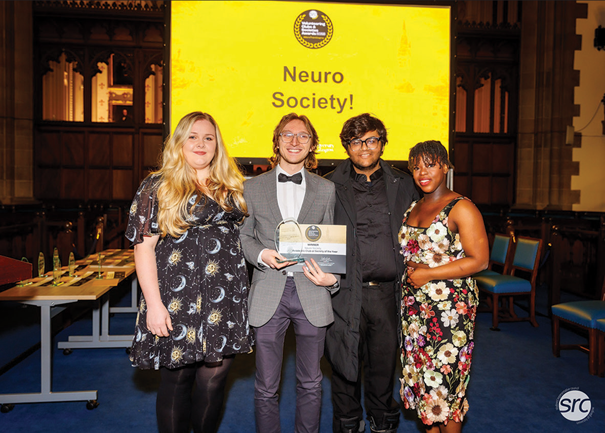
Glasgow Neuro Society Awarded Academic Society of the Year. From left to right: Eilidh Middleton (Vice President), Hassan Ismahel (President), Students’ Representative Council Representative, and Vivienne Evans (Secretary). Photographed at the University of Glasgow Volunteering Clubs and Societies Awards Ceremony, 2023.
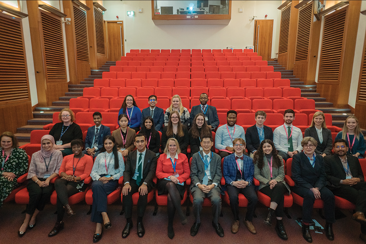
Glasgow Neuro Society Committee 2022–2023, accompanied by conference speakers and faculty. Top Row from left: Emma Ball (Educational Officer), Devansh Shah (Conference Convenor), Eilidh Middleton (Vice President), Mr. Samih Hassan (Consultant Neurosurgeon). Middle row from left: Dr. Amy Davidson (Consultant Neurologist and Honorary President of Glasgow Neuro), Eranga Goonewardena (National Undergraduate Neuroanatomy Competition Marketing Officer), Yihui Cheng (Co-Marketing Officer), Aneesah Bashir (Research Officer), Sophie Hay (Junior Treasurer), Meaghan Nelson-Hughes (Co-Marketing Officer), Gregory Kosisochukwu Anyaegbunam (Junior Representative), Dr. James Ulrich (Neurosurgical registrar), Dr. Nathan McSorley (Neurosurgical registrar), Miss Maya Kommer (Neurosurgical registrar), Meghan Minnis (Senior Representative). Bottom row from left: Dr. Natasha Fullerton (Consultant Diagnostic Neuroradiologist), Ameerah Gardee (National Undergraduate Neuroanatomy Competition Primary Chair), Vivienne Evans (Secretary), Attika Chaudhary (Senior Representative), Mohammad Ashraf (Immediate Past President), Dr. Gail Rosseau (Keynote speaker, Neurosurgeon, George Washington University), Dr. Michael Lim (Guest speaker, Neurosurgeon, Stanford University), Hassan Ismahel (President), Laulwa Al Salloum (Clinical skills officer), Miss Jennifer Brown (Consultant Neurosurgeon), and Mr. Likhith Alakandy (Consultant Neurosurgeon and Honorary President of Glasgow Neuro). Photographed at the Royal College of Physicians and Surgeons of Glasgow.
ABSTRACTS
ABSTRACT 1
Accompanying video discussion found here:
Elucidating the role of regulatory T-cells and CD25 expression in human and mouse glioblastoma using single-cell RNA sequencing
Ericka Mejia Farias1, Felipe Galvez-Cancino2, Gordon Beattie3, Mariela Navarrete Sanchez4, Sergio Quezada5
1Cancer MSc Graduate, University College London, London, UK, 2Postdoctoral Researcher, UCL Cancer Institute, University College London, London, UK, 3Bioinformatician, UCL Cancer Institute, University College London, London, UK, 4Research Assistant, UCL Cancer Institute, University College London, London, UK, 5Group Leader of Immune Regulation and Tumour Immunotherapy Group, UCL Cancer Institute, University College London, London, UK.
Background: Glioblastoma (GBM) is the deadliest and most common brain malignancy. Its tumor microenvironment is poorly understood, but regulatory T-cell (Tregs) presence appears to contribute greatly to GBM’s highly-immunosuppressive nature, likely through CD25-expression. CD25+Tregs have been successfully targeted using a non-IL-2-blocking anti-CD25 monoclonal antibody (RG6292) in mouse studies in other cancers. Here, analysis of single-cell RNA-sequencing (scRNA-seq) data was conducted to characterize Tregs in human and mouse GBM and to quantify the treatment effect of RG6292 in an in vivo GBM mouse model.
Methods: Data analysis was conducted using R, with the package “Seurat.” Two recently-published ×10-scRNA-seq human-GBM datasets[1,2] and 6 ×10-scRNA-seq mouse-GBM samples were analyzed to characterize Treg presence and CD25-expression. The mouse samples, 3 untreated and 3 RG6292-treated, were prepared by the Quezada lab from an orthotopic NPE-IE GBM mouse model. Changes in counts, proportions, and clonal expansion of different cell types were assessed to establish the RG6292-treatment effect on in vivo mouse-GBM. FACS analysis results confirmed scRNA-seq observations at the protein-level.
Results: Data analysis confirmed the presence of similar immune cells, including CD25+Tregs, and a conserved immunosuppressive Treg signature involving Tnsfrsf4, Tnsfrsf18, Foxp3, Batf, Icos, and Entpd1, between the two species. More phagocytic-myeloid cell types expressing Fc-gamma-receptor-III were seen in RG6292-treated mouse samples compared to control, as was marked T-cell clonal expansion, particularly CD8-cytotoxic T-cells. Depletion of both Tregs and tumor cells was confirmed in RG6292-mice compared to control mice, as was increased mouse survival.
Conclusion: scRNa-seq and FACS analysis confirmed RG629-treatment in our mouse model depleted Tregs, likely through an Fc-gamma-receptor-mediated mechanism involving phagocytic-myeloid cells, and depleted tumor cells through enhanced clonal expansion of CD8 T-cells. Conservation of Treg signatures between species suggests that the strong RG6292 treatment effect observed in our GBM mouse model could be translated to clinics in the future and provide much-needed treatment for GBM patients.
REFERENCES
Mathewson ND, Ashenberg O, Tirosh I, Gritsch S, Perez EM, Marx S, et al. Inhibitory CD161 receptor identified in glioma-infiltrating T cells by single-cell analysis. Cell 2021;184:1281-98. Pombo Antunes AR, Scheyltjens I, Lodi F, Messiaen J, Antoranz A, Duerinck J. Single-cell profiling of myeloid cells in glioblastoma across species and disease stage reveals macrophage competition and specialization. Nat Neurosci 2021;24:595-610.
ABSTRACT 2
Accompanying video discussion found here:
Sonodynamic therapy for glioblastoma: Focused ultrasound mapping, in vitro mitochondrial ROS-induced apoptosis, and neurosphere models
Andrew Keenlyside1,2, Theo Marples1,2, Zifan Gao1,3, Hong Hu1,3, Lynden Guy-Nicely2,4, Vasudha Tandon2, Quina Nogales2, Han Li1,3, Lisa Landgraf5, Andreas Melzer1,3,5, Kismet Hossain-Ibrahim6, Zhihong Huang1,3, Sourav Banerjee4, James Joseph1,3
1Centre for Medical Engineering and Technology, University of Dundee, Dundee, UK, 2School of Medicine, Ninewells Hospital and Medical School, University of Dundee, Dundee, UK, 3Department of Biomedical Engineering, School of Science and Engineering, Fulton Building, University of Dundee, UK, 4Department of Cellular and Systems Medicine, Jackie Wood Cancer Centre, School of Medicine, Ninewells Hospital, Dundee, UK, 5Innovation Center Computer Assisted Surgery, Institute at the Faculty of Medicine, Leipzig University, Leipzig, Germany, 6Department of Neurosurgery, Ninewells Hospital and Medical School, NHS Tayside, Dundee, UK.
Background: Sonodynamic therapy (SDT) for glioblastoma is a proposed, novel therapeutic option utilizing the activation of 5-aminolevulinic acid metabolites through focused ultrasound. This paper aims to describe the factors influencing in vitro cell testing and observe the impacts of SDT in pilot 2D and 3D cell tests in this system.
Methods: An automated SDT system was developed to allow the application and mapping of focused ultrasound (FUS) fields under varied conditions; well plate characteristics, frequency, and effect of a continuous fluid column. Pilot testing in cell lines with reactive oxygen species (ROS) and apoptosis immunofluorescence assays and the investigation of neurosphere growth were undertaken within the system.
Results: Standard polystyrene plates reduced peak central FUS intensity, while the reduction in frequency and measures to reduce vertical dose reflection increased FUS intensity. Upper limits of sonication showed a thermal effect of <0.5°C. Simulations of FUS field maps showed concordant patterns to system testing. GBM22 cell lines showed increased signal in SDT 2 h posttreatment with both ROS and Annexin V fluorescent agents, showing the greatest ROS signal matching that of the mitochondria. SDT reduced the growth of newly formed neurospheres by 83% after 21 days (P < 0.001). Further, the growth of pre-formed neurospheres was reduced by 54.9% 22 days posttreatment (P < 0.0001).
Conclusion: SDT selectively induces mitochondrial ROS-mediated apoptosis and significant neurosphere growth inhibition in pilot tests. This system, when optimal parameters are introduced, shows close concordance with simulation for estimating FUS dose imparted to cells. The translatability of in vitro SDT experiments relies heavily on the accurate determination of FUS dose. As such, developing systems to mitigate sources of error may aid in guiding future in vivo studies. In vitro system and tumor model, development provides an opportunity for both mechanistic and translatable research.
ABSTRACT 3
Accompanying video discussion found here:
Analyzing the effectiveness of endoscopic endonasal approaches in the surgical resection of tuberculum sellae meningiomas
Hareesha Rishab Bharadwaj
The University of Manchester, Manchester, United Kingdom.
Background: Tuberculum sellae meningiomas are tumors that develop in the tubercle of the sella turcica. Patients usually present with visual symptoms, such as visual loss and optic atrophy due to compression of the optic chiasm. Treatment of these tumors has been historically carried out through microscopic transcranial approaches (mTCA); however, there has been an advent of innovative surgical resection techniques; endoscopic endonasal approaches, particularly the keyhole superior interhemispheric transfalcine approach. Considering this, we decided to perform a systematic review of the approach, looking at visual outcomes and complication rates to analyze effectiveness.
Methods: In line with Preferred Reporting Items for Systematic Reviews and Meta-Analyses guidelines, a search was conducted on MEDLINE and EMBASE by our trust librarian, which yielded 830 results. The searches were then scrutinized, following which eight were included based on our inclusion criteria.
Results: A total of 63 patients were reported in the eight included studies/papers; 89% of patients were located within Europe/North America. The average age of patients was 64.6 years. Total gross resection rates in patients were 84.8%, with an average tumor volume being reported at 7.8 cm3. Visual improvement was reported in 81% of cases, with deterioration of 3.4%. Leakage of cerebrospinal fluid was reported in 13.2% of cases, which was higher than anticipated. About 1.3% of patients developed other complications (including vascular injury and endocrine disorders).
Conclusion: Our study reports that the approach is safe, and an apt surgical option with low rates of complications and high resection rates. Further studies comparing this approach to mTCAs might prove useful to decide which approach is superior.
ABSTRACT 4
Accompanying video discussion found here:
Analyzing meningiomas in children: A rural Indian study
Hareesha Rishab Bharadwaj
The University of Manchester, Manchester, United Kingdom.
Background: Intracranial meningiomas in children are often considered to be rare, with these tumors accounting for anywhere between 0.4% and 4.6% of all brain tumors in children. As compared to the thoroughly studied pattern of meningioma development in adults, pediatric meningiomas tend to show differing developmental patterns. One study conducted at the University of Iowa reported that pediatric meningiomas tend to have more malignant histological subtypes while presenting with greater rates of recurrence. Studies on the histological spectrum of brain tumors in India report a higher frequency of pediatric craniopharyngiomas in India as compared to the Western world.
Methods: A retrospective study and analysis of 26 children presenting with meningiomas (of the posterior fossa, sphenoid, and tuberculum sellae) in a rural government–run hospital in rural southwestern India (near Bangalore) was done. Children were retrospectively analyzed on sex, age, symptoms, signs, radiological findings, treatment, and prognosis. Data collected have been summarized in the results section.
Results: The range of patients reported ranged from 1 to 17 years, with the ratio of males to females being 1.8:1. About 23% of reported meningiomas occurred in the posterior fossa, 45% in the sphenoid and 21% in the tuberculum sellae, with the remaining developing in other regions for which precise data on location and subtype were not available. Out of the 26 cases, atypical/malignant meningiomas were reported in 4 patients. Complete tumor excision was achieved in 18 patients (69.2%), with postoperative mortality seen in three patients (11.5%).
Conclusion: Within this cohort of patients, the rates of complete tumor excision were lower than the national and worldwide average, with a higher mortality rate. This could potentially indicate a worse prognosis and outcomes in children as compared to adults – however, greater research is required to make a firm conclusion.
ABSTRACT 5
Accompanying video discussion found here:
Comparison of Alzheimer’s disease-indicated drugs on neurons generated from Alzheimer’s disease patient-derived iPSCs
Muzuki Ueda1, Takayuki Kondo2, Haruhisa Inoue2
1MBBS Stage 2, Newcastle University, Newcastle on Tyne, United Kingdom,
2Centre for iPS Cell Research and Application, Kyoto University, Kyoto, Japan.
Background: Neurons derived from human-induced pluripotent stem cells (iPSCs), named induced neurons (iNs) are recently being used to screen therapeutic candidates for Alzheimer’s disease (AD). Kondo et al.[1] screened a library of 1280 drugs approved by the Food and Drug Administration in America against AD patient-derived iNs and identified bromocriptine as having the most potent anti-Aβ effect for AD patients with mutations in the presenilin 1 (PSEN1) gene. This study aimed to compare the effect of bromocriptine and four other AD -indicated drugs on iNs generated from AD patient-derived iPSCs using immunofluorescence staining.
Methods: The iPSCs were generated from a familial AD patient with a mutation in the PSEN1 G384A gene. The iPSCs were cultured and differentiated into iNs. The following five AD-indicated drugs were introduced into the iN wells: β-secretase inhibitor IV, semagacestat, JNJ-40418677, bromocriptine, and ergotamine. The iNs were stained with 14 different primary antibodies and images were taken using a fluorescence microscope.
Results: The 14 primary antibodies either did not stain, only stained cell bodies, only stained neurites, or stained both the cell bodies and neurites of the iNs. There were three primary antibodies for which the staining pattern differed depending on the AD-indicated drug that it was treated with. Anti-APH1A showed the most prominent staining on β-secretase inhibitor IV treated iNs compared to iNs treated with other drugs. Anti-BACE1 exhibited more prominent staining on β-secretase inhibitor IV, semagacestat, and JNJ-40418677 in comparison to bromocriptine and ergotamine. Anti-presenilin 1 antibodies showed more prominent staining around the cell bodies of the negative control compared to the drug-treated iNs.
Conclusion: Further, investigation is required to explore the reason for the differences in immunofluorescence staining patterns of anti-APH1A, anti-BACE1, and anti-presenilin 1 antibodies.
REFERENCE
1. Kondo T, Imamura K, Funayama M, Tsukita K, Miyake M, Ohta A, et al. iPSC-based compound screening and in vitro trials identify a synergistic anti-amyloid β combination for Alzheimer’s disease. Cell Rep 2017;21:2304-12.
ABSTRACT 6
Accompanying video discussion found here:
Comparison of surgical procedures in treatment of carotid artery stenosis
Abdullah Aziz, Paul Z. Yang
School of Medicine, University of Glasgow, Glasgow, Scotland.
Background: At the forefront of global mortality are cardiovascular diseases, among which ischemic stroke—a result of atherosclerosis of the carotid arteries—stands as one of the leading causes of death. When carotid stenosis reaches critical levels, medical therapies often prove insufficient, making surgical intervention necessary. The purpose of this study is to perform a literature review comparing carotid endarterectomy (CEA), carotid artery stenting (CAS), and transcarotid arterial revascularization (TCAR) and identify the most effective surgical intervention for the treatment of carotid artery disease.
Methods: A primary literature search was carried out. Multiple search engines were used. The Boolean logic method was used in conjunction with key search terms to further refine the search. Inclusion and exclusion criteria were applied. Twenty-eight papers were found and three papers were further selected for analysis.
Results: The results show that CEA had the highest mortality rate (1.2%) and highest stroke rate (5.6%) yet had the lowest rate of myocardial infarction (0.18%) at 30 days postprocedure compared to CAS and TCAR. In addition, CAS was shown to have a mortality rate and stroke rate of 0.6% and 3.1%, respectively, at 30 days post procedure which is an improvement on CEA but not TCAR. CAS also resulted in the highest rate of myocardial infarction (1.9%) compared to both CEA and TCAR. The results found TCAR to have the lowest mortality rate and stroke rate of 0.2% and 0.6%, respectively, at 30 days post procedure compared to both CEA and CAS. TCAR also resulted in a myocardial infarction rate of 0.9% which is lower than CAS but not CEA.
Conclusion: My paper has shown that although CEA and CAS are both effective interventions, due to its highly optimistic preliminary results, on completion of further trials, TCAR has the potential to become the most effective surgical intervention in the treatment of CAD.
ABSTRACT 7
Accompanying video discussion found here:
Effect of xanthine oxidase inhibitor on post-stroke cognitive status: A post hoc analysis of XILO-FIST randomized control trial
Yun Yan Wong1, Michele Robertson2, Niall Broomfield3,4, Krishna Dani5, David Alexander Dickie4,6, Alex Doney7, Kirsten Forbes8, Graeme Houston9, Alex McConnachie2, Terry Quinn4, Allan Struthers9, Phillip Bath10, Matthew Walters4, Jesse Dawson4
1School of Medicine, Dentistry and Nursing, College of Medical, Veterinary and Life Sciences, University of Glasgow, UK, 2Robertson Centre for Biostatistics, Institute of Health and Wellbeing, College of Medical, Veterinary and Life Sciences, University of Glasgow, Glasgow, UK, 3Stroke Psychology, West Glasgow Ambulatory Care Hospital, Scotland, UK, 4Institute of Cardiovascular and Medical Sciences, College of Medical, Veterinary and Life Sciences, Queen Elizabeth University Hospital, University of Glasgow, Glasgow, UK, 5Department of Neurology, Institute of Neurological Sciences Glasgow, Queen Elizabeth University Hospital, Glasgow, 6DD Analytics Cubed Ltd, Bishopton, 7Medicine Monitoring Unit (MEMO), School of Medicine, University of Dundee, Ninewells Hospital, Dundee, 8Department of Neuroradiology, Institute of Neurological Sciences, Queen Elizabeth University Hospital, Glasgow, 9Division of Molecular and Clinical Medicine, School of Medicine, Ninewells Hospital, Dundee, 10Stroke Trials Unit, Mental Health and Clinical Neuroscience, University of Nottingham, Nottingham.
Background: Allopurinol, a xanthine oxidase inhibitor, reduces serum uric acid, which may have a role in the development of post-stroke cognitive impairment. We aimed to assess the effect of allopurinol on cognition following ischemic stroke and transient ischemic attack (TIA).
Methods: We performed a post hoc analysis of data from the XILO-FIST trial. Participants with recent ischemic stroke or TIA aged ≥50 years who had detailed cognitive assessments were included in the study. Participants were randomized to allopurinol or placebo for 2 years. The primary outcome was the change in Montreal Cognitive Assessment (MoCA) at 2 years. Secondary outcomes were the change in global cognitive function, measured as standardized Z-scores calculated from a detailed neuropsychological test, and the change in MoCA Z-score. Z-scores were calculated using age and education-adjusted normative data. Outcomes were compared between groups using a two-sample t-test.
Results: A total of 370 participants were included (mean age 66 years ± 8.6, 68.8% male, mean years of education 12.4 years ± 2.6). The change in MoCA was 0.22 in the allopurinol group and 0.43 in the placebo group (between-group difference 0.20; [95% confidence interval, CI −0.36–0.76]; P = 0.473). The change in MoCA Z-score was 0.13 with allopurinol and 0.16 with placebo (between-group difference 0.03; [95% CI −0.14–0.20]; P = 0.703). The change in Global cognitive Z-score with allopurinol was 0.04 and −0.08 with placebo (between-group difference 0.12; [95% CI −0.30–0.62]; P = 0.198).
Conclusion: We saw no evidence of an effect of allopurinol on cognitive outcome following a recent ischemic stroke or TIA.
ABSTRACT 8
Accompanying video discussion found here:
Recent advancements in robotic skull base neurosurgery – A review
Agilandiswari Arumuga Jothi
School of Medicine, University of Glasgow, Glasgow, Scotland.
Background: The endoscopic endonasal approach (EEA) is the standard surgical approach used for midline skull base pathologies. However, the EEA has its own limitations such as the need for a second surgeon, constrained sinonasal corridor, lack of sufficient dexterity to close large skull base defects, and long learning curves. Robotic surgery has garnered widespread use in neurosurgical applications due to its high degree of accuracy and could potentially overcome these limitations. Hence, this review aims to present recent advancements in robotic skull base neurosurgery.
Methods: A comprehensive literature search of PubMed, Medline, and Embase databases was conducted, in accordance with Preferred Reporting Items for Systematic Reviews and Meta-Analyses guidelines. Search criteria included keywords such as robot, robotic, surgery, neurosurgery, precision, minimally invasive, and stereotactic. Selection criteria included any studies published from 2018 onward, which report new advancements in robotic neurosurgery. Only full-length text and papers published in the English language were selected. Laboratory, clinical, and cadaveric studies were included in the study. Covidence was used for abstract and full-text screening. Studies with only otorhinolaryngological applications or robotic radiosurgery were excluded from the study.
Results: A total of 99 studies were identified from the initial search, of which six studies were included after a full-text review. The recent advancements in robotic neurosurgery that were identified include concentric tube robot and robotic handle prototypes; Endoscope Robot®; micro continuum robot; Versius® Robotic System; and semi-autonomous integrated robotic system using RAVEN™ II Surgical System. Each robotic system has its own advantages. Nevertheless, the overall advantages of these robotic systems combined were improved performance, high dexterity, and modularity. However, these systems do have limitations such as tissue deformation and validation of the reliability of the robots.
Conclusion: This review article provides an overview of the advantages and limitations of recent advancements in robotic skull base neurosurgery. Future work on the improvement of these robotic systems should include improvements in design and large-scale studies.
ABSTRACT 9
Accompanying video discussion found here:
Perioperative pain assessment and management in the institute of neurological sciences: An audit
Mhairi Hunter
School of Medicine, University of Glasgow, Glasgow, Scotland.
Background: Effective postoperative analgesia influences surgical morbidity and quality of care. Neurosurgical patients frequently report severe pain postoperatively. Mounting evidence suggests that the quality of analgesia following neurosurgery is often suboptimal. Therefore, the aim of this audit was to evaluate the quality of postoperative analgesic management at the Institute of Neurological Sciences (INS), Glasgow. It aimed to determine whether this management is in accordance with local (NHSGGC), and national (Royal College of Anesthetists) guidelines, to a level that grants patient satisfaction.
Methods: This prospective clinical audit was conducted over 4 weeks in September 2022. All individuals undergoing inpatient surgery in the INS were considered for inclusion, which provided that verbal consent was gained. Those who were confused or overly sedated were excluded from the study. A pro forma was conducted preoperatively, on Day 1, and Day 3 postoperatively. Collected data included patient demographic data, operative details, and pain scores (0–10) through the numerical rating score. Perioperative analgesic prescriptions were noted, along with patient satisfaction with their pain control.
Results: Complete data were collected from 13 of the 19 patients included in the study. Adherence to guidelines was observed in the management of 62% (n = 8) of cases. Where guidelines were not followed (n = 5), the average reported pain score on Day 1 was 7.6, compared to 4.6 in the guideline-compliant group. This discrepancy in pain scores continued on D3. Higher scores were noted in patients with a history of chronic pain and complex analgesic requirements.
Conclusion: The postoperative analgesic management of the majority of patients in the INS follow current guideline recommendations. Where guidelines were not followed, this was most commonly due to the co-prescription of weak and strong opioids and inaccuracies in pain score documentation. Regular re-education and emphasis on the importance of available guidance may reduce these errors. Earlier referrals to acute pain services will be beneficial in the management of complex patients.
ABSTRACT 10
Insight into physiologically normal ranges of intracranial pressure in three body positions from 188 patients with telemetric ICP monitors
Ptolemy D. W. Banks, Anand S. Pandit, Eleanor M. Moncur, Simon Thompson, Lewis W. Thorne, Laurence D. Watkins, Ahmed K. Toma
Department of Neurosurgery, National Hospital for Neurology and Neurosurgery, London, UK.
Background: Maintaining intracranial pressure (ICP) within physiologically normal ranges is critical to preserving cerebral perfusion and structure. Despite their clinical importance, “normal” ranges remain unclear. Implantable telemetric ICP monitors afford routine, non-invasive ICP measurement in patients with and without symptoms, offering new insight into optimal ICP values.
Methods: This was a single-center and retrospective study of shunt-dependent hydrocephalus patients with implantable telemetric ICP monitors at the National Hospital for Neurology and Neurosurgery. Symptoms and ICP recordings were taken from electronic patient records. The ICP recordings of patients presenting without pressure-related symptoms were extracted and analyzed.
Results: Across the 188 included patients (69% female), 31 patients were identified as asymptomatic. Their ICP recordings (in mmHg) averaged −9.3 (±5.6) standing, −8.8 (±4.9) sitting, and 10.2 (±6.8) supine. There was an average change of 19.3 (±4.7) between standing and supine. Males had higher ranges (25.3 ± 8.3) compared to females (21.8 ± 6.6).
Conclusion: Although symptoms are a proxy measure of functional ICP, these asymptomatic patients represent pressure values where pressure-related symptoms do not occur. These values could be used to identify high/ low pressure patients and serve as a target in valve adjustments.
ABSTRACT 11
Rates of thromboembolic events in neuropathic patients on long-term intravenous immunoglobulin therapy
Yu Ying Chia1*,2, Eranga Goonewardena1*,2
1* - Yu Ying Chia and Eranga Goonewardena are joint first authors.
1School of Medicine, University of Glasgow, Glasgow, 2Queen Elizabeth University Hospital, Glasgow.
Background: Intravenous immunoglobulins (IVIg) are an established treatment for neurological disorders such as multifocal motor neuropathy (MMN) and chronic inflammatory demyelinating polyneuropathy (CIDP). While generally well-tolerated, there have been concerns about the increased risk of thromboembolic events (TEE) among patients on long-term IVIg therapy. Numerous studies have been carried out assessing the same, but they were not specific to neurological disorders, were not carried out in the West of Scotland, and yielded varying results. Here, we show the prevalence of IVIg-related TEEs at a regional level in patients with neurological disorders on long-term treatment and analyzed possible associations with suspected risk factors.
Methods: A descriptive study was conducted, using data from the medical records of 106 patients with neurological disorders who were currently or were previously on long-term IVIg therapy at the Queen Elizabeth University Hospital, Glasgow. Patients with and without IVIg-related TEEs were compared in relation to age, sex, smoking history, number of comorbidities, and duration of treatment.
Results: We found that 12 of the 106 patients had a TEE within 30 days of their IVIg infusion treatment (11.3%). Smoking and a male gender doubled the risk of a TEE, while patients with 3 or more comorbidities had a 25% chance of developing a TEE. Our findings also showed a linear increase in TEEs as the duration of IVIg treatment increased as well.
Conclusion: Our findings confirmed that there is a high rate of TEEs in neuropathic patients on long-term IVIg treatment, as 11.3% of the study population had a TEE within 30 days of an IVIg infusion. We hope that this study provides greater focus and awareness within medical practice on the risk of TEEs relating to IVIg therapy and can be used as a starting point in determining effective and targeted intervention to safeguard those on long-term IVIg therapy.
ABSTRACT 12
Multimodal management in primary central nervous system lymphomas – A systematic review and update of the literature
J. Wellington1, A. Wellington2
1Cardiff University School of Medicine, Cardiff University, Cardiff, UK,
2Cardiff University School of Biosciences, Cardiff University, Cardiff, UK.
Background: Rare and often sporadic, primary central nervous system lymphoma (PCNSL) accounts for 2.2% of central nervous system (CNS) cancers.[1,2] PCNSL comprises 4–6% of extranodal lymphomas, wherein 90–95% are diffuse large B-cell lymphomas exclusively confined to the CNS.[3] Structures involved may include the cerebrum, meninges, and spinal cord.[4] Effective treatment stratagems include single-modality whole-brain radiotherapy (WBRT) to multi-chemotherapeutic, high-dose methotrexate (HD-MTX)-based regimens with WBRT reticent in disease relapse.[5] However, PCNSL is a rarity in oncological practice due to limited study populations.
Methods: We reviewed current literature concerning multimodal PCNSL management, including the chemoradiotherapeutic paradigm. Pre-decided inclusion criteria included studies adhering to Preferred Reporting Items for Systematic Reviews and Meta-Analyses guidelines. Letters to the editor, abstract-only publications, meta-analyses, and systematic reviews were excluded. An OVID search comprising databases MEDLINE (1946–2022), Embase (1947–2022), and Global Health (1910–2022) was conducted, yielding 190 original articles.
Results: Penetrative neoplasms with multiple malignant foci circumnavigating discernible disease margins bared poor surgical efficacy.[6] Select studies demonstrated PCNSL persisting disproportionate mortality in affected elderly populations, with MTX, temozolomide, and rituximab therapy following hyperfractionated WBRT consolidation conceding complete remission and partial response rates of 51% and 34%, respectively.[7,8]
Conclusion: Primary or consolidatory WBRT remains contentious in affected populations postinduction chemotherapy. International concerns toward standalone immune chemotherapy have sparked randomized phase-2 trials, for example, progression-free survival rates undergoing sequential rituximab, methotrexate, procarbazine, and vincristine (R-MPV)/ high-dose (HD)-cytarabine therapy against post-R-MPV/HD-cytarabine WBRT.[9] In addition, incorporating autologous stem cell transplantation following rituximab, carmustine, etoposide, HD-cytarabine, and MTX therapy.[10] The so-called “relative radioresistance” enigma regarding PCNSL is ongoing.
REFERENCES
Dolecek TA, Propp JM, Stroup NE, Kruchko C. CBTRUS statistical report: Primary brain and central nervous system tumors diagnosed in the United States in 2005-2009. Neuro Oncol 2012;14 Suppl 5:v1-49. Primary CNS Lymphoma (PCNSL) - Brain tumour research. Available from: Villano JL, Koshy M, Shaikh H, Dolecek TA, McCarthy BJ. Age, gender, and racial differences in incidence and survival in primary CNS lymphoma. Br J Cancer 2011;105:1414-8. Grommes C, DeAngelis LM. Primary CNS lymphoma. J Clin Oncol 2017;35:2410-8. Henry JM, Heffner RR Jr., Dillard SH, Earle KM, Davis RL. Primary malignant lymphomas of the central nervous system. Cancer 1974;34:1293-302. Jahr G, Da Broi M, Holte H Jr., Beiske K, Meling TR. The role of surgery in intracranial PCNSL. Neurosurg Rev 2018;41:1037-44. Nelson DF, Martz KL, Bonner H, Nelson JS, Newall J, Kerman HD, et al. Non-Hodgkin’s lymphoma of the brain: Can high dose, large volume radiation therapy improve survival? Report on a prospective trial by the Radiation Therapy Oncology Group (RTOG):RTOG8315. Int J Radiat Oncol Biol Phys 1992;23:9-17. Glass J, Won M, Schultz CJ, Brat D, Bartlett NL, Suh JH, et al. Phase I and II study of induction chemotherapy with methotrexate, rituximab, and temozolomide, followed by whole-brain radiotherapy and postirradiation temozolomide for primary CNS lymphoma: NRG oncology RTOG0227. J Clin Oncol 2016;34:1620-5. Rituximab, methotrexate, vincristine sulfate, procarbazine hydrochloride, and cytarabine with or without radiation therapy in treating patients with primary central nervous system lymphoma - ClinicalTrials.gov. Available from: Institute Curie, Ministry of Health, France, Hoffmann-La Roche, Amgen, & Pierre Fabre Laboratories. Prospective, multicentric, randomized Phase II study, evaluating the role of cranial radiotherapy or intensive chemotherapy with hematopoietic stem cell rescue after conventional chemotherapy for primary central nervous system in young patients (< 60 y). Clinicaltrials.gov; 2019. Available from:
ABSTRACT 13
Influence of magnetic vestibular stimulation on anxiety and depersonalization/ derealization symptoms
Amit Arora
Queen Square Institute of Neurology, University College London, London, United Kingdom.
Background: This pilot aimed to evaluate the feasibility of a novel approach to establish a correlation between magnetic vestibular stimulation (MVS) and anxiety and depersonalization/derealization (Dp/Dr) symptoms, which are often experienced by individuals undergoing MRI.
Methods: Six healthy volunteers were exposed to increasing magnetic field strengths and were predicted to display vestibular stimulation induced by the MR static field and, hence, experience intensifying anxiety and Dp/ Dr symptoms. MVS-induced nystagmus was used as a surrogate measure of vestibular activation. Eye tracking revealed greater intensity of vestibular nystagmus with increasing field strength.
Results: An overall non-significant increase in the severity of symptoms with exposure to a stronger magnetic field was observed; however, due to the complexity of symptoms and the small sample size, we observed a high level of variability in subjective responses. The increase in anxiety demonstrated a linear relationship with deteriorating nystagmus.
Conclusion: These preliminary findings indicate intimate neural interrelations between vestibular function and anxiety and Dp/ Dr symptoms. The implications of this novel initiative and a possible relationship are discussed.
Disclaimer
The views and opinions expressed in this article are those of the authors and do not necessarily reflect the official policy or position of the Journal or its management. The information contained in this article should not be considered to be medical advice; patients should consult their own physicians for advice as to their specific medical needs.

