- Department of Neurosurgery, Neurosurgery Teaching Hospital, Baghdad, Iraq,
- Department of Neurosurgery, King Fahad Hospital of the University, Imam Abdulrahman Alfaisal University, Dammam, Saudi Arabia,
- Department of Neurosurgery, University of Washington, Seattle, Washington,
- Department of Neurosurgery, Johns Hopkins Hospital, Baltimore, Maryland, United States,
- School of Medicine, Baghdad University, Baghdad, Iraq.
Correspondence Address:
Zahraa F. Al-Sharshahi, Department of Neurosurgery, Neurosurgery Teaching Hospital, Baghdad, Iraq.
DOI:10.25259/SNI_1158_2021
Copyright: © 2021 Surgical Neurology International This is an open-access article distributed under the terms of the Creative Commons Attribution-Non Commercial-Share Alike 4.0 License, which allows others to remix, tweak, and build upon the work non-commercially, as long as the author is credited and the new creations are licensed under the identical terms.How to cite this article: Hoz SS1, Al-Jehani H2, Aljuboori Z3, Muhsen BA4, Algburi HA5, Neamah AM5, Al-Sharshahi ZF1. The role of the orbitofrontal artery in the clipping of superiorly projecting anterior communicating artery aneurysms. Surg Neurol Int 30-Dec-2021;12:627
How to cite this URL: Hoz SS1, Al-Jehani H2, Aljuboori Z3, Muhsen BA4, Algburi HA5, Neamah AM5, Al-Sharshahi ZF1. The role of the orbitofrontal artery in the clipping of superiorly projecting anterior communicating artery aneurysms. Surg Neurol Int 30-Dec-2021;12:627. Available from: https://surgicalneurologyint.com/surgicalint-articles/11313/
BACKGROUND
Anterior communicating artery (ACoA) aneurysms are the most common type of cerebral aneurysms 23–40%.[
MICROSURGICAL ANATOMY
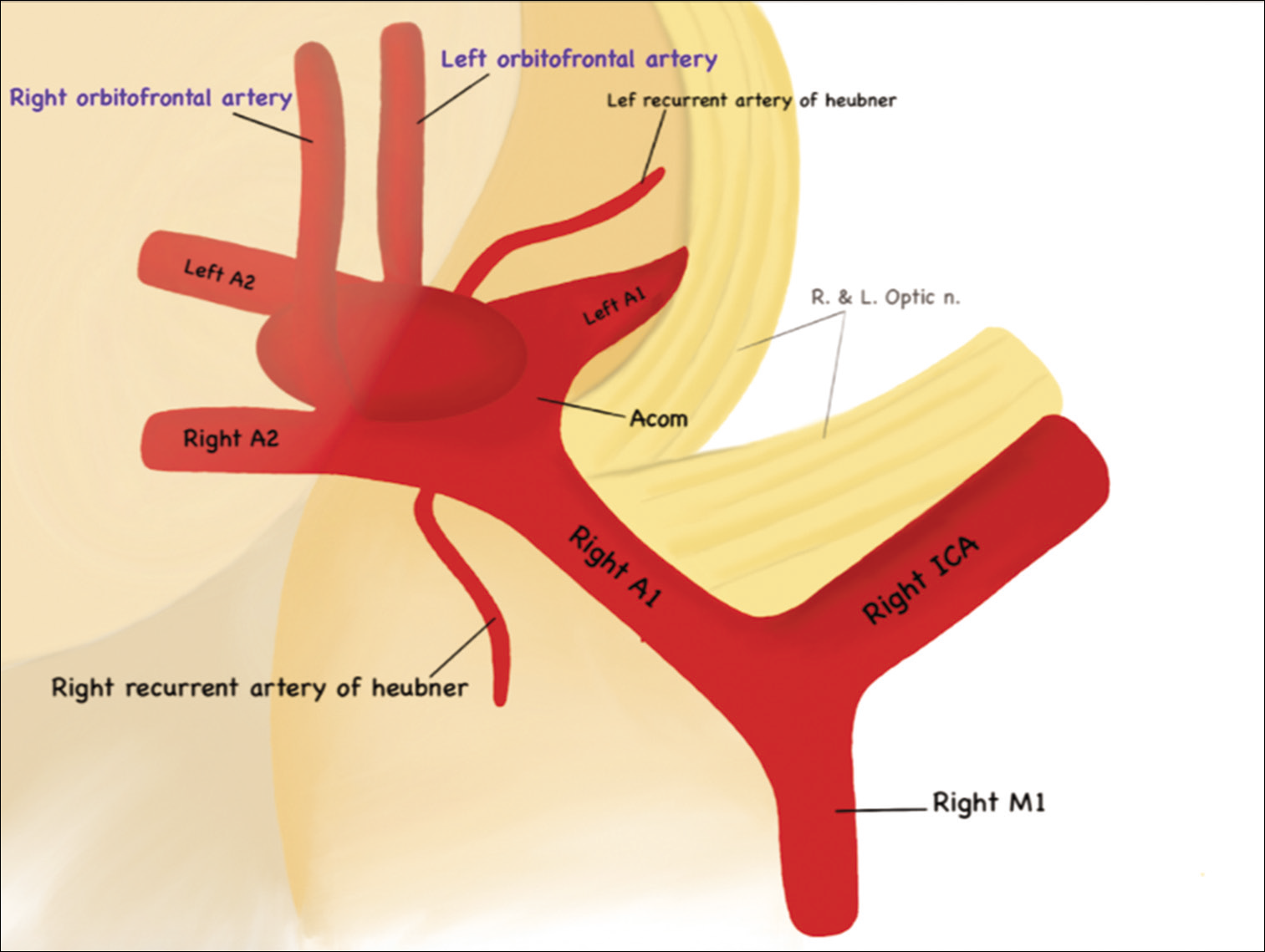
The A2 (a.k.a. postcommunicating) segment of the ACA begins at the ACoA junction and continues along the rostrum of the corpus callosum – hence the term subcallosal segment – until it reaches the rostrum-genu juncture of the corpus callosum. The A2 segment of the ACA gives three main branches, two of which (RAH and OF artery) are closely related to ACoA aneurysm clipping.[
The RAH is the first branch of the A2 and is the largest and longest branch, frequently originating within 4 mm of the ACoA junction, with an average length of 23.4 mm (range, 12–38 mm).[
The OF artery is a term commonly applied for the medial OF artery of the A2 (a.k.a. frontobasal). The lateral OF artery is a branch of the superior trunk of the middle cerebral artery (M2 segment) and is outside the scope of this discussion. The OF artery is the first cortical branch from the A2; it may originate as a common trunk with the frontopolar (FP) branch (18%) or, rarely, from the A1 segment of the ACA. The OF artery originates within 5 mm (range, 4–9 mm) from the ACoA junction and runs anteriorly and inferiorly, passing perpendicularly over the gyrus rectus and across the olfactory tract en route to the OF cortex. The course of the OF artery may resemble that of a RAH, but it diverges distally from the A1 segment. It supplies the gyrus rectus, the olfactory bulb, and the medial orbital surface of the frontal lobe, producing numerous perforators (Brodmann’s areas 25,11).[
THE OF ARTERY IN ACOA CLIPPING SURGERY
The OF artery is a component of the core vascular complex that must be identified and preserved during the surgical clipping of ACoA aneurysms, especially the superiorly projecting type.
It is imperative to underline an existing discrepancy between our 2D understanding of the OF and its intraoperative orientation in the setting of ACoA clipping surgery [
In superiorly projecting ACoA aneurysms, the aneurysm dome is located between the bilateral A2 segments, effectively blocking the view of the contralateral A2. As a result, access to the contralateral proximal A2 requires deep dissection around and above the dome and up through the interhemispheric fissure. In certain scenarios, the presence of the OF artery can add more technical challenges to the exposure as it would be located at the level of the aneurysm. In such a case, the OF artery represents a real obstacle, complicating the dissection and raising the question of what are the potential immediate and long-term consequences of intentional vessel sacrifice or inadvertent injury [
Figure 3:
Intraoperative images of a ruptured, superiorly projecting anterior communicating artery (ACoA) clipping through the right pterional approach. (a) Initial dissection: the right A1, the ipsilateral A2, as well as the ACoA aneurysm are visible. (b) The orbitofrontal (OF) artery runs in proximity relation to the aneurysm dome (black arrow). (c) The preserved OF artery appears superior to the clip at the end of surgery.
The clinical significance of OF artery sacrifice is not well understood with a noticeable gap between microanatomical and surgical-clinical studies. A search of the Medline at PubMed database from its inception to the present using the following search algorithm; ((OF) OR (Fronto-orbital) OR (Frontobasal) AND (ACoA)) AND (aneurysm), revealed no papers discussing the functional significance of the OF artery. The existing research was either cadaver based or primarily focused on the origin, course, territories, and anastomoses of the OF artery, with no studies connecting the functional, neurological, or cognitive outcomes of patients undergoing ACoA clipping surgery to the preservation or sacrifice of the OF artery. At our institution, the OF had to be sacrificed in 6 out of 50 patients with clip secured ruptured ACoA aneurysms. All six patients had gyrus rectus resection necessitated by the ruptured status of the aneurysm, resulting in the aneurysm dome being adherent to the surrounding parenchyma. All six patients had no noticeable neurological deficits with a mean follow-up of two years; this phenomenon may be explained in part by probable collateralization between medial and lateral OF arteries, which acts to mitigate neurological damage in the event of OF artery sacrifice.
CONCLUSION
The OF artery is a critical component in the surgical clipping of superiorly projecting ACoA aneurysms. It can obscure the surgeon’s view of the aneurysm and the contralateral A2, which can increase the risk of surgical dissection and aneurysm rupture. Future snalysis of the clinical repercussions resulting from OF artery scarifice are required to guide intraoperative decisions of vessel preservation or sacrifice.
Declaration of patient consent
Patient’s consent not required as patients identity is not disclosed or compromised.
Financial support and sponsorship
Nil.
Conflicts of interest
There are no conflicts of interest.
References
1. Bhattarai R, Liang CF, Chen C, Wang H, Huang TC, Guo Y. Factors determining the side of approach for clipping ruptured anterior communicating artery aneurysm via supraorbital eyebrow keyhole approach. Chin J Traumatol. 2020. 23: 20-4
2. Chen J, Li M, Zhu X, Chen Y, Zhang C, Shi W. Anterior communicating artery aneurysms: Anatomical considerations and microsurgical strategies. Front Neurol. 2020. 11: 1020
3. Hernesniemi J, Dashti R, Lehecka M, Niemelä M, Rinne J, Lehto H. Microneurosurgical management of anterior communicating artery aneurysms. Surg Neurol. 2008. 70: 8-28
4. Perlmutter D, Rhoton AL. Microsurgical anatomy of the distal anterior cerebral artery. J Neurosurg. 1978. 49: 204-28


