

- Department of Neurological Surgery, São José Hospital, Santa Casa de Misericórdia de Porto Alegre Hospital Complex, Porto Alegre, Brazil
- School of Medicine, Catholic University of Pelotas, Pelotas, Brazil
- Department of Pathology, Santa Rita Hospital, Santa Casa de Misericórdia de Porto Alegre Hospital Complex, Porto Alegre, Brazil
Correspondence Address:
Albert Vincent B. Brasil
Department of Pathology, Santa Rita Hospital, Santa Casa de Misericórdia de Porto Alegre Hospital Complex, Porto Alegre, Brazil
DOI:10.4103/sni.sni_90_18
Copyright: © 2018 Surgical Neurology International This is an open access journal, and articles are distributed under the terms of the Creative Commons Attribution-NonCommercial-ShareAlike 4.0 License, which allows others to remix, tweak, and build upon the work non-commercially, as long as appropriate credit is given and the new creations are licensed under the identical terms.How to cite this article: Albert Vincent B. Brasil, Ruy Gil Rohrmoser, Guilherme Gago, Eduardo Cambruzzi. Atypical spinal epidural capillary hemangioma: Case report. 03-Oct-2018;9:198
How to cite this URL: Albert Vincent B. Brasil, Ruy Gil Rohrmoser, Guilherme Gago, Eduardo Cambruzzi. Atypical spinal epidural capillary hemangioma: Case report. 03-Oct-2018;9:198. Available from: http://surgicalneurologyint.com/surgicalint-articles/9032/
Date of Submission
20-Mar-2018
Date of Acceptance
16-Apr-2018
Date of Web Publication
03-Oct-2018
Abstract
Background:Hemangiomas are benign vascular malformations that can involve the spine. Pure epidural hemangiomas are rare and represent only 4% of all epidural lesions. Most hemangiomas are of the cavernous type; the capillary variant is atypical, and only ten cases have been reported in the literature.
Case Description:A 69-year-old female presented with nonspecific dorsal pain. Magnetic resonance imaging (MRI) showed a spinal epidural tumor at the T9-T10 level. Following a T9-T11 laminectomy, the lesion was completely resected en bloc. Histopathologic analysis showed a pure epidural capillary hemangioma with adipose tissue mesenchyma.
Conclusions:Although epidural capillary hemangiomas are extremely rare, they should be considered among the differential diagnoses of extradural, extramedullary spinal lesions. Further, they must be differentiated from other more common lesions such as meningiomas and schwannomas. The recommended surgical management is en bloc gross total excision.
Keywords: Epidural capillary hemangioma, primary spinal tumors, spinal vascular lesion
INTRODUCTION
Ten percent of hemangiomas, benign vascular malformations, involve the spinal column.[
CASE REPORT
A 69-year-old female presented with nonspecific dorsal pain for several months without any myelopathy/radiculopathy. She exhibited only mild pyramidal signs in the lower extremities (e.g., mild patellar and Achilles hyperreflexia but no Babinski signs).
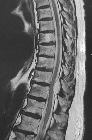
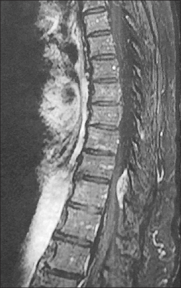
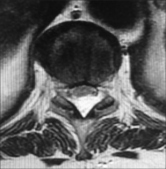
Sagittal magnetic resonance imaging (MRI) revealed a hyperintense dorsal epidural lesion (on T1 and T2-weighted images) at the T9-T10 levels, which homogeneously enhanced with intravenous gadolinium contrast. There was mild/moderate compression of the spinal cord without bony involvement or invasion [Figures
Surgery
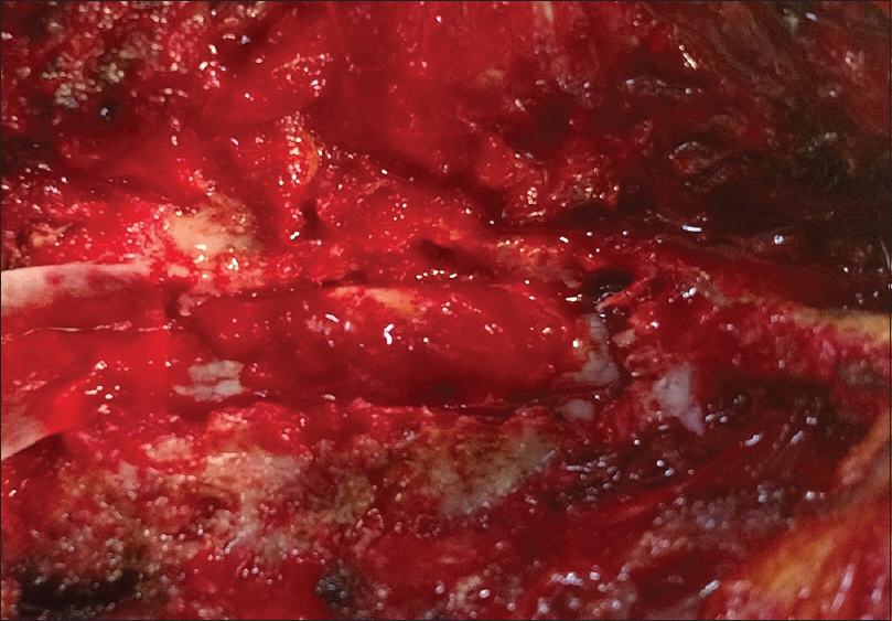
Under fluoroscopic guidance a T9-T11 laminectomy revealed a reddish highly vascularized soft lesion easily dissected away from the dura; a total en bloc excision was achieved without a dural fistula [
Histopathology
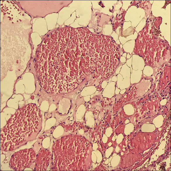
The sample on gross inspection revealed a benign mesenchymal neoplasm with blood vessel differentiation [
DISCUSSION
Capillary hemangiomas are ubiquitous hamartomatous malformations resulting from the proliferation of vascular endothelial cells. Their histopathological characteristics include thin irregular capillary-sized vessels captured in fibrous low attenuating, lobular architecture, and the presence of a continuous basal lamina coating.[
CONCLUSION
Pure epidural capillary hemangiomas are extremely rare lesions but should be considered in the differential diagnosis of highly vascular spinal epidural lesions. They must be differentiated from other more common vascular lesions. Gross total surgical excision is the procedure of choice to avoid recurrence and perioperative hemorrhagic complications.
Financial support and sponsorship
No funding was received for this research.
Conflicts of interest
All authors certify that they have no affiliations with or involvement in any organization or entity with any financial interest, or non-financial interest in the subject matter or materials discussed in this manuscript.
References
1. Alakandy LM, Hercules S, Balamurali G, Reid H, Herwadkar A, Holland JP. Thoracic intradural extramedullary capillary haemangioma. Br J Neurosurg. 2006. 20: 235-8
2. Babu R, Owens TR, Karikari IO, Moreno J, Cummings TJ, Gottfried ON. Spinal cavernous and capillary hemangiomas in adults. Spine (Phila Pa 1976). 2013. 38: E423-30
3. Badinand B, Morel C, Kopp N, Tran Min VA, Cotton F. Dumbbell-shaped epidural capillary hemangioma. AJNR Am J Neuroradiol. 2003. 24: 190-2
4. Egu K, Mounadi M, Maaqili MR El, Abbadi N El. Hémangiome capillaire épidural lombosacré mimant un neurinome en sablier: à propos d’un cas et revue de la littérature. Neurochirurgie. 2015. 62: 113-7
5. Gencpinar P, Acıkbas SC, Nur BG, Karaali K, Arslan M, Gurer EI. Epidural capillary hemangioma: A review of the literature. Clin Neurol Neurosurg. 2014. 126: 99-102
6. Hasan A, Guiot MC, Torres C, Marcoux J. A case of a spinal epidural capillary hemangioma: Case report. Neurosurgery. 2011. 68: E850-3
7. Rajeev MP, Waykule PY, Pavitharan VM, Nandeesh BN. Spinal epidural capillary hemangioma: A rare case report with a review of literature. Surg Neurol Int. 2017. 8: 123-
8. Seferi A, Alimehmeti R, Vyshka G, Bushati T, Petrela M. Case study of a spinal epidural capillary hemangioma: A 4-year postoperative follow-up. Glob Spine J. 2014. 4: 55-8

