

Modified pure endoscopic approach (MAPEnd) in neurosurgery
Date of publication: 15-Jan-2019

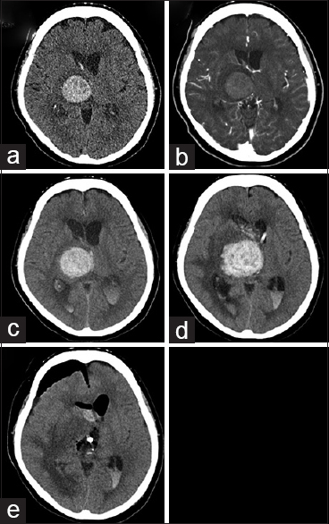
A case of metastatic brain tumor mimicking an expanding thalamic hematoma
Date of publication: 15-Jan-2019
Background:Brain tumor are a major etiology of secondary intracranial hemorrhage (ICH) because ICH in patients with cancer often occurs from an intratumoral hemorrhage. However, it is sometimes difficult to detect a tumor when it is tiny and buried, especially during initial examination.

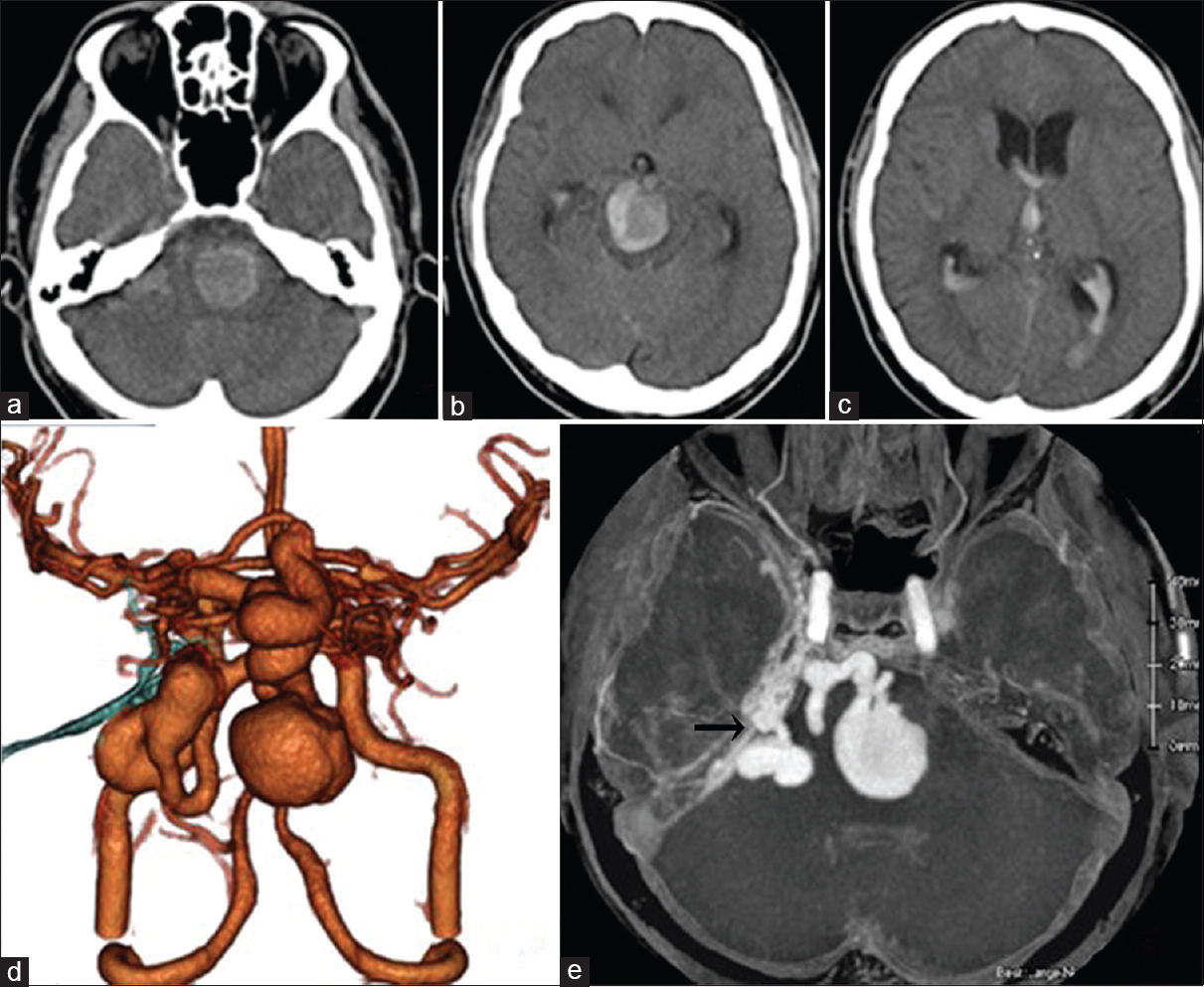
Successful treatment of a case of tentorial dural arteriovenous fistula causing subarachnoid hemorrhage with invagination of the brainstem by huge and multiple venous pouches
Date of publication: 15-Jan-2019
Background:We present a case of tentorial dural arteriovenous fistula (TDAVF) causing subarachnoid hemorrhage with mass effect of large venous pouches, which was struggling to diagnosis and management due to complex vasculature and severe general condition.
A case of metastatic brain tumor mimicking an expanding thalamic hematoma
Date of publication: 15-Jan-2019
Background:Brain tumor are a major etiology of secondary intracranial hemorrhage (ICH) because ICH in patients with cancer often occurs from an intratumoral hemorrhage. However, it is sometimes difficult to detect a tumor when it is tiny and buried, especially during initial examination.
The American Association of Neurological Surgeons (AANS) Suspends Surgeon for Arguing Against Unnecessarily Extensive Spine Surgery; Was this Appropriate?
Date of publication: 27-Dec-2018
Does the American Association of Neurological Surgeons seek to limit members from testifying for patients/plaintiffs through proceedings resembling a kangaroo court and/or star chamber?
Date of publication: 27-Dec-2018
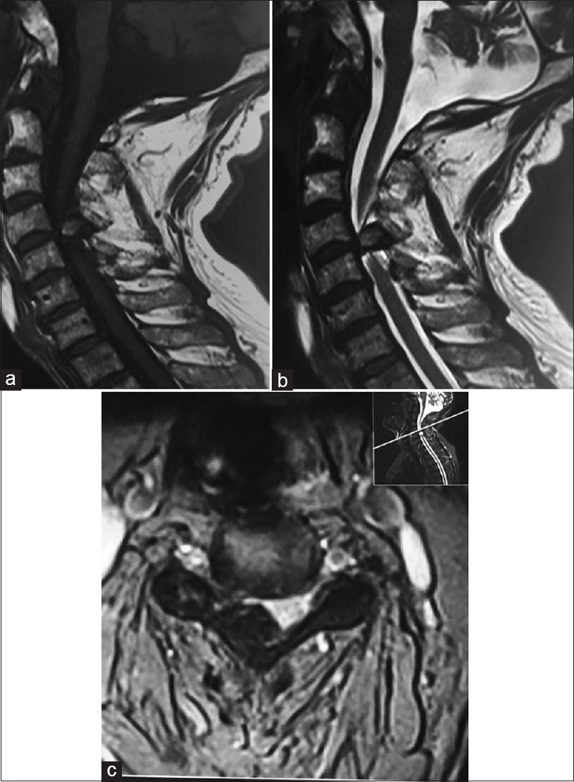
Ossification of the cervical ligamentum flavum and case report with myelopathy
Date of publication: 24-Dec-2018
Background:Ossification of the ligamentum flavum (OLF) occurs mostly in adult males, typically in the thoracolumbar spine where it may contribute to neurological deficits. Here we reviewed 68 cases of cervical OLF resulting in progressive quadriparesis.
Unedited microneurosurgery of a mixed germ cell tumor of the pineal region
Date of publication: 24-Dec-2018
Background:Germ cell tumors comprise a heterogeneous group of neoplasms, classified as germinomas and nongerminomatous germ cell tumors based on clinicopathological features. The nongerminomatous group of tumors includes embryonal carcinoma, endodermal sinus tumor (yolk sac tumor), choriocarcinoma, mature and immature teratoma, and mixed germ cell tumors with more than one element. While germinomas are radiation-sensitive tumors, all other tumors have less response to radiotherapy, and it is suggested that gross total resection improves their overall survival and tumor-free survival rates. Herein, we present the microsurgical management of a histologically confirmed mixed-germ cell of the pineal region.
Unedited microneurosurgery of a pineal region ependymoma
Date of publication: 24-Dec-2018
Background:Ependymomas are rarely located in the pineal region. The 2016 WHO classification of tumors of the central nervous system includes five ependymal tumors, the grade I subependymoma and mixopapillary ependymoma, the grade II ependymoma, the grade II–III ependymoma RELA fusion-positive, and the grade III anaplastic ependymoma. However, this grading system has been controversial with respect to its reproducibility and clinical significance and it is estimated that further studies of the molecular characteristics of ependymoma will provide more precise and objective classification. Herein, we present an unedited microneurosurgery of a gross total removed WHO grade II ependymoma.

