

Unedited microneurosurgery of a fourth ventricular ependymoma
Date of publication: 10-Sep-2018
Background:In this video abstract, we present an unedited microsurgical resection of a fourth ventricular ependymoma performed by a senior author (JH). Currently, the goal of a standard treatment of a fourth ventricular ependymoma is based on microsurgical resection followed by radiochemotherapy. Our aim is to demonstrate the efficiency and safety of our microsurgical technique in deep brain territories under the principle “simple, clean, and preserving the normal anatomy.” For this, a midline suboccipital approach and a proper praying sitting position are essential.
Anterior clinoidectomy for paraclinoid aneurysms in Helsinki Neurosurgery
Date of publication: 10-Sep-2018
Background:In this video abstract, we present an intradural anterior clinoidectomy for management of some paraclinoid aneurysms. Quick adenosine cardiac arrest performed instead of an anterior clinoidectomy and proximal temporary clipping usually allows us a proximal control of aneurysms in Helsinki Neurosurgery. However, when the neck of the aneurysm remains hidden under the anterior clinoid process, or when some complex aneurysms have reduced space for placing temporary clips obstructing the definitive clipping, anterior clinoidectomy is the most available option.
Focused opening of the Sylvian fissure for the management of middle cerebral artery aneurysms
Date of publication: 10-Sep-2018
Background:A wide opening of the Sylvian fissure (SF) regarding the treatment of middle cerebral artery (MCA) aneurysm allows us to ensure early proximal control by the proximal start of Sylvian dissection and enough comfort for the microsurgical manipulation and aneurysm clipping. However, major mechanical manipulation of arteries associated with blood oozing into the surgical field may increase the incidence of postoperative vasospasm. The risk of Sylvian venous injury is bigger, and the damage of the superior temporal gyrus increases the risk of postoperative epilepsy as well. A focused opening of the SF based on 18 years experience of a senior author is an alternative technique we present in this video abstract.
One burr-hole craniotomy: Posterior interhemispheric approach in Helsinki Neurosurgery
Date of publication: 10-Sep-2018
Background:In this video abstract, we present a one burr-hole craniotomy for the posterior interhemispheric approach developed in Helsinki Neurosurgery to access posteriorly the medial surface of cerebral hemispheres, falx cerebri, and deep midline cerebrovascular structures. Therefore, preoperative imaging is essential to achieve an optimal operative corridor for a safest and more effcient approach.
One burr-hole craniotomy: Modified presigmoid approach in Helsinki Neurosurgery
Date of publication: 10-Sep-2018
Background:In this video abstract, we present a one burr-hole craniotomy for a modified presigmoid approach developed in Helsinki Neurosurgery to access the space extended to both middle and posterior fossa. Thus, indications for this approach are lesions that extend to both middle and posterior fossa, petroclival tumors, basilar tip aneurysms located extremely low below the posterior clinoid process, trunk basilar aneurysms, and bypass procedures from the P2 segment of the posterior cerebral artery. The procedure is composed by three stages: a temporal and presigmoid craniotomy, a partial petromastoidectomy, and the dura opening with section of the superior petrosal sinus (SPS) and the tentorium. Even though some risks related to the opening of the mastoid cells or cut of the SPS may exist, benefits of this optimized craniotomy are higher compared with the complications.
Seizure freedom from temporal lobe epilepsy with mesial temporal lobe tumor by tumor removal alone without hippocampectomy despite remaining abnormal discharges on intraoperative electrocorticography: Report of two pediatric cases and reconsideration of the surgical strategy
Date of publication: 10-Sep-2018
Background:In the surgical treatment of temporal lobe epilepsy with mesial temporal lobe tumor, whether to remove the hippocampus aiming for a better seizure outcome in addition to removing the tumor is a dilemma. Two pediatric cases treated successfully with tumor removal alone are presented.
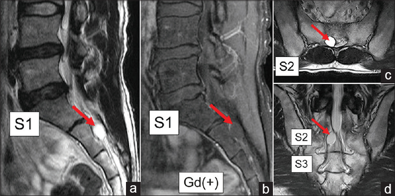
Resection and imbrication of symptomatic sacral Tarlov cysts: A case report and review of the literature
Date of publication: 04-Sep-2018
Background:Symptomatic Tarlov cysts are extremely rare, and there is no consensus regarding their optimal surgical management. Here, we encountered a patient with a symptomatic sacral Tarlov cyst and reviewed the appropriate literature.
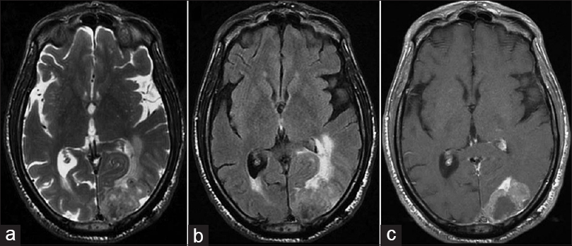
Prognostic factors in brain metastases from laryngeal squamous cell carcinoma: Case report and review
Date of publication: 04-Sep-2018
Background:Brain metastases from laryngeal squamous cell carcinoma (SCC) are rare, and there are no standardized treatments. Here we reported on a case of brain metastasis from laryngeal SCC and performed a literature review on these cases. Moreover, by plotting Kaplan–Meier curves, we carried out a survival analysis to provide an estimation of overall survival (OS) and to find possible prognostic factors.
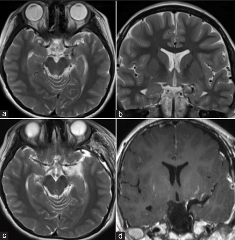
Rare case of meningeal tuberculoma mimicking meningioma in term pregnancy and its management
Date of publication: 03-Sep-2018
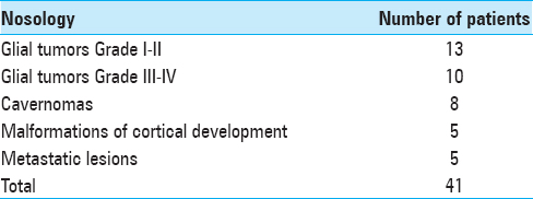
Awake craniotomy without sedation in treatment of patients with lesional epilepsy
Date of publication: 03-Sep-2018
Background:The use of awake craniotomy for surgical treatment of epilepsy was applied in surgery of convexital tumors, arteriovenous malformations, some superficial aneurysms, and stereotactic neurosurgery. The aim of this study was to show the advantages of awake craniotomy without sedation, accompanied by intraoperative neurophysiological monitoring in patients with symptomatic epilepsy.

