

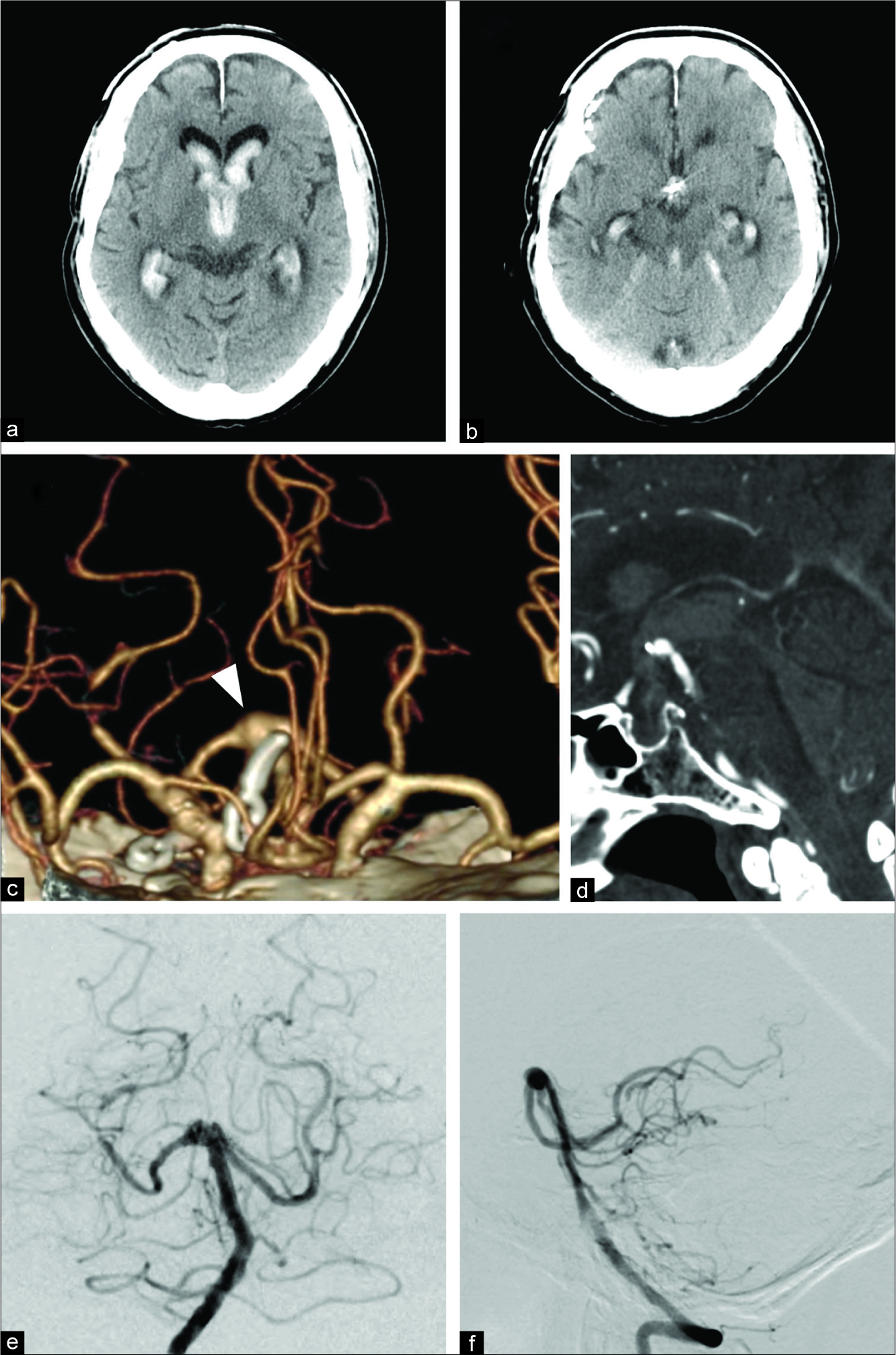
Trans-lamina terminalis approach assisted by endovascular temporary basilar artery occlusion for high-positioned, recurrent, basilar tip aneurysm: A technical case report
Date of publication: 24-Jan-2020
Background: Coil embolization is increasingly becoming the surgical intervention of choice for cerebral aneurysms, particularly for those in the posterior circulation. However, in cases where it is difficult to perform coil embolization, microsurgical clipping is still required.

A novel temporary cranial fixation device for awake cranial surgery: Technical report of 14 cases
Date of publication: 24-Jan-2020
Background: Awake craniotomy has become the gold standard in various cranial procedures. As part of the awake technique, three-point pin fixation of the patient’s head is important. One of the issues we encountered is the problem of matching the scalp infiltration site with the final pin position. To overcome this problem, we developed a flat plunger type fixator that adapts to the Mayfield holder.
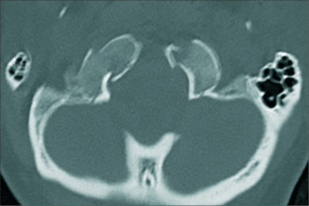
Contemporaneous avulsion fractures of the inferior clivus and bilateral occipital condyles with injury of the tectorial membrane
Date of publication: 17-Jan-2020
Background: Craniocervical junction (CCJ) injuries are highly variable, and there are only limited data to suggest their optimal treatment. Here, we present a rare case of concomitant bilateral occipital condyle fractures with an inferior clival avulsion fracture with concomitant focal injury of the tectorial membrane.
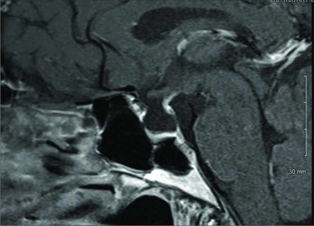
Orbitofrontal approach for the fenestration of a symptomatic sellar arachnoid cyst
Date of publication: 17-Jan-2020
Background: Sellar arachnoid cysts (SACs) are rare lesions and incidentally found on brain imaging. The pathophysiology is poorly understood. Some authors suggested that SACs develop as a herniation of arachnoid membrane through the diaphragma sellae followed by cyst formation. Furthermore, Meyer et al. postulated that SACs are formed by splitting of the arachnoid layers. Symptomatic SACs present with headache, visual field deficit, or pituitary dysfunction. The data are limited on the indications and timing for intervention. We present a case of symptomatic SAC that was fenestrated using orbitofrontal approach.
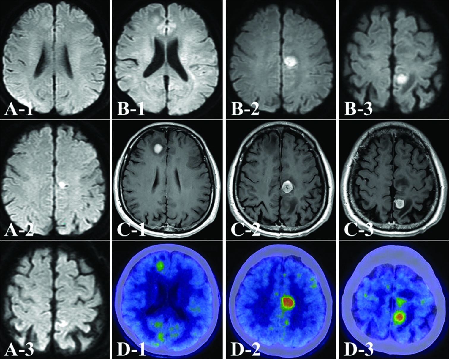
Epithelioid glioblastoma presenting as multicentric glioma: A case report and review of the literature
Date of publication: 17-Jan-2020
Background: Epithelioid glioblastoma is a rare aggressive variant of glioblastoma multiforme (GBM), which was formally recognized by the World Health Organization classification of the central nervous system in 2016. Clinically, epithelioid GBMs are characterized by aggressive features, such as metastases and cerebrospinal fluid dissemination, and an extremely poor prognosis. A rare case of epithelioid GBM that was discovered as a multicentric glioma with different histopathology is reported.
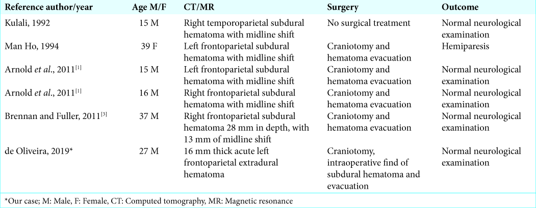
Rare acute idiopathic subdural hematoma: A case report and literature review
Date of publication: 17-Jan-2020
Background: Acute spontaneous subdural hematoma is rare. For patients under 40 years of age, we found only five previous reports. Here, we have presented a sixth case study.
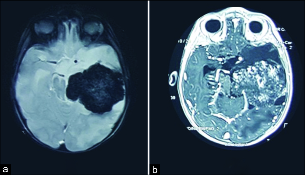
Rare case of giant pediatric cavernous angioma of the temporal lobe: A case report and review of the literature
Date of publication: 10-Jan-2020
Background: Giant cavernous malformations of the central nervous system are quite rare. They are more common in children and may be misdiagnosed as other intracranial neoplasms. Here, we presented a very rare giant cavernous angioma mimicking a neoplastic temporal lobe lesion in an 18-month-old male.
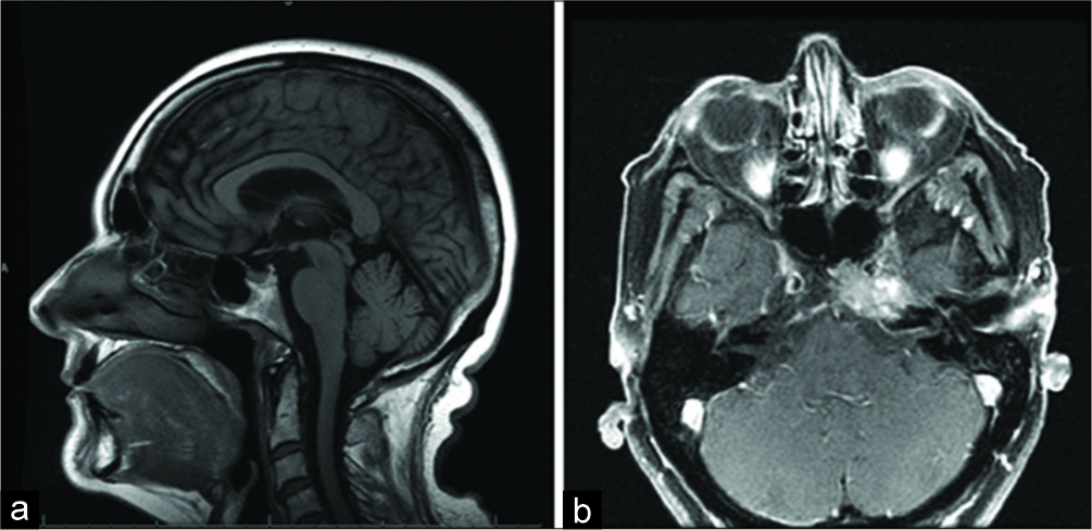
Primary Ewing’s sarcoma of the petroclival bone: A case report and literature review
Date of publication: 10-Jan-2020
Background: Primary Ewing’s sarcoma (ES) is typically seen within the long bones, vertebrae, or pelvis. Uncommonly, it can be found within the cranium among the rarest locations for primary ES are the skull base, in particular, the petroclival bone.
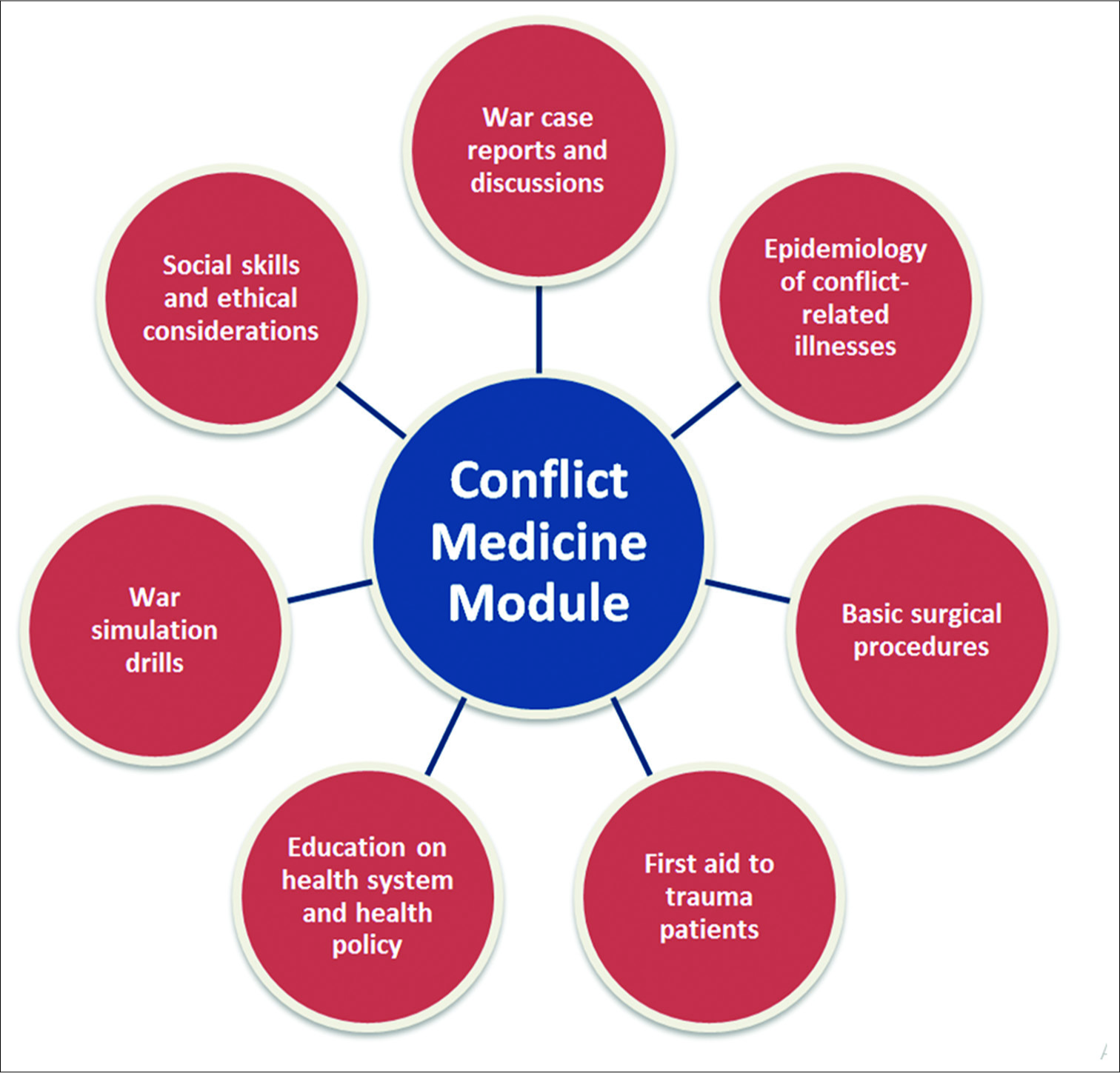
Medical schools in times of war: Integrating conflict medicine in medical education
Date of publication: 03-Jan-2020
Abstract
Amid the rise in conflict and war and their ensuing repercussions, traumatic injuries, psychological distress, and communicable diseases spread widely. Today, health-care providers in the Middle East are faced with new and unfamiliar cases resulting from the use of new and advanced types of weapons. In addition, there has not been enough emphasis on hands-on experiences in medical school, which can be imperative in times of war. Lack of academia is another inadequacy that limits the transmission of knowledge onto the newer generations. Here, we will shed light on the inadequacies in medical curricula in the Middle East when it comes to addressing patients of war. We also call for action to advance medical education in war-ridden areas by incorporating “conflict medicine” as an integral module in medical curricula.
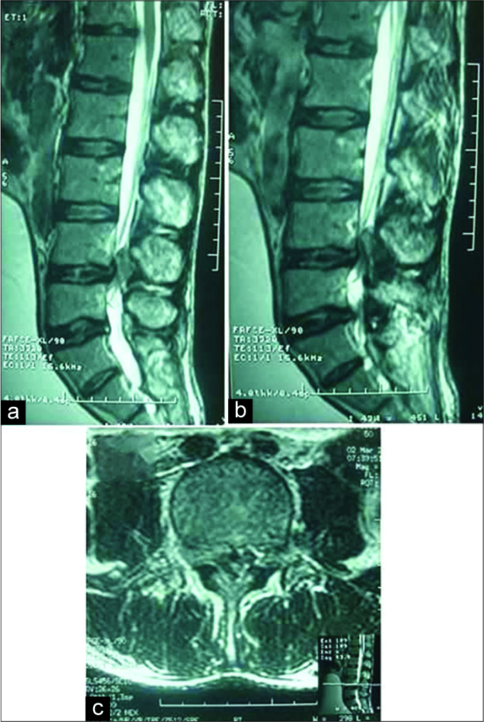
Posterior epidural migration of a lumbar disc herniation
Date of publication: 03-Jan-2020
Background: Posterior epidural migration of a lumbar disc fragment (PEMLDF) refers to the dorsal migration of disc material around the thecal sac that can lead to radiculopathy and/or cause a cauda equina syndrome. It is rare and the diagnosis is often just established intraoperatively.


