

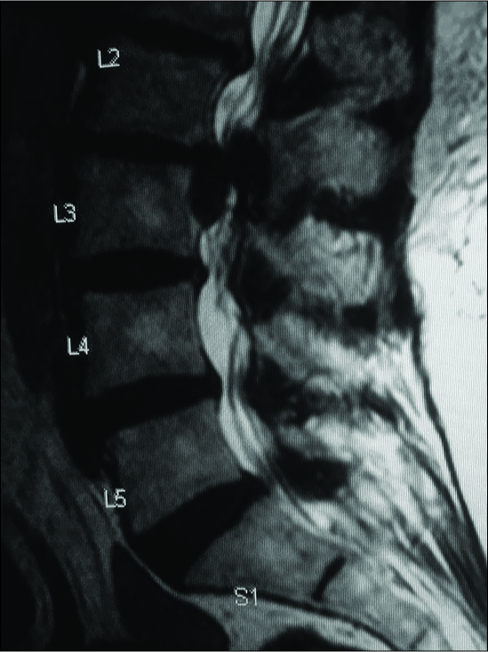
Case of the Week: Preoperative MR/CT Diagnosis of Left L2-L3 Disc Surgically Documented As Massive Synovial Cyst
Date of publication: 30-Aug-2019
Background: The diagnosis of a lumbar herniated disc, stenosis, and other degenerative findings are typically established preoperatively with MR scans, supplemented with non-contrast CT studies. Here, a 77-year-old female, diagnosed as having L2-S1 stenosis and a large left-sided L2-L3 herniated disc was found at surgery to have a massive left-sided L2-L3 synovial cyst.
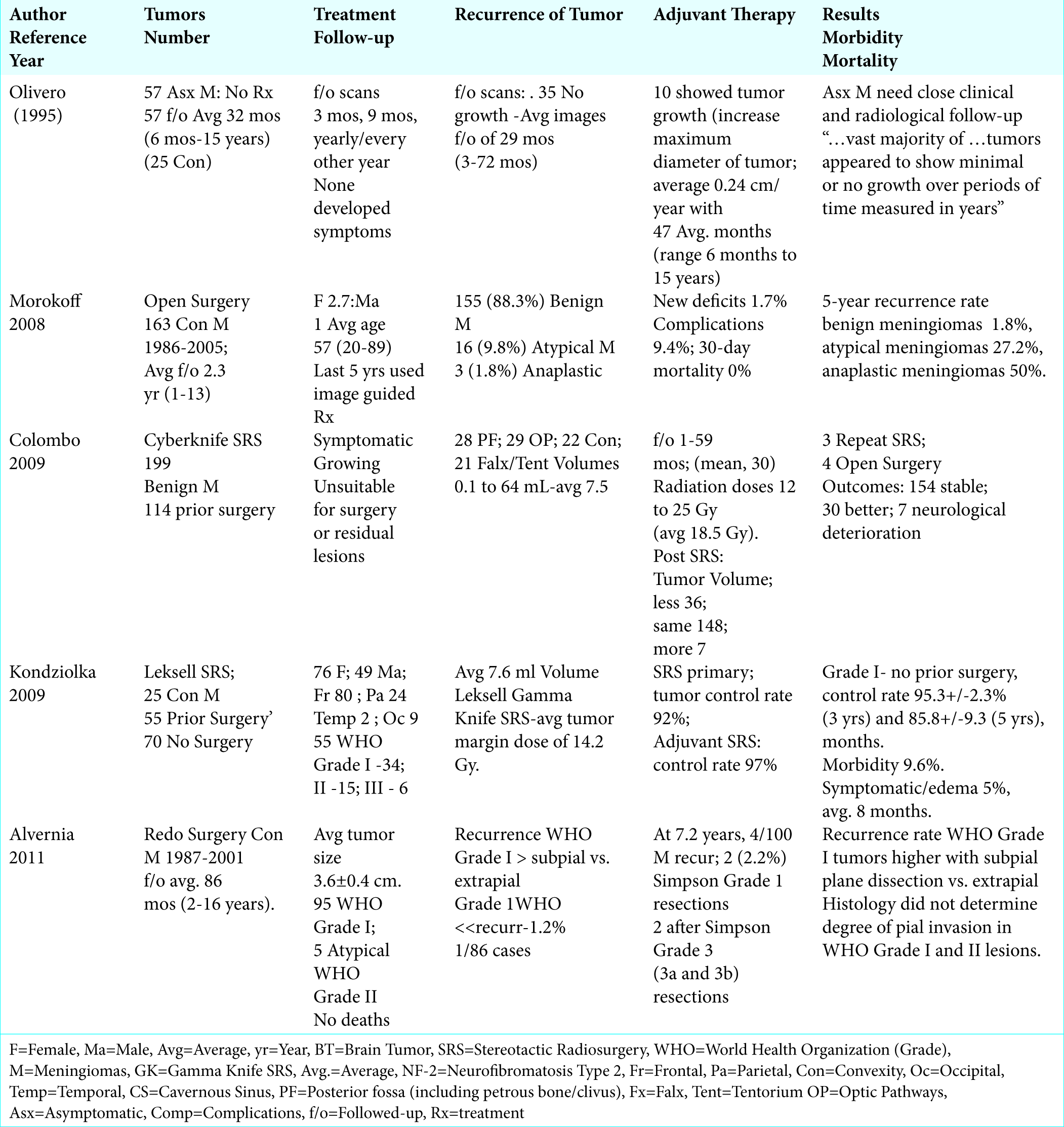
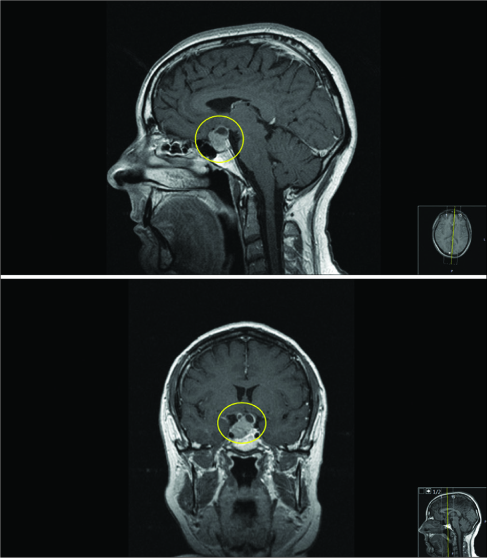
Review of Treatment Options for Smaller Benign Cranial Meningiomas: Observation, Stereotactic Radiosurgery, and Rarely, Open Surgery
Date of publication: 23-Aug-2019
Background: MR/CT documented smaller cranial meningiomas in asymptomatic patients are often followed for years without requiring any intervention. Only a subset of patients who become symptomatic attributed to significant tumor growth, edema and/or mass effect may require stereotactic radiosurgery (SRS), and rarely, open surgery. Clearly, the decision for choosing any treatment modality must be made on a case by case basis and include an analysis of risks vs. benefits to the individual patient.
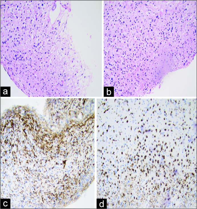
A challenging case of concurrent multiple sclerosis and anaplastic astrocytoma
Date of publication: 23-Aug-2019
Background: Cases of gliomas coexisting with multiple sclerosis (MS) have been described over the past few decades. However, due to the complex clinical and radiological traits inherent to both entities, this concurrent phenomenon remains difficult to diagnose. Much has been debated about whether this coexistence is incidental or mirrors a poorly understood neoplastic phenomenon engaging glial cells in the regions of demyelination.
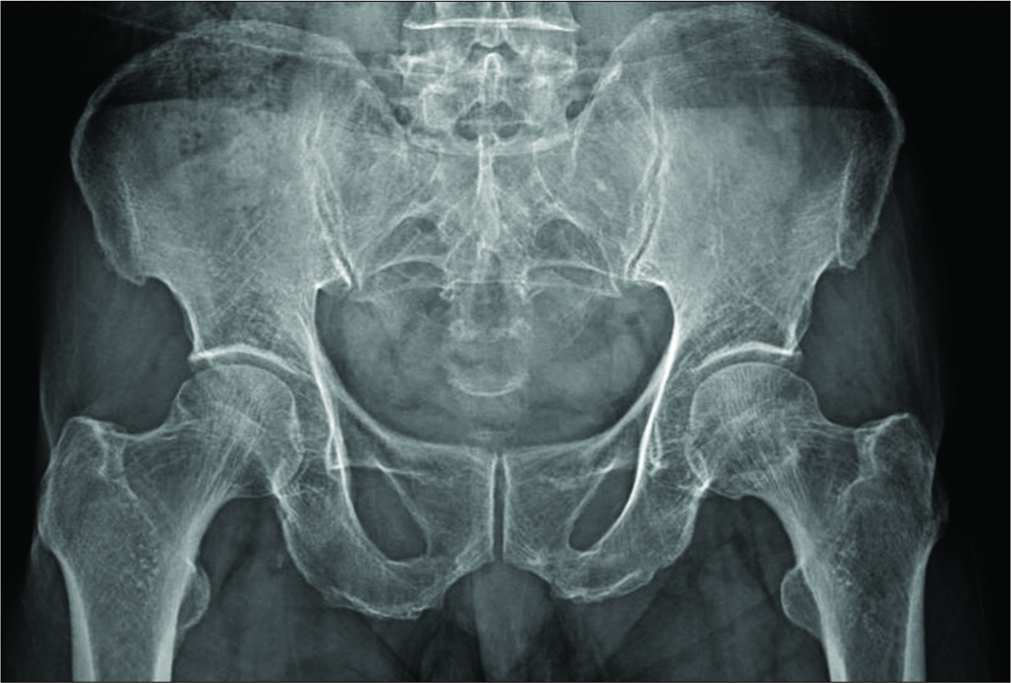
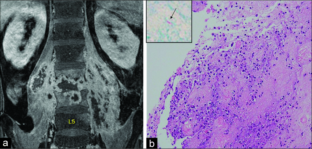
X-rays and scans can fail to differentiate hip pathology from lumbar spinal stenosis: Two case reports
Date of publication: 23-Aug-2019
Background: Occasionally, hip pathologies may present alone or combined with lumbar spine pathology, especially lumbar stenosis. Although the history and clinical examination may help differentiate between the two, hip X-rays alone without accompanying magnetic resonance imaging (MRI) studies may prove unreliable.
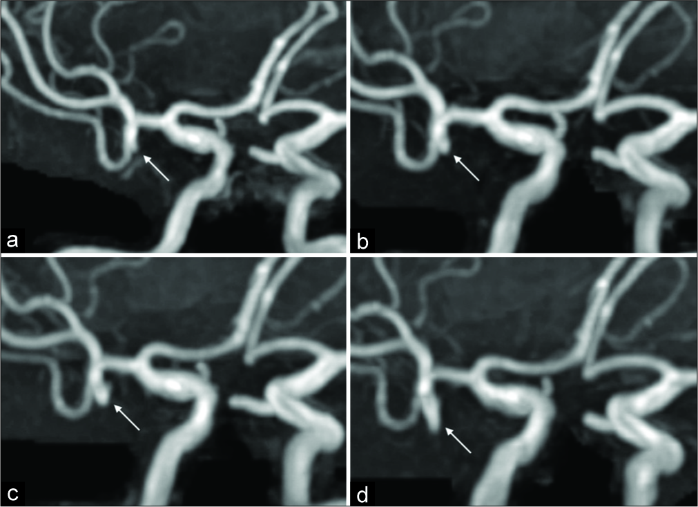
Rupture immediately after growth of unruptured intracranial aneurysms during follow-up
Date of publication: 23-Aug-2019
Background: The annual rupture rate of small unruptured intracranial aneurysms (UIAs) <5 mm is generally low; further, small UIAs are often treated conservatively. While the growth of aneurysms during follow-up is associated with a high risk of rupture, the urgency for surgical treatment of asymptomatic enlarged UIAs remains controversial. We experienced two patients in whom UIAs ruptured shortly after asymptomatic growth during follow-up.
Professional Medical Associations Exert Undue Influence Despite Conflicts of Interest
Date of publication: 09-Aug-2019
A surgical case of cerebellar tuberculoma caused by a paradoxical reaction while on therapy for tuberculosis spondylitis
Date of publication: 09-Aug-2019
Background: A paradoxical reaction (PR) is a phenomenon in which the primary tuberculous lesion worsens or another de novo tuberculous lesion appears while on anti-tuberculosis therapy. Here, we report a rare case of cerebellar tuberculoma caused by a PR during therapy for lumbar tuberculous spondylitis (Pott’s disease).
Pituitary carcinomas: Rare and challenging
Date of publication: 09-Aug-2019
Background: Pituitary carcinomas (PCs) are defined as adenohypophyseal tumors with metastatic activity within and outside the boundaries of the central nervous system (CNS). The condition is rare and therefore seldom reported; most lesions are hormone producing and have a tendency for complex evolution. As such, the management of PCs remains difficult. We present an illustrative case of PC with a brief review of the recent medical literature.
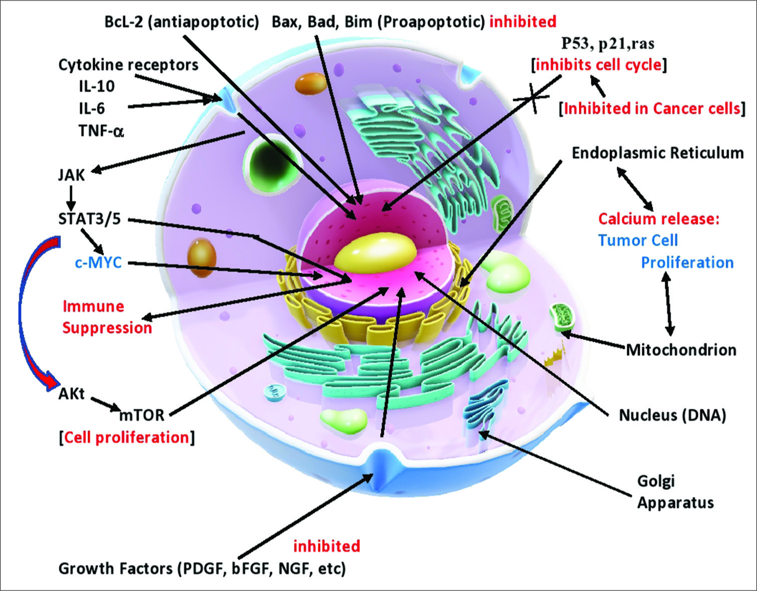
Viruses and tumor cell microenvironment: A brief summary
Date of publication: 09-Aug-2019
Abstract
An infectious etiology for a number of cancers has been entertained for over 100 years and modern studies have confirmed that a number of viruses are linked to cancer induction. While a large number of viruses have been demonstrated in a number of types of cancers, most such findings have been dismissed in the past as opportunistic infections, especially with persistent viruses with high rates of infectivity of the world’s populations. More recent studies have clearly shown that while not definitely causing these cancers, these viruses appear capable of affecting the biology of these tumors in such a way as to make them more aggressive and more resistant to conventional treatments. The term oncomodulatory viruses have been used to describe this phenomenon. A number of recent studies have shown a growing number of ways; these oncomodulatory viruses can alter the pathology of these tumors by affecting cell signaling, cell metabolism, apoptosis mechanisms, cell-cell communication, inflammation, antitumor immunity suppression, and angiogenesis. We are also learning that much of the behavior of tumors depends on cancer stem cells and stromal cells within the tumor microenvironment, which participate in extensive, dynamic crosstalk known to affect tumor behavior. Cancer stem cells have been found to be particularly susceptible to infection by human cytomegaloviruses. In a number of studies, it has been shown that while only a select number of cells are actually infected with the virus, numerous viral proteins are released into the cancer and stromal cells in the microenvironment and these viral proteins are known to affect tumor behavior and aggressiveness.
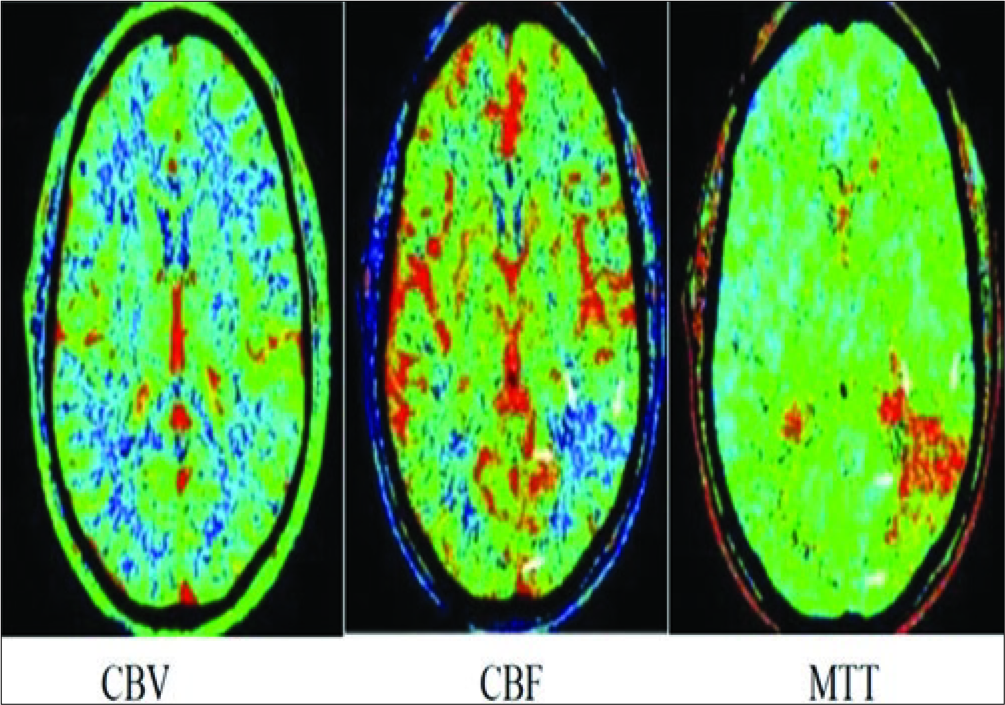
Computed tomography perfusion in detecting malignant middle cerebral artery infarct
Date of publication: 09-Aug-2019
Background: Computed tomography perfusion (CTP) is an emerging modality which produces maps of time-to- peak (TTP), cerebral blood flow (CBF), and cerebral blood volume (CBV), with a computerized automated map of the infarct and penumbra. This modality provides a better evaluation of the extent of infarction, making it a potential method for assessing patients suffering from large middle cerebral artery (MCA) infarctions.

