

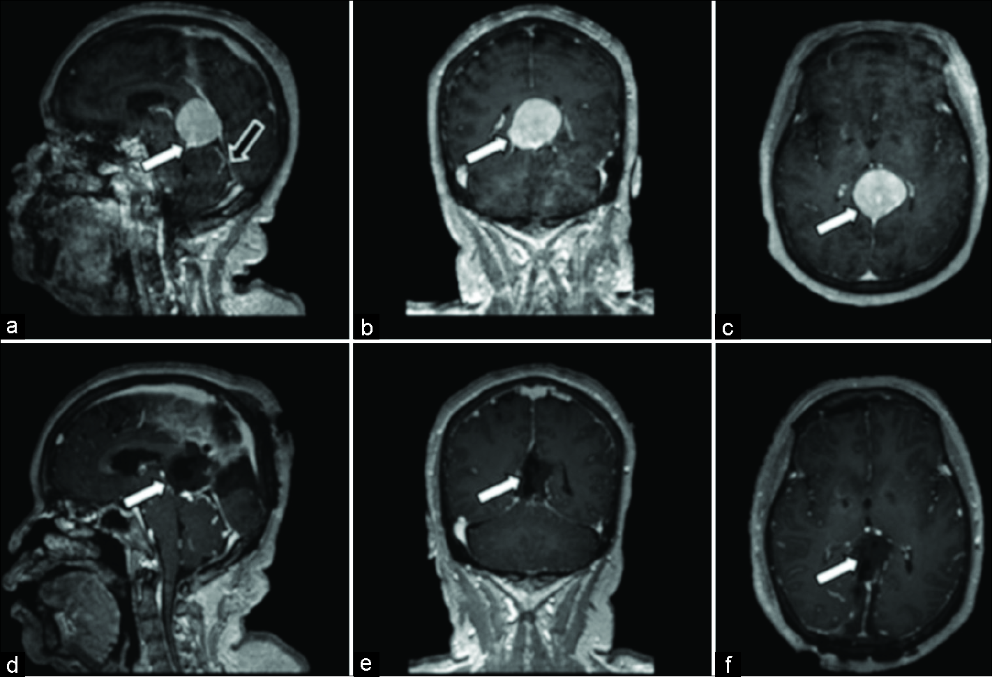
Cranial migration of lumboperitoneal shunt: A case report and review of literature
Date of publication: 28-Jun-2019
Background: Lumboperitoneal shunt is an easy and effective way of managing benign intracranial hypertension (BIH) and other causes of increased intracranial pressure. Yet, it is associated with a relative high failure rate. Proximal migration of the shunt is rare and only few cases have been reported.
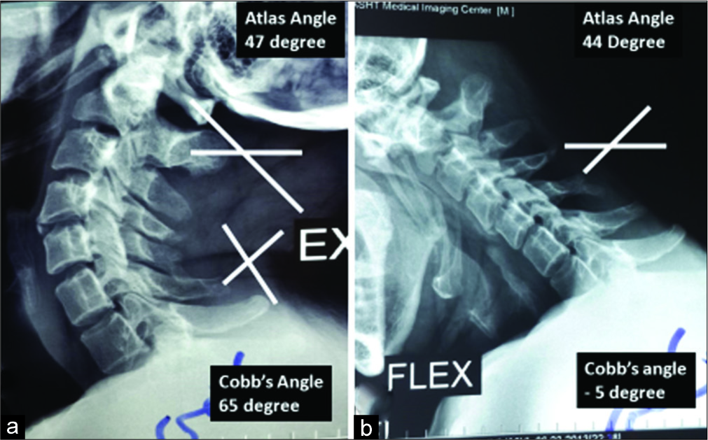
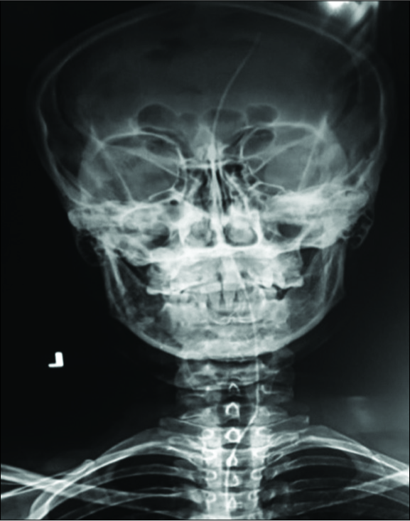
Unstable os odontoideum contributing to cervical myelopathy and obstructive sleep apnea
Date of publication: 28-Jun-2019
Background: Sleep apnea is characterized by repetitive cessation of breathing during sleep. It may be attributed to obstructive, central, or mixed pathologies close to the upper airway resulting in a decreased diameter of the oropharyngeal tract.
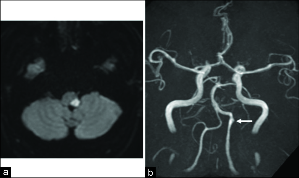
A case of unilateral vertebral artery dissection progressing in a short time period to bilateral vertebral artery dissection
Date of publication: 28-Jun-2019
Background: Vertebral artery dissection (VAD) is an important cause of stroke in young and middle- aged people. Bilateral occurrence of VAD is generally considered rare, but the number of reports of bilateral VAD has been increasing in recent years. In this paper, we report a case of de novo VAD on the contralateral side presenting with subarachnoid hemorrhage in the acute stage of cerebral infarction due to unilateral VAD.
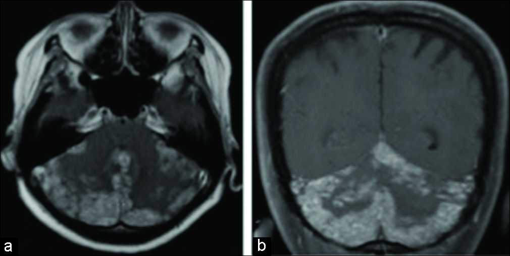
A case of leptomeningeal metastases of human epidermal growth factor receptor 2-positive breast cancer that responded well to lapatinib plus capecitabine
Date of publication: 28-Jun-2019
Background: Leptomeningeal metastases (LM) pose the most difficult form of cancer metastasis to treat and portend a poor prognosis. Standard treatment has yet to be established, and intrathecal chemotherapy and whole- brain radiotherapy are administered on an empirical basis.
Retractorless interhemispheric transtentorial approach for large lesions in the posterior incisural space
Date of publication: 28-Jun-2019
Background: Surgical resection of lesions in the posterior incisural space presents a significant surgical challenge, which may result in postoperative visual complications and other neurological deficits. We, therefore, describe a retractorless interhemispheric transtentorial approach that avoids surrounding brain structures with positive outcomes and no complications or visual damage.
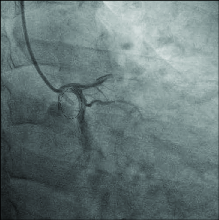
Catheter fragment retrieved from an arterial branch of the right middle cerebral artery
Date of publication: 28-Jun-2019
Background: Cerebral emboli is a rare complication of endovascular procedures and foreign bodies in the cerebrovascular system can lead to stroke. When an intravascular foreign body is identified, endovascular retrieval should be attempted due to its high success rate and minimal morbidity.
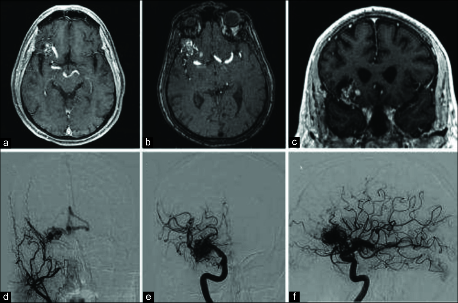
Oculomotor neuropathy from an unruptured arteriovenous malformation in the frontal operculum: A case report
Date of publication: 28-Jun-2019
Background: Cerebral arteriovenous malformations (AVMs) are vascular lesions with a network of dysplastic vessels between an arterial and a venous tree with no intervening capillary bed. They most commonly present with an acute hemorrhage, seizures, or persistent headaches.
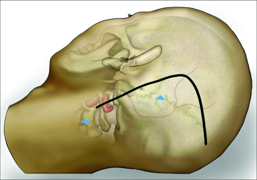
Surgical microanatomy of the occipital artery for suboccipital muscle dissection and intracranial artery reconstruction
Date of publication: 28-Jun-2019
Background: The occipital artery (OA) is an important donor artery for posterior fossa revascularization. Harvesting the OA is difficult in comparison to the superficial temporal artery because the OA runs between suboccipital muscles. Anatomical knowledge of the suboccipital muscles and OA is essential for harvesting the OA during elevation of the splenius capitis muscle (SPL) for reconstruction of the posterior inferior cerebellar artery. We analyzed the running pattern of the OA and its anatomic variations using preoperative and intraoperative findings.
Based upon 7.2% of the Eligible Voting Members, the American Association of Neurological Surgeons (AANS) Suspended Dr. Nancy E. Epstein for Arguing Against Unnecessarily Extensive Spine Surgery
Date of publication: 28-Jun-2019
Vein of Galen arteriovenous malformation: Unedited microneurosurgery
Date of publication: 25-Jun-2019
Background: Vein of Galen arteriovenous malformations (VGAVMs) are vascular malformations of the pineal region between a persistent embryological median prosencephalic vein of Markowski and the arterial choroidal system by a direct (mural type) or indirect (choroidal type) communication. Angiographic evaluation of VGAVMs usually describes a limbic arch between the anterior and posterior cerebral arteries throughout a pericallosal artery, and the classic “ε” shape configuration of the thalamostriate veins drainage into a subtemporal or a lateral mesencephalic vein due to the underdevelopment of the straight sinus, sigmoid sinus, and jugular bulbs. Moreover, falcine dural channels join the pouch of the malformation with the posterior third of the superior sagittal sinus and less frequently with the cavernous sinus, inferior petrosal sinus, and facial veins. At present, endovascular therapy is the standard management for these lesions. However, under failure of endovascular procedures such in this case, microsurgical management of VGAVMs under an experienced neurosurgical team might be paramount.

