

Comparing deep brain stimulation in the ventral intermediate nucleus versus the posterior subthalamic area in essential tremor patients
Date of publication: 04-Dec-2018
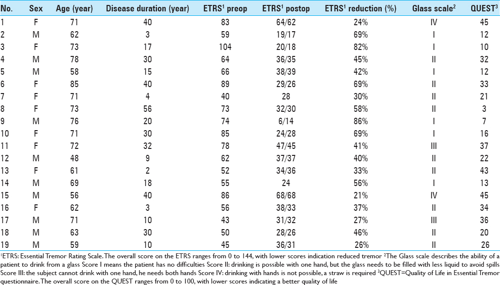
Background:The ventral intermediate nucleus (VIM) is the most commonly used target for deep brain stimulation (DBS) in patients with essential tremor (ET). Recent evidence suggests that the posterior subthalamic area (PSA) might be a better target for tremor reduction. We compared the outcome of VIM DBS with PSA DBS in our cohort of patients.
Primary meningeal melanocytoma of the cerebellopontine angle associated with ipsilateral nevus of Ota: A case report
Date of publication: 04-Dec-2018
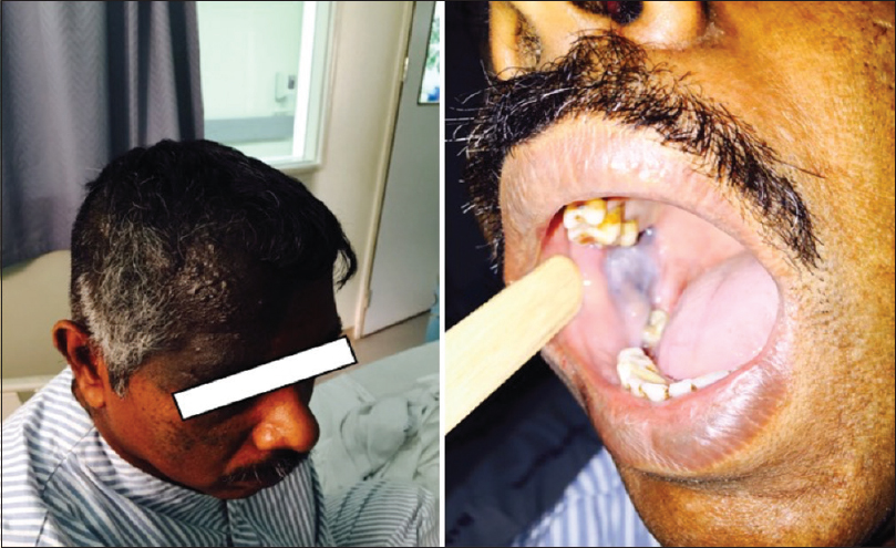
Background:Cerebellopontine angle represents a complex anatomical area of the brain. A cerebellopontine angle lesion could be a vestibular schwannoma, meningioma, epidermoid cyst, or less likely, arachnoid cyst, metastasis, lower cranial nerves schwannoma, lipoma, hemangioma, paraganglioma, or vertebra-basilar dolichoectasia. Primary meningeal melanocytoma is a rare neoplasm, especially when it occurs at the cerebellopontine angle. Nevus of Ota (aka oculodermal melanocytosis) is a hyperpigmentation along the distribution of the ophthalmic and maxillary branches of trigeminal nerve; it occurs due to entrapment of melanocytes at the upper third of the dermis. It may not present at birth and may show up at puberty.
Frontoethmoidal encephalocele presenting in concert with schizencephaly
Date of publication: 04-Dec-2018
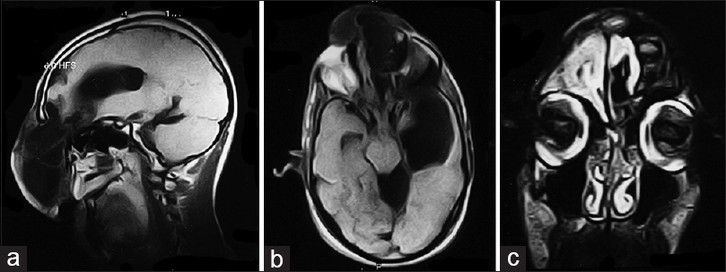
Background:Schizencephaly is a rare defect which is identified as clefts that are lined with grey matter extending from the ependyma of the cerebral ventricles to the pia mater. An encephalocele occurs due to failure of neural tube closure resulting in a gap through which cerebrospinal fluid and meninges can bulge into a pouch. There have been rare instances when these two defects have presented simultaneously.
Microsurgical clipping of a ruptured A1 segment aneurysm
Date of publication: 04-Dec-2018
Background:Proximal anterior cerebral artery aneurysms are usually rare small aneurysms, mostly arising at the origin of perforating arteries on the A1 segment. They account for <1% of all intracranial aneurysms and may be treated by microsurgical or endovascular procedures. The microsurgical approach requires careful evaluation of the imaging. The 3D configuration and orientation of the aneurysm related with the anatomical landmarks (optic chiasm and the adjacent structures of the skull base) might be useful for the navigation. The dominancy, length, deep, and course of the ipsilateral A1 are also important features for planning the temporary and definitive clipping. The presence of vascular abnormalities and space-occupying hematoma should be also evaluated. Intraoperatively, the identification of the medial lenticulostriate branches and the recurrent artery of Heubner, which can originate from the distal A1 in around 10% of cases, might be essential aiming to carry a safe procedure.
Unedited microneurosurgery of a high-grade pineal parenchymal tumor of intermediate differentiation
Date of publication: 04-Dec-2018
Background:WHO Grade II–III pineal parenchymal tumors of intermediate differentiation (PPTIDs) were included in the 2007 World Health Organization Classification of Central Nervous System Tumors as pineal parenchymal tumors between pineocytomas and pineoblastomas. PPTIDs comprise more than 20–60% of all pineal parenchymal tumors (PPT) s and are characterized by moderately high cellularity, mild-to-moderate nuclear atypia, and low-to-moderate mitotic activity. Moreover, PPTID includes transitional cases in which pineocytomatous and pineoblastoma features are associated. Synaptophysin and neuron-specific enolase are usually positive, with variable reactivity to neurofilament protein, chromogranin A, retinal S-antigen, and S-100 protein. PPTID Grades II and III can be distinguished on the basis of mitotic activity (higher in high-grade PPTID) and neurofilament protein immunoreactivity (higher in low-grade PPTID). Herein, we present the microsurgical management of a histologically confirmed high-grade PPTID.
A large primary orbital lymphoma with proptosis: A case report and review
Date of publication: 04-Dec-2018
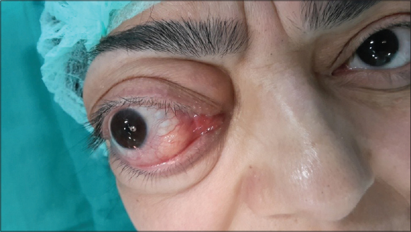
Background:Primary orbital lymphomas are a rare subset of tumors constituting 1–2% of non-Hodgkin's lymphoma. They are mostly indolent B-cell lymphomas presenting with gradual progressive proptosis, decreased visual acuity, restricted ocular mobility, and diplopia. The role of surgery is mainly for obtaining a biopsy. Most of these tumors require multimodality treatment including chemotherapy, radiation, or both, which have major role.
Intramedullary and intratumoral hemorrhage in spinal hemangioblastoma: Case report and review of literature
Date of publication: 04-Dec-2018
Background:Intramedullary hemorrhages involving spinal hemangioblastomas are rare. They are frequently associated with devastating neurologic outcomes, despite with emergent surgical intervention. Here, we presented an example of an intramedullary hemorrhage occurring in a spinal hemangioblastoma, where the patient markedly improved with surgery. Additionally, the appropriate literature was reviewed (including intraoperative video).
Comprehensive review of neuromyelitis optica and clinical characteristics of neuromyelitis optica patients in Puerto Rico
Date of publication: 03-Dec-2018
Abstract
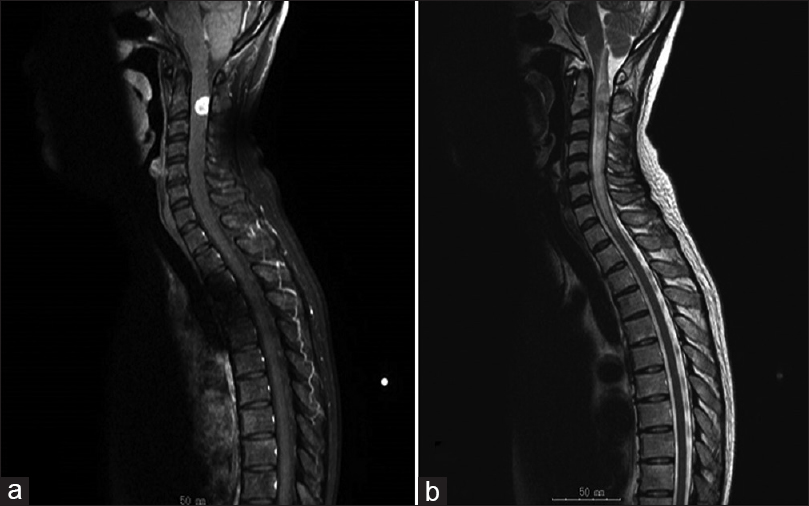
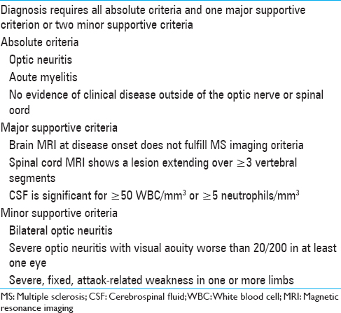
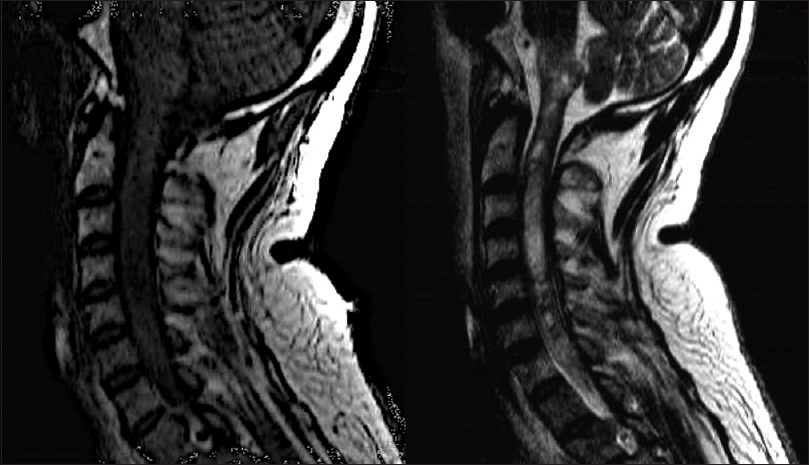
Neuromyelitis optica (NMO) is an immune-mediated inflammatory disorder of the central nervous system. It is characterized by concurrent inflammation and demyelination of the optic nerve (optic neuritis [ON]) and the spinal cord (myelitis). Multiple studies show variations in prevalence, clinical, and demographic features of NMO among different populations. In addition, ethnicity and race are known as important factors on disease phenotype and clinical outcomes. There are little data on information about NMO patients in underserved groups, including Puerto Rico (PR). In this research, we will provide a comprehensive overview of all aspects of NMO, including epidemiology, environmental risk factors, genetic factors, molecular mechanism, symptoms, comorbidities and clinical differentiation, diagnosis, treatment, its management, and prognosis. We will also evaluate the demographic features and clinical phenotype of NMO patients in PR. This will provide a better understanding of NMO and establish a basis of knowledge that can be used to improve care. Furthermore, this type of population-based study can distinguish the clinical features variation among NMO patients and will provide insight into the potential mechanisms that cause these variations.
Cervicomedullary primitive neuroectodermal tumor of the spine: Case report
Date of publication: 28-Nov-2018
Background:Intramedullary primitive neuroectodermal tumors (PNETs) are tumors found rarely in the cervical region, with only five such cases described in the literature. The available literature contains only one report regarding cervicomedullary junction PNET.
Odontoid fracture with missed diagnosis of Transverse Atlantal Ligament (TAL) injury resulting in late-onset instability
Date of publication: 28-Nov-2018
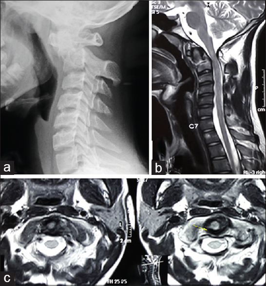
Background:Concurrent injuries to both the odontoid and transverse atlantal ligament are rare and can be easily missed. Failure to diagnose both lesions potentially leads to the late onset of sagittal plane instability and acute myelopathy. Here, we present a patient with an odontoid fracture whose transverse atlantal ligament (TAL) injury was originally missed on magnetic resonance imaging (MRI) and computed tomography (CT) scans. He later developed atlantoaxial instability requiring surgery.

