

An aggressive multidisciplinary approach reduces mortality in rhinocerebral mucormycosis
Date of publication: 25-May-2016
Background:Rhinocerebral mucormycosis occurs in immunocompromised hosts with uncontrolled diabetes, solid organ transplants, and hematologic malignancies. Primary disease is in the paranasal sinuses but often progresses intracranially, via direct extension or angioinvasion. Rhinocerebral mucormycosis is rapidly fatal with a mortality rate of 85%, even when maximally treated with surgical debridement, antifungal therapy, and correction of underlying processes.
Spontaneous shrinkage of vestibular schwannoma
Date of publication: 19-May-2016
Background:“Watch, wait, and rescan” (WWR) has an established place as a successful management option for a significant proportion of vestibular schwannomas (VS) as an alternative to microsurgical removal or stereotactic radiotherapy. VS may grow slowly and continuously, followed by stagnation or even shrinkage. We present two case reports of spontaneous shrinkage of VS along with a review of the literature.
Erratum: A rare intracranial tumor consisting of malignant anaplastic and papillary meningioma subtypes
Date of publication: 19-May-2016
Intramedullary spinal cord metastasis arising from papillary thyroid carcinoma: A case report and review of literature
Date of publication: 17-May-2016
Background:Intramedullary spinal cord metastases (IMSCM) are typically drop lesions from intracranial metastases and are a rare manifestation of systemic malignancy (8.5% of central nervous system metastases). They arise from primaries such as the lungs, breast, kidney, melanoma, or lymphoma. On the other hand, they arise very rarely from papillary thyroid carcinoma (PTC), even though it is the most common type of primary thyroid malignancy.
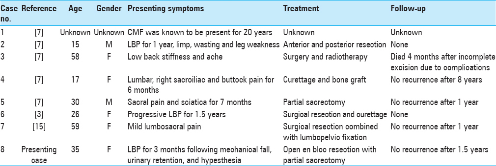
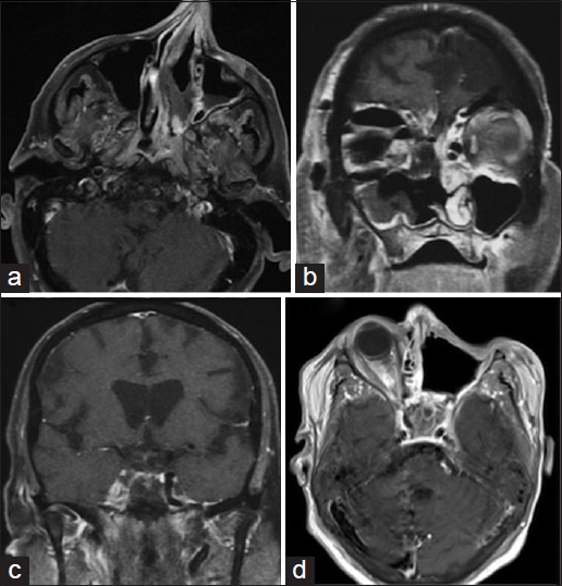
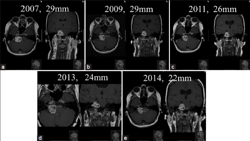
Chondromyxoid fibroma of the sacrum: A case report and literature review
Date of publication: 17-May-2016
Background:Chondromyxoid fibroma (CMF) is an extremely rare, benign cartilaginous tumor that makes up <0.5% of all bone tumors, typically presenting in the second or third decade of life. CMF of the sacrum is exceedingly rare, with only seven documented cases reported in the neurosurgical literature.
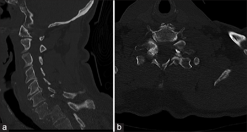
Traumatic spondyloptosis at the cervico-thoracic junction without neurological deficits
Date of publication: 17-May-2016
Background:There have been rare cases of traumatic cervical spondyloptosis without neurological compromise. We report another case and provide a review of the literature, with a focus on appropriate management.

Imaging characteristic analysis of metastatic spine lesions from breast, prostate, lung, and renal cell carcinomas for surgical planning: Osteolytic versus osteoblastic
Date of publication: 17-May-2016
Background:Surgeons treating metastatic spine disease can use computed tomography (CT) imaging to determine whether lesions are osteolytic, osteoblastic, or mixed. This enables treatment that considers the structural integrity of the vertebral body (VB), which is impaired with lytic lesions but not blastic lesions. The authors analyzed CT imaging characteristics of spine metastasis from breast, lung, prostate, and renal cell carcinomas (RCCs) to determine the metastasis patterns of each of these common tumors.
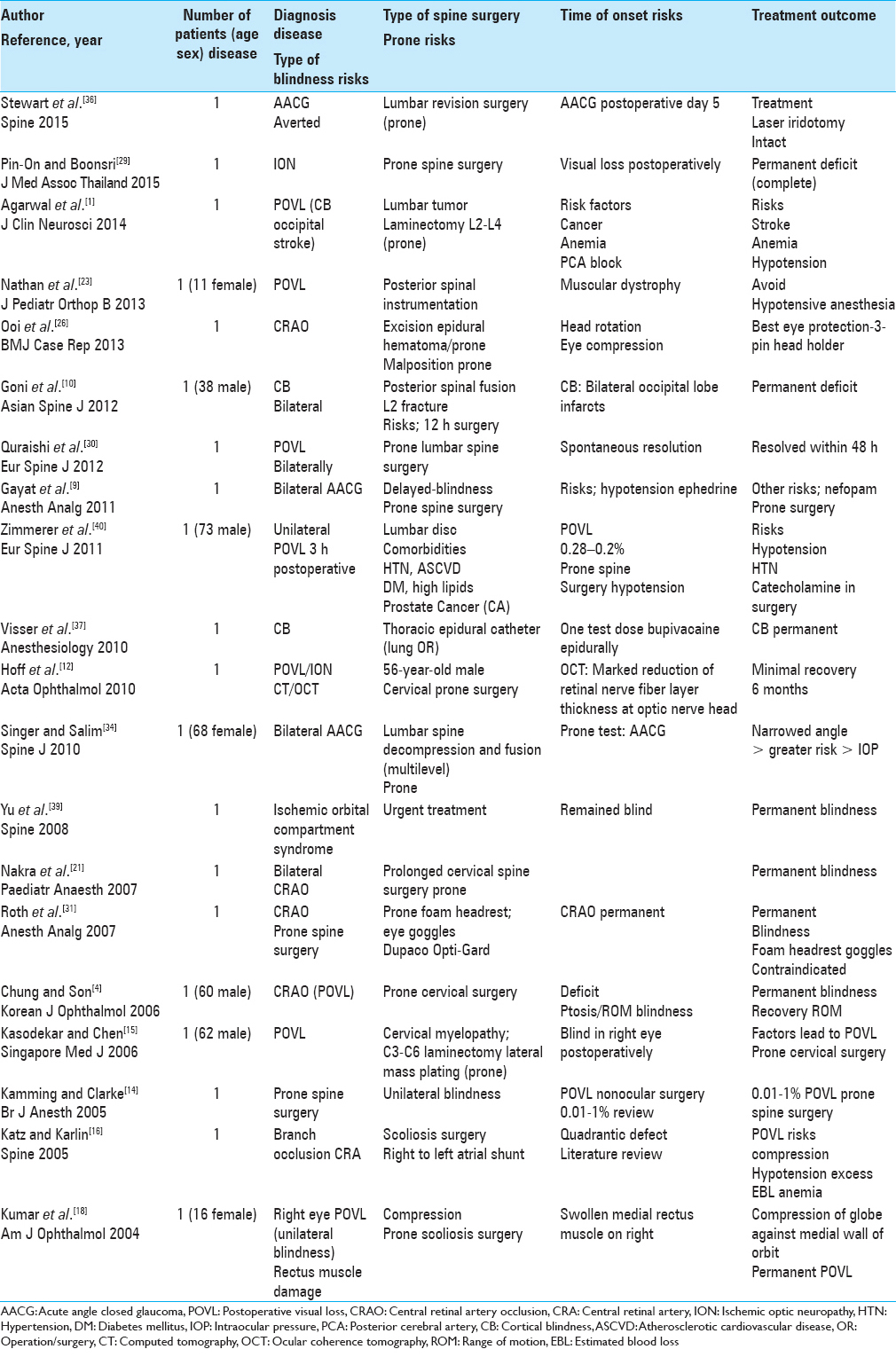
Perioperative visual loss following prone spinal surgery: A review
Date of publication: 17-May-2016
Background:Postoperative visual loss (POVL) following prone spine surgery occurs in from 0.013% to 1% of cases and is variously attributed to ischemic optic neuropathy (ION: anterior ION or posterior ION [reported in 1.9/10,000 cases: constitutes 89% of all POVL cases], central retinal artery occlusion [CRAO], central retinal vein occlusion [CRVO], cortical blindness [CB], direct compression [horseshoe, prone pillows, and eye protectors Dupaco Opti-Gard]), and acute angle closure glaucoma (AACG).

