

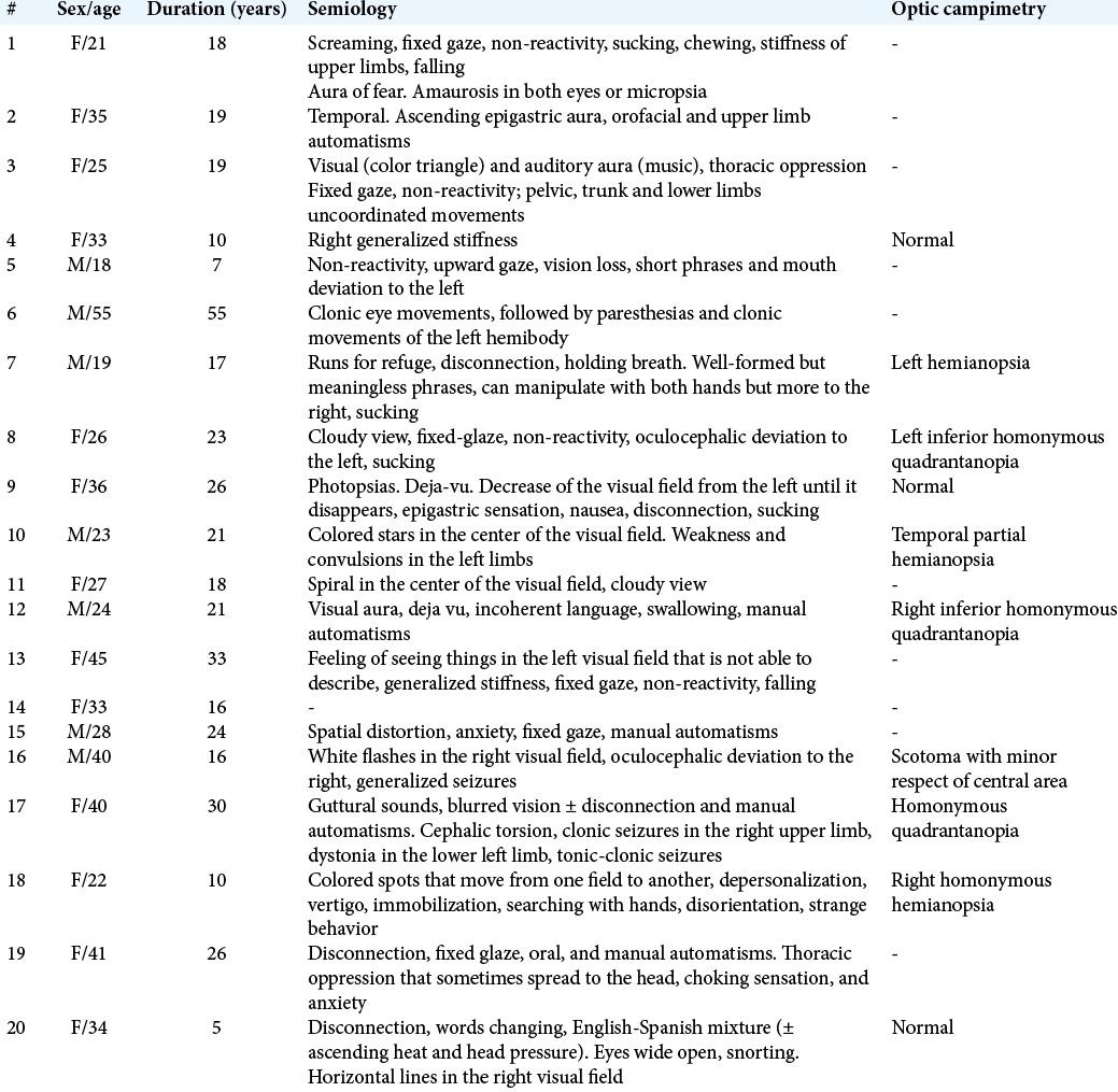
Evolution of patients with surgically treated drug-resistant occipital lobe epilepsy
Date of publication: 01-Aug-2020
Background: This study was to describe the evolution of patients who underwent surgical treatment of drug- resistant occipital lobe epilepsy (OLE) at our institution.
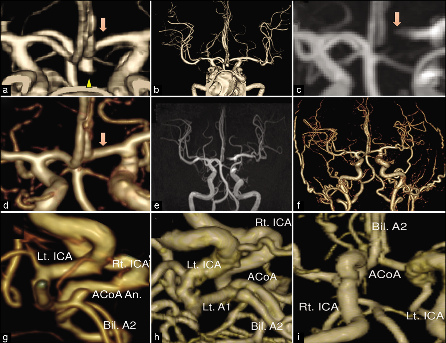
Microsurgical embolectomy with superficial temporal artery-middle cerebral artery bypass for acute internal carotid artery dissection: A technical case report
Date of publication: 01-Aug-2020
Background: Dissection of the internal carotid artery (ICA) is an important cause of stroke. Intravenous alteplase administration and mechanical thrombectomy have been strongly recommended for selected patients with acute ischemic stroke. However, the efficacy and safety of these treatments for ischemic stroke due to ICA dissection remain unclear. Here, we report a case of acute ICA dissection successfully treated by microsurgical embolectomy.
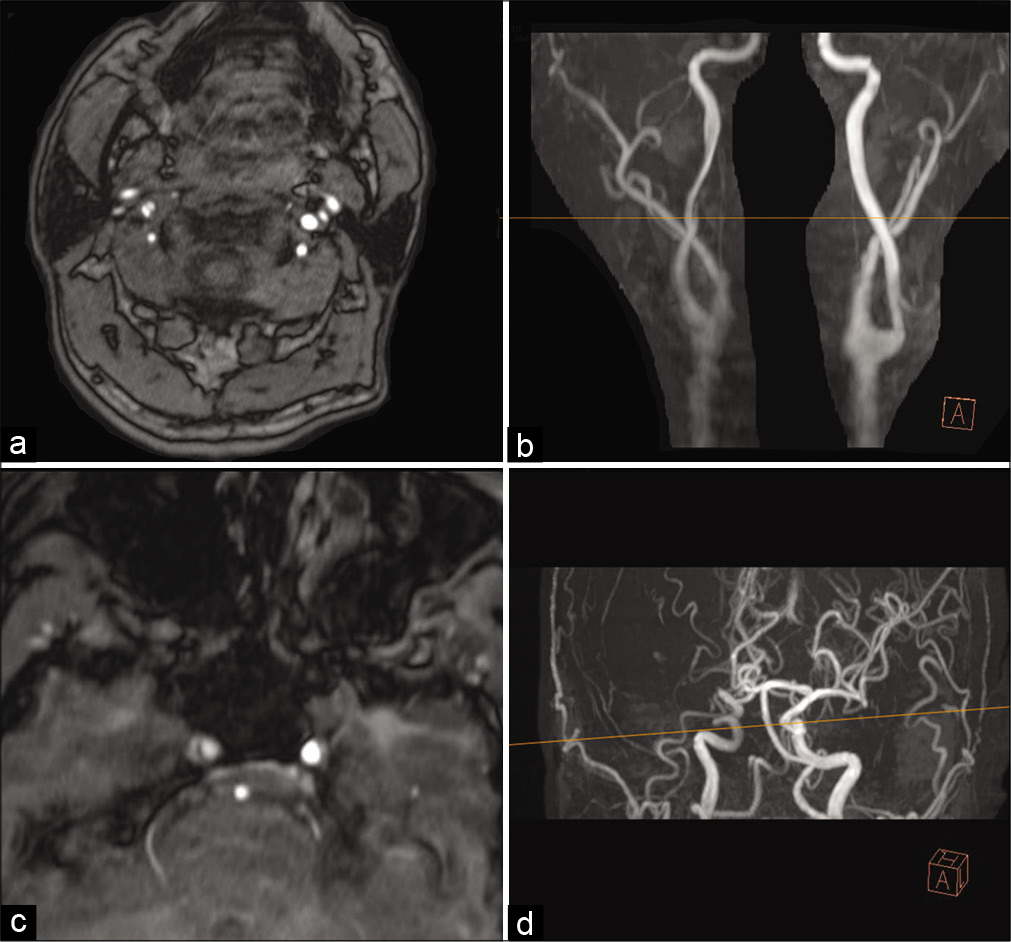
Three tesla magnetic resonance angiography with ultrashort echo time describes the arteries near the cerebral aneurysm with clip and the peripheral cerebral arteries
Date of publication: 01-Aug-2020
Background: The assessment of the clipped cerebral aneurysm and the cerebral arteries after the treatment of subarachnoid hemorrhage (SAH) is important to find aneurysm regrowth or postoperative cerebral vasospasm. Usually, contrast-enhanced computed tomography angiography is performed for the evaluation of the arteries, but it has side effects of contrast medium. Time-of-flight magnetic resonance angiography (MRA) is a fast and non-invasive method, but clip-induced artifact limits assessment of the artery in the vicinity of the clip. 1.5T MRA with ultrashort echo time (UTE) reduces metal artifact, but the obtained image is too rough to evaluate the aneurysm remnant, and the description range is too narrow to assess the cerebral vasospasm. We routinely use SIGNA Pioneer 3.0T (GE Healthcare Life Sciences, Buckinghamshire, England) and perform SILENT SCAN with UTE-MRA for the postoperative assessment of the clipped aneurysm and cerebral arteries for SAH patients treated by clipping. It has better image quality and describes arteries with a wide description range, so it possesses the potential to overcome the disadvantages of 1.5T UTE-MRA.
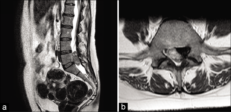
Cauda equina syndrome caused by lumbar leptomeningeal metastases from lung adenocarcinoma mimicking a schwannoma
Date of publication: 01-Aug-2020
Background: Cauda equina syndromes (CESs) due to leptomeningeal metastases from primitive lung tumors are rare. Despite recent advancements in neuro-oncology and molecular biology, the prognosis for these patients remains poor. Here, we present a case in which a patient developed lumbar leptomeningeal metastases from lung carcinoma that contributed to a CES and reviewed the appropriate literature.
Causes of hospital readmissions within 7 days from the neurosurgical service of a quaternary referral hospital
Date of publication: 01-Aug-2020
Background: Evaluation of readmission rates as a proxy metric of health-care quality in neurological surgery has grown to become a prevalent area of investigation in the last several years. Significant attention has been paid to 30-day readmission rates due to the financial incentive to health-care providers following the enforcement of the penalties created by the Affordable Care Act. However, relatively little attention has been paid to patients readmitted within 7 days of discharge to large quaternary neurological surgery services. This study was conducted to examine the causes and unique characteristics of 7-day readmission rates from a neurosurgical service at a large quaternary referral hospital.
Pure endoscopic transsphenoidal treatment of skull base ameloblastoma with intracranial extension: Case report and literature review
Date of publication: 01-Aug-2020
Background: Ameloblastoma is a benign locally invasive lesion that represents 1% of all oral tumors. Epidemiological characteristics are variable in the literature. The most common origin sites are mandible and maxilla. Rarely presents metastasis, but the skull base, lymph nodes, and the lung are described as metastatic sites. Low recurrence rates were reported by the authors when surgical treatment achieved complete resection.
Double neurophysiological certification of the filum terminale during sectioning surgery in pediatric population
Date of publication: 01-Aug-2020
Background: Surgery of thickened-fibrolipoma filum terminale (FT) is performed routinely and without conflict but is not a risk-free surgical procedure. Intraoperative neurophysiological monitoring with mapping techniques can help to certify the FT before sectioning. However, a tailored surgical approach to cauda equina and a low threshold of surrounding nerve roots can confuse the final surgical decision. The aim is to demonstrate the usefulness of this double methodology for FT certification.
Surgical removal of a spinal intrathecal projectile led to a significant improvement of cauda equina syndrome
Date of publication: 01-Aug-2020
Background: Penetrating gunshot wounds of the spine are common and can cause severe neurological deficits. However, there are no guidelines as to their optimal treatment. Here, we present a penetrating injury to the lower thoracic spine at the T12 level that lodged within the canal at L1, resulting in a cauda equina syndrome. Notably, the patient’s deficit resolved following bullet removal.
Elective inferior temporal lobe resection as an adjunct to subtemporal approach for a case of tentorial meningioma arising from the middle part of the free edge of the tentorium: A case report
Date of publication: 01-Aug-2020
Background: Tentorial meningiomas attached to the inner edge of the tentorium are difficult to excise due to their deep location. Sufficient space may not be always available through a subtemporal approach. Thus, the aim of not retracting the brain is not fulfilled.
Subdural empyema caused by Morganella morganii
Date of publication: 01-Aug-2020
Background: Morganella morganii is a species of Gram-negative enteric rod found in normal human gut flora. Pathologically, this most often presents as urinary tract infections, wound infections, and bacteremia. It is highly uncommon for M. morganii to be implicated in a central nervous system infection, with only 12 reported cases of parenchymal abscesses or meningitis.

