

Cerebrospinal fluid drains reduce risk of spinal cord injury for thoracic/thoracoabdominal aneurysm surgery: A review
Date of publication: 23-Feb-2018
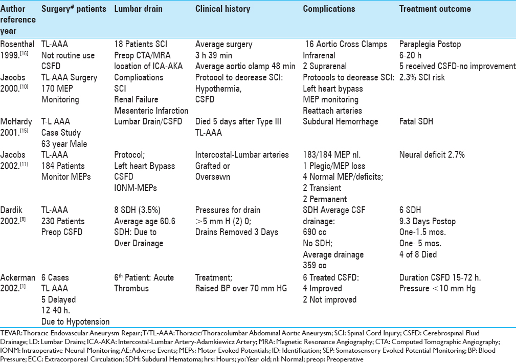
Background:The risk of spinal cord injury (SCI) due to decreased cord perfusion following thoracic/thoracoabdominal aneurysm surgery (T/TL-AAA) and thoracic endovascular aneurysm repair (TEVAR) ranges up to 20%. For decades, therefore, many vascular surgeons have utilized cerebrospinal fluid drainage (CSFD) to decrease intraspinal pressure and increase blood flow to the spinal cord, thus reducing the risk of SCI/ischemia.
Randomized clinical trials in carpal tunnel release: A double-edged sword
Date of publication: 21-Feb-2018
Acute-on-chronic subdural hematoma in a patient taking Red Clover herbal supplement: A case report
Date of publication: 21-Feb-2018
Background:Herbal supplements are commonly used, however, their side-effect profiles are poorly understood and not subject to the same scrutiny as prescribed medications. Some herbal supplements such as St Johns’ Wort are accepted to interfere with clotting pathways, however others, including Red Clover have theoretical bleeding risks based on coumarin content with very little underlying evidence.
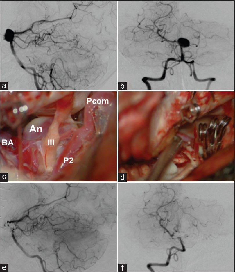
Should we still consider clips for basilar apex aneurysms? A critical appraisal of the literature
Date of publication: 21-Feb-2018
Background:Basilar apex aneurysms constitute 5–8% of all intracranial aneurysms, and their treatment remains challenging for both microsurgical and endovascular approaches. The perceived drawback of the microsurgical approach is its invasiveness leading to increased surgical morbidity. However, many high-volume centers have shown excellent clinical results with better occlusion rates compared to endovascular treatment. With endovascular therapy taking a larger role in the management of cerebral aneurysms, the future role of microsurgery for basilar apex aneurysm treatment is unclear.
Primary intracerebral INI1-deficient rhabdoid tumor with CD34 immunopositivity in a young adult
Date of publication: 21-Feb-2018
Background:Primary CNS malignant rhabdoid tumors are very rare in adults and much less is known about their biological behavior than in children. Recently, two adult cases of SMARCB1 (also known as INI1)-deficient tumor with rhabdoid cells have been described, suggesting an emerging group of primary meningeal SMARCB1-deficient tumors. We have recently encountered a case of INI1-deficient tumor with similar histology and immunophenotype to the above cases, but with a superficial cerebral, yet apparent intra-axial origin.
Axial and oblique C2 pedicle diameters and feasibility of C2 pedicle screw placement: Technical note
Date of publication: 16-Feb-2018
Background:For C2 pedicle screw placement/instrumentation, it is critical to adequately measure the axial and oblique C2 pedicle diameters utilizing the intraoperative O-arm.
Nursing review of spinal meningiomas
Date of publication: 16-Feb-2018
Background:Spinal meningiomas are found in patients typically between the ages of 75 and 84: some report the average age to be 50. They occur with an incidence of approximately 1000 patients per year in the US, are mostly single (90%) rather than multiple (10%), and arise from the spinal meninges (arachnoid/dura). Tumors are typically posterior/posterolateral (70%) in location, leaving the remaining 30% in the anterior/anterolateral spinal canal. They produce symptoms and signs of radiculopathy (nerve root) and/or myelopathy (cord compression) depending on their site of origin.
IDH1, ATRX, and BRAFV600E mutation in astrocytic tumors and their significance in patient outcome in north Indian population
Date of publication: 14-Feb-2018
Background:According to the current World Health Organization (WHO) classification of central nervous system (CNS) tumors (2016), histological diagnosis of gliomas should be supplemented by molecular information. This study was carried out to determine the frequency of isocitrate dehydrogenase 1 (IDH1), ATRX, and BRAF V600E mutations in different grade astrocytomas and their prognostic value.
Enlargement of an incidental internal carotid artery aneurysm embedded in pituitary adenoma associated with medical shrinkage of the tumor: Case report
Date of publication: 14-Feb-2018
Background:Currently, transsphenoidal surgery (TSS) is the preferred method for surgical treatment of intrasellar pituitary adenomas. However, it carries some risk of intraoperative arterial injuries, which is mainly attributed to direct iatrogenic rupture of the internal carotid artery (ICA). There is anecdotal evidence suggesting that intracranial aneurysms are coincidentally found significantly more frequently in the setting of pituitary adenomas than when the incidence is compared to other intracranial neoplasms. The exact cause of this discrepancy remains unclear, but it certainly raises concerns about the potential existence of an ICA aneurysm, which might be encountered during TSS and in some cases may cause hemorrhagic complications.
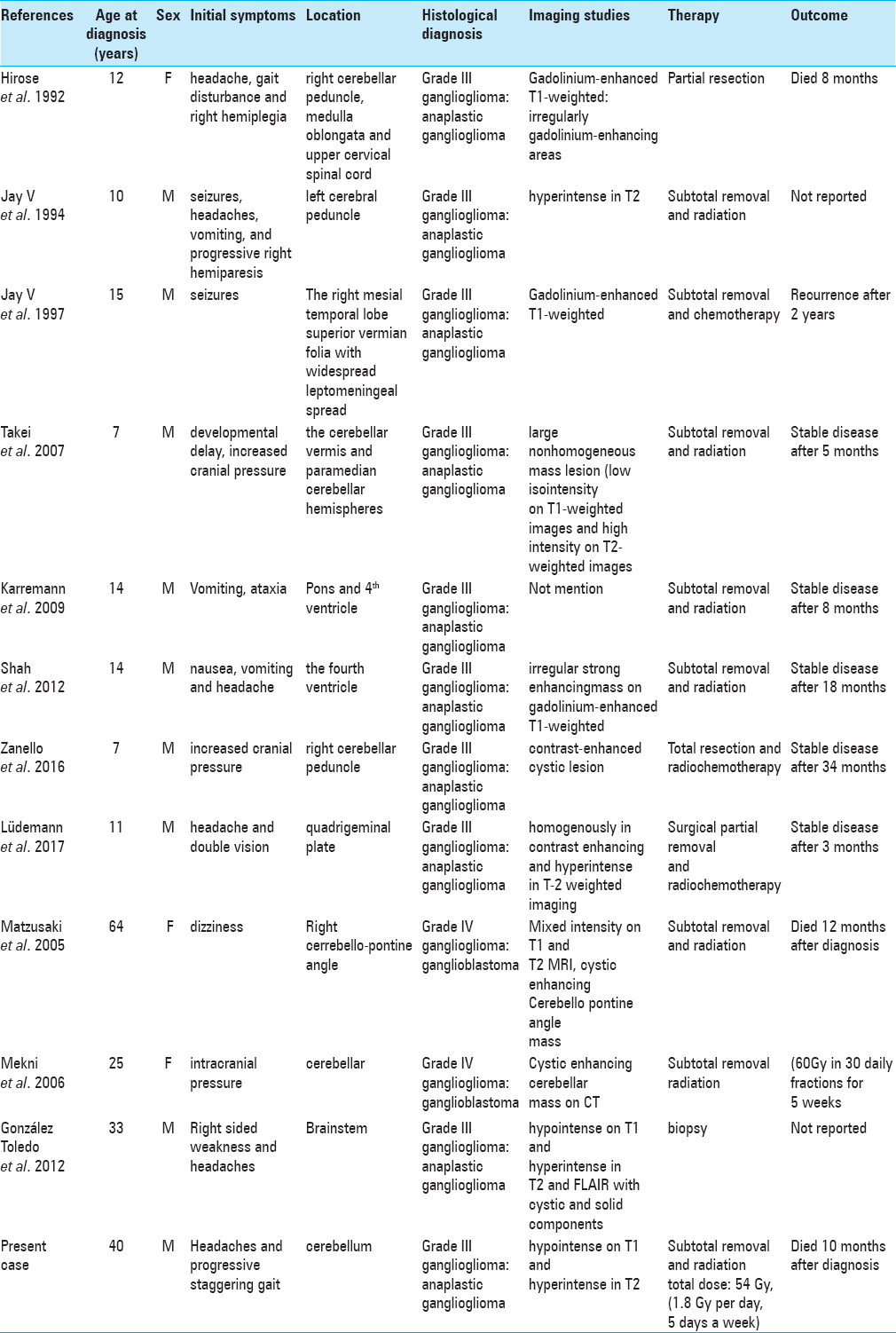
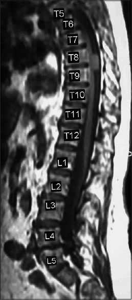
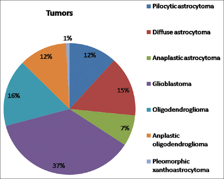
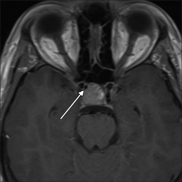
A case of adult anaplastic cerebellar ganglioglioma
Date of publication: 14-Feb-2018
Background:Anaplastic posterior fossa ganglioglioma in adults is exceedingly rare. To date, only one case of adult anaplastic posterior fossa ganglioglioma has been reported in the English literature and none has been described at the cerebellum. To our knowledge, this report is the third case of malignant posterior fossa ganglioglioma in adults and the first at the cerebellum. In general, this entity can be misdiagnosed preoperatively as a primary posterior fossa neoplasm, and by reporting our clinical and radiographic observations we want to add to the existing literature on this rare entity.

