

Erratum: Exploring the Virchow–Robin spaces function: A unified theory of brain diseases
Date of publication: 28-Nov-2016
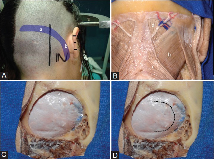
Abordaje a la Cisterna Ambiens
Date of publication: 21-Nov-2016
Objetivo:Describir paso a paso el abordaje a la cisterna ambiens por la vía suboccipital retrosigmoidea supracerebelosa infratentorial (SRSI).
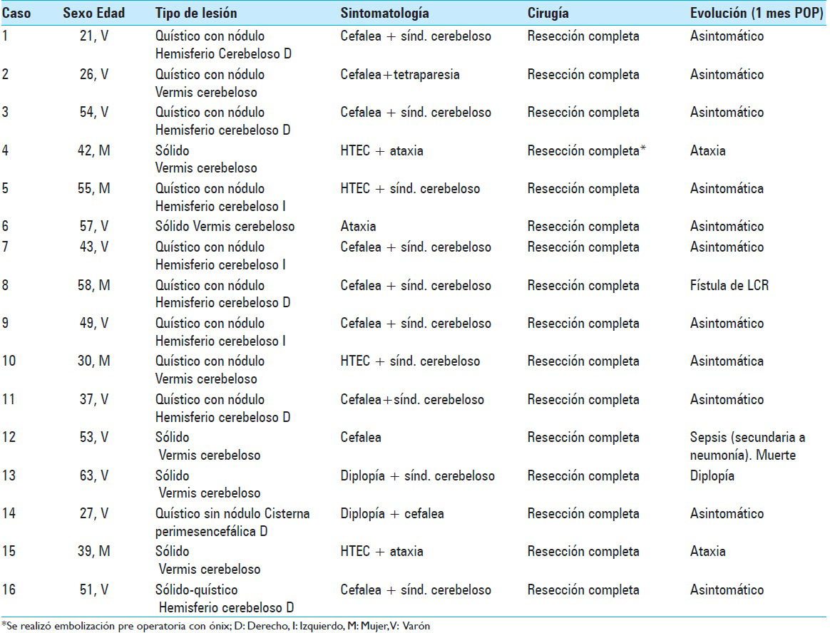
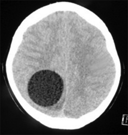
Hemangioblastomas de fosa posterior: Reporte de 16 casos y revisión de la literatura
Date of publication: 21-Nov-2016
Objetivo:El propósito del presente trabajo es presentar los resultados de 16 pacientes con diagnóstico de hemangioblastoma de fosa posterior (HBFP), operados con técnicas microquirúrgicas.
Secretory paraspinal paraganglioma of thoracolumar spine: Case report and review of literature
Date of publication: 21-Nov-2016
Background:Pheochromocytomas are catecholamine secreting tumors of the adrenal medulla chromaffin cells, however, when present extra-adrenally they are called paragangliomas. Paragangliomas rarely produce catecholamine in excess, which is evident by clinical symptoms, urine, and blood biochemistry. Total resection of these tumors can lead to complete clinical and biochemical resolution. This case report presents the clinical features, radiological findings, and neurological outcome in a middle-aged female with a secretory paraganglioma.
Hypervascular glioblastoma multiforme or arteriovenous malformation associated Glioma? A diagnostic and therapeutic challenge: A case report
Date of publication: 21-Nov-2016
Background:Simultaneous presentation of arteriovenous malformation (AVM) and glioblastoma multiforme (GBM) is rarely reported in the literature and needs to be differentiated from “angioglioma”, a highly vascular glioma and other differential diagnosis such as hypervascular glioblastoma. Incorporating critical features of both, malignant glioma and AVM, such lesions lack a standard algorithm for diagnosis and therapy due to their rare incidence as well as their complex radiological and highly individualized clinical presentation.
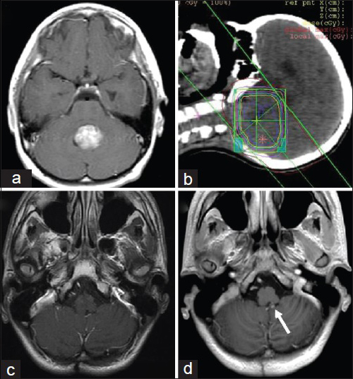
Intracranial aneurysm formation after radiotherapy for medulloblastoma
Date of publication: 21-Nov-2016
Background:The development of an intracranial aneurysm after radiotherapy is rare but secondary effect of cranial irradiation in a primary disease treatment.
Fibrin glue injection into the hematoma cavity for refractory chronic subdural hematoma: A case report
Date of publication: 21-Nov-2016
Background:Repeat burr hole irrigation and drainage has been effective in most cases of recurrent chronic subdural hematoma (CSDH), however, refractory cases require further procedures or other interventions.
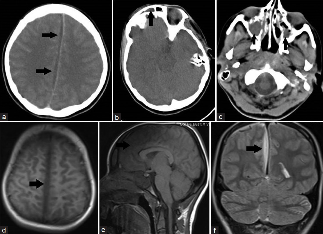
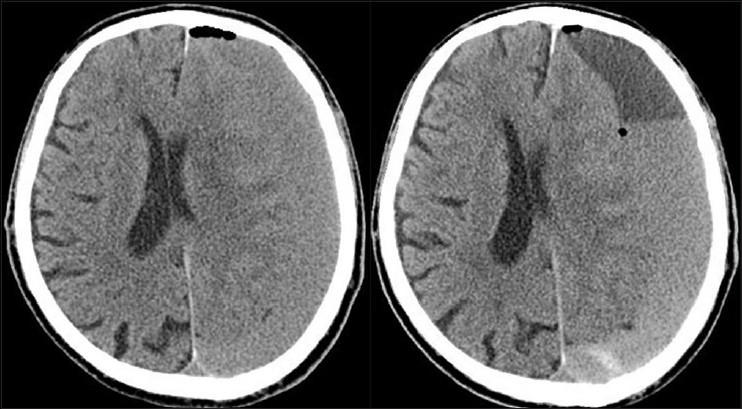
Rapidly progressing interhemispheric subdural empyema showing a three-fold increase in size within 12 hours: Case report
Date of publication: 21-Nov-2016
Background:Subdural empyema is a rare form of intracranial infection. It is described as accumulation of purulent infective material between the inner layer of dura mater and outer layer of arachnoid membrane.
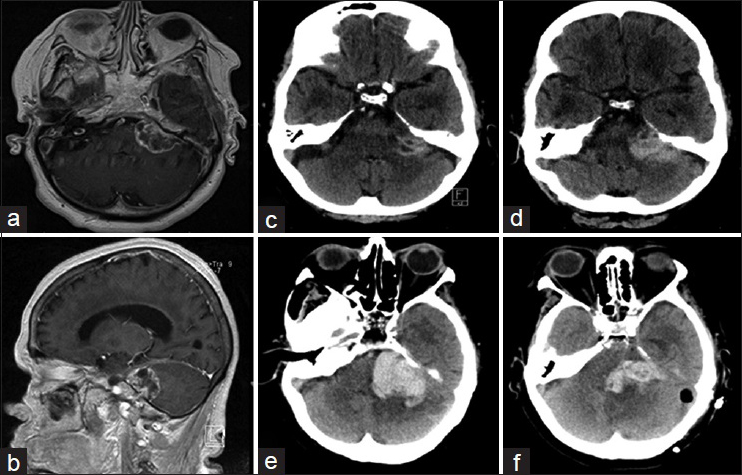
An unusual case of repeated intracranial hemorrhage in vestibular schwannoma
Date of publication: 21-Nov-2016
Background:Symptomatic intratumoral hemorrhage (ITH) in vestibular schwannoma (VS) is rare. A repeated hemorrhage is, therefore, even more exceptional. Repeated ITH has been reported in four cases thus far in English literature. Here, we describe a patient with a Koos grade D VS who presented to our Skull Base team with repeated ITH and an unexpected disease course.
Primary cerebral echinoccocosis in a child: Case report – Surgical technique, technical pitfalls, and video atlas
Date of publication: 21-Nov-2016
Background:Hydatid disease is a life-threatening parasitic infestation caused by Echinococcus granulosus. Infection with E. granulosus typically results in the formation of hydatid cysts in the liver, lungs, kidney, and spleen. Primary intracranial hydatid cyst disease is extremely rare. Here, we are reporting an unusual case of Echinococcus, where the only identifiable lesion was a hydatid cyst in the brain without liver or lung involvement. We are also providing a description for the surgical technique used to remove the cyst, highlighting the possible surgical pitfalls.

